Dorsal root ganglion

Last updated
Dorsal root ganglion
DRG Chicken e7.jpg
A dorsal root ganglion (DRG) from a chicken embryo (around stage of day 7) after incubation overnight in NGF growth medium stained with anti-neurofilament antibody. Neurites growing out of the ganglion are visible.
Gray675.png
A spinal nerve with its ventral and dorsal roots. The dorsal root ganglion is the "spinal ganglion", following the dorsal root.
Details
Precursor Neural crest
Identifiers
Latin ganglion sensorium nervi spinalis
MeSH D005727
TA98 A14.2.00.006
TA2 6167
FMA 5888
Anatomical terminology

A dorsal root ganglion (or spinal ganglion; also known as a posterior root ganglion [1] ) is a cluster of neurons (a ganglion) in a dorsal root of a spinal nerve. The cell bodies of sensory neurons known as first-order neurons are located in the dorsal root ganglia. [2]

Contents

The axons of dorsal root ganglion neurons are known as afferents. In the peripheral nervous system, afferents refer to the axons that relay sensory information into the central nervous system (i.e. the brain and the spinal cord).

Structure

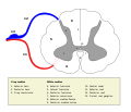
The neurons comprising the dorsal root ganglion are of the pseudo-unipolar type, meaning they have a cell body (soma) with two branches that act as a single axon, often referred to as a distal process and a proximal process.

Unlike the majority of neurons found in the central nervous system, an action potential in posterior root ganglion neuron may initiate in the distal process in the periphery, bypass the cell body, and continue to propagate along the proximal process until reaching the synaptic terminal in the posterior horn of spinal cord.

Distal section

The distal section of the axon may either be a bare nerve ending or encapsulated by a structure that helps relay specific information to nerve. Two examples where the nerve ending of the distal process is encapsulated as such are, Meissner's corpuscles, which render the distal processes of mechanosensory neurons sensitive to stroking only, and Pacinian corpuscles, which make neurons more sensitive to vibration. [3]

Location

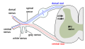
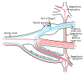
The dorsal root ganglia lie in the intervertebral foramina. The anterior and posterior spinal nerve roots join just beyond (lateral) to the location of the dorsal root ganglion.

Development

The dorsal root ganglia develop in the embryo from neural crest cells, not neural tube. Hence, the spinal ganglia can be regarded as gray matter of the spinal cord that became translocated to the periphery.

Function

Nociception

Proton-sensing G protein-coupled receptors are expressed by DRG sensory neurons and might play a role in acid-induced nociception. [4]

Mechanosensitive channels

The nerve endings of dorsal root ganglion neurons have a variety of sensory receptors that are activated by mechanical, thermal, chemical, and noxious stimuli. [5] In these sensory neurons, a group of ion channels thought to be responsible for somatosensory transduction have been identified. Compression of the dorsal root ganglion by a mechanical stimulus lowers the voltage threshold needed to evoke a response and causes action potentials to be fired. [6] This firing may even persist after the removal of the stimulus. [6]

Two distinct types of mechanosensitive ion channels have been found in the posterior root ganglion neurons. The two channels are broadly classified as either high-threshold (HT) or low threshold (LT). [5] As their names suggest, they have different thresholds as well as different sensitivities to pressure. These are cationic channels whose activity appears to be regulated by the proper functioning of the cytoskeleton and cytoskeleton associated proteins. [5] The presence of these channels in the posterior root ganglion gives reason to believe that other sensory neurons may contain them as well.

High-threshold mechanosensitive channels

High-threshold channels have a possible role in nociception. These channels are found predominantly in smaller sensory neurons in the dorsal root ganglion cells and are activated by higher pressures, two attributes that are characteristic of nociceptors. [5] Also, the threshold of HT channels was lowered in the presence of PGE2 (a compound that sensitizes neurons to mechanical stimuli and mechanical hyperalgesia) which further supports a role for HT channels in the transduction of mechanical stimuli into nociceptive neuronal signals. [5] [6] [7]

Presynaptic control

The presynaptic regulation of the dorsal nerve ending discharge in the spinal cord can occur through certain types of GABAA receptors but not through the activation of glycine receptors which are absent from these types of terminals. Thus GABAA receptors but not glycine receptors can presynaptically control nociception and pain transmission. [8]

See also

Related Research Articles

In physiology, nociception, also nocioception; from Latin nocere 'to harm/hurt') is the sensory nervous system's process of encoding noxious stimuli. It deals with a series of events and processes required for an organism to receive a painful stimulus, convert it to a molecular signal, and recognize and characterize the signal to trigger an appropriate defensive response.

<span class="mw-page-title-main">Trigeminal nerve</span> Cranial nerve responsible for the faces senses and motor functions

In neuroanatomy, the trigeminal nerve (lit. triplet nerve), also known as the fifth cranial nerve, cranial nerve V, or simply CN V, is a cranial nerve responsible for sensation in the face and motor functions such as biting and chewing; it is the most complex of the cranial nerves. Its name (trigeminal, from Latin tri- 'three', and -geminus 'twin') derives from each of the two nerves (one on each side of the pons) having three major branches: the ophthalmic nerve (V1), the maxillary nerve (V2), and the mandibular nerve (V3). The ophthalmic and maxillary nerves are purely sensory, whereas the mandibular nerve supplies motor as well as sensory (or "cutaneous") functions. Adding to the complexity of this nerve is that autonomic nerve fibers as well as special sensory fibers (taste) are contained within it.

<span class="mw-page-title-main">Afferent nerve fiber</span> Axonal projections that arrive at a particular brain region

Afferent nerve fibers are axons of sensory neurons that carry sensory information from sensory receptors to the central nervous system. Many afferent projections arrive at a particular brain region.

<span class="mw-page-title-main">Grey column</span>

The grey column refers to a somewhat ridge-shaped mass of grey matter in the spinal cord. This presents as three columns: the anterior grey column, the posterior grey column, and the lateral grey column, all of which are visible in cross-section of the spinal cord.

<span class="mw-page-title-main">Nociceptor</span> Sensory neuron that detects pain

A nociceptor is a sensory neuron that responds to damaging or potentially damaging stimuli by sending "possible threat" signals to the spinal cord and the brain. The brain creates the sensation of pain to direct attention to the body part, so the threat can be mitigated; this process is called nociception.

<span class="mw-page-title-main">Spinothalamic tract</span> Sensory pathway from the skin to the thalamus

The spinothalamic tract is a part of the anterolateral system or the ventrolateral system, a sensory pathway to the thalamus. From the ventral posterolateral nucleus in the thalamus, sensory information is relayed upward to the somatosensory cortex of the postcentral gyrus.

<span class="mw-page-title-main">Dorsal column–medial lemniscus pathway</span> Sensory spinal pathway

The dorsal column–medial lemniscus pathway (DCML) is a sensory pathway of the central nervous system that conveys sensations of fine touch, vibration, two-point discrimination, and proprioception from the skin and joints. It transmits information from the body to the primary somatosensory cortex in the postcentral gyrus of the parietal lobe of the brain. The pathway receives information from sensory receptors throughout the body, and carries this in nerve tracts in the white matter of the dorsal column of the spinal cord to the medulla, where it is continued in the medial lemniscus, on to the thalamus and relayed from there through the internal capsule and transmitted to the somatosensory cortex. The name dorsal-column medial lemniscus comes from the two structures that carry the sensory information: the dorsal columns of the spinal cord, and the medial lemniscus in the brainstem.

<span class="mw-page-title-main">Sensory neuron</span> Nerve cell that converts environmental stimuli into corresponding internal stimuli

Sensory neurons, also known as afferent neurons, are neurons in the nervous system, that convert a specific type of stimulus, via their receptors, into action potentials or graded receptor potentials. This process is called sensory transduction. The cell bodies of the sensory neurons are located in the dorsal ganglia of the spinal cord.

<span class="mw-page-title-main">Pseudounipolar neuron</span>

A pseudounipolar neuron is a type of neuron which has one extension from its cell body. This type of neuron contains an axon that has split into two branches. A single process arises from the cell body and then divides into an axon and a dendrite. They develop embryologically as bipolar in shape, and are thus termed pseudounipolar instead of unipolar.

<span class="mw-page-title-main">Dorsal root of spinal nerve</span>

The dorsal root of spinal nerve is one of two "roots" which emerge from the spinal cord. It emerges directly from the spinal cord, and travels to the dorsal root ganglion. Nerve fibres with the ventral root then combine to form a spinal nerve. The dorsal root transmits sensory information, forming the afferent sensory root of a spinal nerve.

<span class="mw-page-title-main">Allodynia</span> Feeling of pain from stimuli which do not normally elicit pain

Allodynia is a condition in which pain is caused by a stimulus that does not normally elicit pain. For example, sunburn can cause temporary allodynia, so that usually painless stimuli, such as wearing clothing or running cold or warm water over it, can be very painful. It is different from hyperalgesia, an exaggerated response from a normally painful stimulus. The term comes from Ancient Greek άλλος (állos) 'other', and οδύνη (odúnē) 'pain'.

<span class="mw-page-title-main">General visceral afferent fiber</span> Part of the visceral nervous system

The general visceral afferent (GVA) fibers conduct sensory impulses from the internal organs, glands, and blood vessels to the central nervous system. They are considered to be part of the visceral nervous system, which is closely related to the autonomic nervous system, but 'visceral nervous system' and 'autonomic nervous system' are not direct synonyms and care should be taken when using these terms. Unlike the efferent fibers of the autonomic nervous system, the afferent fibers are not classified as either sympathetic or parasympathetic.

<span class="mw-page-title-main">Satellite glial cell</span> SINGLE CELL SOMATA

Satellite glial cells, formerly called amphicytes, are glial cells that cover the surface of neuron cell bodies in ganglia of the peripheral nervous system. Thus, they are found in sensory, sympathetic, and parasympathetic ganglia. Both satellite glial cells (SGCs) and Schwann cells are derived from the neural crest of the embryo during development. SGCs have been found to play a variety of roles, including control over the microenvironment of sympathetic ganglia. They are thought to have a similar role to astrocytes in the central nervous system (CNS). They supply nutrients to the surrounding neurons and also have some structural function. Satellite cells also act as protective, cushioning cells. Additionally, they express a variety of receptors that allow for a range of interactions with neuroactive chemicals. Many of these receptors and other ion channels have recently been implicated in health issues including chronic pain and herpes simplex. There is much more to be learned about these cells, and research surrounding additional properties and roles of the SGCs is ongoing.

Cutaneous innervation refers to an area of the skin which is supplied by a specific cutaneous nerve.

<span class="mw-page-title-main">Group C nerve fiber</span> One of three classes of nerve fiber in the central nervous system and peripheral nervous system

Group C nerve fibers are one of three classes of nerve fiber in the central nervous system (CNS) and peripheral nervous system (PNS). The C group fibers are unmyelinated and have a small diameter and low conduction velocity, whereas Groups A and B are myelinated. Group C fibers include postganglionic fibers in the autonomic nervous system (ANS), and nerve fibers at the dorsal roots. These fibers carry sensory information.

Mechanosensation is the transduction of mechanical stimuli into neural signals. Mechanosensation provides the basis for the senses of light touch, hearing, proprioception, and pain. Mechanoreceptors found in the skin, called cutaneous mechanoreceptors, are responsible for the sense of touch. Tiny cells in the inner ear, called hair cells, are responsible for hearing and balance. States of neuropathic pain, such as hyperalgesia and allodynia, are also directly related to mechanosensation. A wide array of elements are involved in the process of mechanosensation, many of which are still not fully understood.

<span class="mw-page-title-main">Spinal cord</span> Long, tubular central nervous system structure in the vertebral column

The spinal cord is a long, thin, tubular structure made up of nervous tissue that extends from the medulla oblongata in the brainstem to the lumbar region of the vertebral column (backbone) of vertebrate animals. The center of the spinal cord is hollow and contains a structure called the central canal, which contains cerebrospinal fluid. The spinal cord is also covered by meninges and enclosed by the neural arches. Together, the brain and spinal cord make up the central nervous system.

Group A nerve fibers are one of the three classes of nerve fiber as generally classified by Erlanger and Gasser. The other two classes are the group B nerve fibers, and the group C nerve fibers. Group A are heavily myelinated, group B are moderately myelinated, and group C are unmyelinated.

<span class="mw-page-title-main">Outline of the human nervous system</span> Overview of and topical guide to the human nervous system

The following diagram is provided as an overview of and topical guide to the human nervous system:

<span class="mw-page-title-main">Presynaptic inhibition</span>

Presynaptic inhibition is a phenomenon in which an inhibitory neuron provides synaptic input to the axon of another neuron to make it less likely to fire an action potential. Presynaptic inhibition occurs when an inhibitory neurotransmitter, like GABA, acts on GABA receptors on the axon terminal. Or when endocannabinoids act as retrograde messengers by binding to presynaptic CB1 receptors, thereby indirectly modulating GABA and the excitability of dopamine neurons by reducing it and other presynaptic released neurotransmitters. Presynaptic inhibition is ubiquitous among sensory neurons.

References

  1. "Ganglion". Physiopedia. Retrieved 2021-05-15.
  2. Purves, Dale; Augustine, George J.; Fitzpatrick, David; Katz, Lawrence C.; LaMantia, Anthony-Samuel; McNamara, James O.; Williams, S. Mark (2001). "The Major Afferent Pathway for Mechanosensory Information: The Dorsal Column-Medial Lemniscus System". Neuroscience. 2nd edition. Retrieved 30 May 2018.
  3. Kandel ER, Schwartz JH, Jessell TM. Principles of Neural Science , 4th ed., p.431–433. McGraw-Hill, New York (2000). ISBN   0-8385-7701-6
  4. Huang CW, Tzeng JN, Chen YJ, Tsai WF, Chen CC, Sun WH (2007). "Nociceptors of dorsal root ganglion express proton-sensing G-protein-coupled receptors" (PDF). Mol. Cell. Neurosci. 36 (2): 195–210. doi:10.1016/j.mcn.2007.06.010. PMID   17720533. S2CID   38351962.
  5. 1 2 3 4 5 Cho, H.; Shin, J.; Shin, C. Y.; Lee, S. Y.; Oh, U. (2002). "Mechanosensitive ion channels in cultured sensory neurons of neonatal rats". The Journal of Neuroscience. 22 (4): 1238–1247. doi:10.1523/JNEUROSCI.22-04-01238.2002. PMC   6757581 . PMID   11850451.
  6. 1 2 3 Sugawara, O.; Atsuta, Y.; Iwahara, T.; Muramoto, T.; Watakabe, M.; Takemitsu, Y. (1996). "The effects of mechanical compression and hypoxia on nerve root and dorsal root ganglia. An analysis of ectopic firing using an in vitro model". Spine. 21 (18): 2089–2094. doi:10.1097/00007632-199609150-00006. PMID   8893432. S2CID   23961565.
  7. Syriatowicz, J. P.; Hu, D.; Walker, J. S.; Tracey, D. J. (1999). "Hyperalgesia due to nerve injury: Role of prostaglandins". Neuroscience. 94 (2): 587–594. doi:10.1016/S0306-4522(99)00365-6. PMID   10579219. S2CID   31565617.
  8. Lorenzo LE, Godin AG, Wang F, St-Louis M, Carbonetto S, Wiseman PW, Ribeiro-da-Silva A, De Koninck Y (June 2014). "Gephyrin Clusters Are Absent from Small Diameter Primary Afferent Terminals Despite the Presence of GABAA Receptors". J. Neurosci. 34 (24): 8300–17. doi: 10.1523/JNEUROSCI.0159-14.2014 . PMC   6608243 . PMID   24920633.

Additional images