Active zone

Last updated
Active zone
Neuron synapse.svg
A diagram of a typical central nervous system synapse. The proteins of the active zone are represented as dark brown pyramids on the upper neuron terminal
Details
Identifiers
Latin zona activa
TH H2.00.06.2.00012
Anatomical terms of microanatomy

The active zone or synaptic active zone is a term first used by Couteaux and Pecot-Dechavassinein in 1970 to define the site of neurotransmitter release. Two neurons make near contact through structures called synapses allowing them to communicate with each other. As shown in the adjacent diagram, a synapse consists of the presynaptic bouton of one neuron which stores vesicles containing neurotransmitter (uppermost in the picture), and a second, postsynaptic neuron which bears receptors for the neurotransmitter (at the bottom), together with a gap between the two called the synaptic cleft (with synaptic adhesion molecules, SAMs, holding the two together [1] ). When an action potential reaches the presynaptic bouton, the contents of the vesicles are released into the synaptic cleft and the released neurotransmitter travels across the cleft to the postsynaptic neuron (the lower structure in the picture) and activates the receptors on the postsynaptic membrane.

Contents

The active zone is the region in the presynaptic bouton that mediates neurotransmitter release and is composed of the presynaptic membrane and a dense collection of proteins called the cytomatrix at the active zone (CAZ). The CAZ is seen under the electron microscope to be a dark (electron dense) area close to the membrane. Proteins within the CAZ tether synaptic vesicles to the presynaptic membrane and mediate synaptic vesicle fusion, thereby allowing neurotransmitter to be released reliably and rapidly when an action potential arrives.

Function

The function of the active zone is to ensure that neurotransmitters can be reliably released in a specific location of a neuron and only released when the neuron fires an action potential. [2] As an action potential propagates down an axon it reaches the axon terminal called the presynaptic bouton. In the presynaptic bouton, the action potential activates calcium channels (VDCCs) that cause a local influx of calcium. The increase in calcium is detected by proteins in the active zone and forces vesicles containing neurotransmitter to fuse with the membrane. This fusion of the vesicles with the membrane releases the neurotransmitters into the synaptic cleft (space between the presynaptic bouton and the postsynaptic membrane). The neurotransmitters then diffuse across the cleft and bind to ligand gated ion channels and G-protein coupled receptors on the postsynaptic membrane. The binding of neurotransmitters to the postsynaptic receptors then induces a change in the postsynaptic neuron. The process of releasing neurotransmitters and binding to the postsynaptic receptors to cause a change in the postsynaptic neuron is called neurotransmission.

Structure

A diagram of the proteins found in the active zone AZ detail.png
A diagram of the proteins found in the active zone

The active zone is present in all chemical synapses examined so far and is present in all animal species. The active zones examined so far have at least two features in common, they all have protein dense material that project from the membrane and tethers synaptic vesicles close to the membrane and they have long filamentous projections originating at the membrane and terminating at vesicles slightly farther from the presynaptic membrane. The protein dense projections vary in size and shape depending on the type of synapse examined. One striking example of the dense projection is the ribbon synapse (see below) which contains a "ribbon" of protein dense material that is surrounded by a halo of synaptic vesicles and extends perpendicular to the presynaptic membrane and can be as long as 500 nm. [3] The glutamate synapse contains smaller pyramid like structures that extend about 50 nm from the membrane. [4] The neuromuscular synapse contains two rows of vesicles with a long proteinaceous band between them that is connected to regularly spaced horizontal ribs extending perpendicular to the band and parallel with the membrane. These ribs are then connected to the vesicles which are each positioned above a peg in the membrane (presumably a calcium channel). [5] Previous research indicated that the active zone of glutamatergic neurons contained a highly regular array of pyramid shaped protein dense material and indicated that these pyramids were connected by filaments. This structure resembled a geometric lattice where vesicles were guided into holes of the lattice. [4] This attractive model has come into question by recent experiments. Recent data shows that the glutamatergic active zone does contain the dense protein material projections but these projections were not in a regular array and contained long filaments projecting about 80 nm into the cytoplasm. [6]

There are at least five major scaffold proteins that are enriched in the active zone; UNC13B/Munc13, RIMS1 (Rab3-interacting molecule), Bassoon, Piccolo/aczonin, ELKS, and liprins-α. These scaffold proteins are thought to be the constituents of the dense pyramid like structures of the active zone and are thought to bring the synaptic vesicles into close proximity to the presynaptic membrane and the calcium channels. The protein ELKS binds to the cell adhesion protein, β-neurexin, and other proteins within the complex such as Piccolo and Bassoon. [7] β-neurexin then binds to cell adhesion molecule, neuroligin located on the postsynaptic membrane. Neuroligin then interacts with proteins that bind to postsynaptic receptors. Protein interactions like that seen between Piccolo/ELKS/β-neurexin/neuroligin ensures that machinery that mediates vesicle fusion is in close proximity to calcium channels and that vesicle fusion is adjacent to postsynaptic receptors. This close proximity vesicle fusion and postsynaptic receptors ensures that there is little delay between the activation of the postsynaptic receptors and the release of neurotransmitters.

Neurotransmitter release mechanism

The vesicle release machinery. Exocytosis-machinery.jpg
The vesicle release machinery.

The release of neurotransmitter is accomplished by the fusion of neurotransmitter vesicles to the presynaptic membrane. Although the details of this mechanism are still being studied there is a consensus on some details of the process. Synaptic vesicle fusion with the presynaptic membrane is known to require a local increase of calcium [9] from as few as a single, closely associated calcium channels [10] and the formation of highly stable SNARE complexes. One prevailing model of synaptic vesicle fusion is that SNARE complex formation is catalyzed by the proteins of the active zone such as Munc18, Munc13, and RIM. The formation of this complex is thought to "prime" the vesicle to be ready for vesicle fusion and release of neurotransmitter (see below: releasable pool). After the vesicle is primed then complexin binds to the SNARE complex this is called "superprimed". The vesicles that are superprimed are within the readily releasable pool (see below) and are ready to be rapidly released. The arrival of an action potential opens voltage gated calcium channels near the SNARE/complexin complex. Calcium then binds to change the conformation of synaptotagmin. This change in conformation of allows synaptotagmin to then dislodge complexin, bind to the SNARE complex, and bind to the target membrane. When synaptotagmin binds to both the SNARE complex and the membrane this induces a mechanical force on the membrane so that it causes the vesicle membrane and presynaptic membrane to fuse. This fusion opens a membrane pore that releases the neurotransmitter. The pore increases in size until the entire vesicle membrane is indistinguishable from the presynaptic membrane. [11] [12] [13]

Synaptic vesicle cycle

The presynaptic active zone and the synaptic vesicle cycle Active zone3.JPG
The presynaptic active zone and the synaptic vesicle cycle

The presynaptic bouton has an efficiently orchestrated process to fuse vesicles to the presynaptic membrane to release neurotransmitters and regenerate neurotransmitter vesicles. This process called the synaptic vesicle cycle maintains the number of vesicles in the presynaptic bouton and allows the synaptic terminal to be an autonomous unit. The cycle begins with (1) a region of the golgi apparatus is pinched off to form the synaptic vesicle and this vesicle is transported to the synaptic terminal. At the terminal (2) the vesicle is filled with neurotransmitter. (3) The vesicle is transported to the active zone and docked in close proximity to the plasma membrane. (4) During an action potential the vesicle is fused with the membrane, releases the neurotransmitter and allows the membrane proteins previously on the vesicle to diffuse to the periactive zone. (5) In the periactive zone the membrane proteins are sequestered and are endocytosed forming a clathrin coated vesicle. (6) The vesicle is then filled with neurotransmitter and is then transported back to the active zone.

The endocytosis mechanism is slower than the exocytosis mechanism. This means that in intense activity the vesicle in the terminal can become depleted and no longer available to be released. To help prevent the depletion of synaptic vesicles the increase in calcium during intense activity can activate calcineurin which dephosphorylate proteins involved in clathrin-mediated endocytosis. [14]

Vesicle pools

The synapse contains at least two clusters of synaptic vesicles, the readily releasable pool and the reserve pool. The readily releasable pool is located within the active zone and connected directly to the presynaptic membrane while the reserve pool is clustered by cytoskeletal and is not directly connected to the active zone.

Releasable pool

The releasable pool is located in the active zone and is bound directly to the presynaptic membrane. It is stabilized by proteins within the active zone and bound to the presynaptic membrane by SNARE proteins. These vesicles are ready to release by a single action potential and are replenished by vesicles from the reserve pool. The releasable pool is sometimes subdivided into the readily releasable pool and the releasable pool.

Reserve pool

The reserve pool is not directly connected to the active zone. The increase in presynaptic calcium concentration activates calcium–calmodulin-dependent protein kinase (CaMK). CaMK phosphorylates a protein, synapsin, that mediates the clustering of the reserve pool vesicles and attachment to the cytoskeleton. Phosphorylation of synapsin mobilizes vesicles in the reserve pool and allows them to migrate to the active zone and replenish the readily releasable pool. [15] [16]

Periactive zone

The periactive zone surrounds the active zone and is the site of endocytosis of the presynaptic terminal. In the periactive zone, scaffolding proteins such as intersectin 1 recruit proteins that mediate endocytosis such as dynamin, clathrin and endophilin. [17] In Drosophila the intersectin homolog, Dap160, is located in the periactive zone of the neuromuscular junction and mutant Dap160 deplete synaptic vesicles during high frequency stimulation. [18]

Ribbon synapse active zone

The ribbon synapse is a special type of synapse found in sensory neurons such as photoreceptor cells, retinal bipolar cells, and hair cells. Ribbon synapses contain a dense protein structure that tethers an array of vesicles perpendicular to the presynaptic membrane. In an electron micrograph it appears as a ribbon like structure perpendicular to the membrane. Unlike the 'traditional' synapse, ribbon synapses can maintain a graded release of vesicles. In other words, the more depolarized a neuron the higher the rate of vesicle fusion. The Ribbon synapse active zone is separated into two regions, the archiform density and the ribbon. The archiform density is the site of vesicle fusion and the ribbon stores the releasable pool of vesicles. The ribbon structure is composed primarily of the protein RIBEYE, about 64–69% of the ribbon volume, and is tethered to the archiform density by scaffolding proteins such as Bassoon. [19]

Proteins

ProteinStructure/Function
Structural Proteins
Piccolo
Bassoon
RIMs
ELKS (ERCs or CAST)
CASK
Mint
Liprin-alpha-1
Docking and Priming
Munc-13
Munc-18
SNAREs
SNAP25
VAMP2
syntaxin Located on the synaptic membrane and binds to SNAP-25 and synaptobrevin to mediate vesicle fusion.
Cytoskeletal Proteins
Actin
Tubulin
myosin Multiple myosin II molecules generate force in skeletal muscle through a power stroke mechanism fuelled by the energy released from ATP hydrolysis
spectrin
β-catenin
Calcium Channel
Voltage-dependent calcium channel (VDCC)Allows the rapid influx of calcium during an action potential.

Measuring neurotransmitter release

A diagram showing the change in membrane capacitance before (top) and after (middle and bottom) vesicle fusion. Membrane capacitance.JPG
A diagram showing the change in membrane capacitance before (top) and after (middle and bottom) vesicle fusion.

Neurotransmitter release can be measured by determining the amplitude of the postsynaptic potential after triggering an action potential in the presynaptic neuron. Measuring neurotransmitter release this way can be problematic because the effect of the postsynaptic neuron to the same amount of released neurotransmitter can change over time. Another way is to measure vesicle fusion with the presynaptic membrane directly using a patch pipette. A cell membrane can be thought of as a capacitor in that positive and negative ions are stored on both sides of the membrane. The larger the area of membrane the more ions that are necessary to hold the membrane at a certain potential. In electrophysiology this means that a current injection into the terminal will take less time to charge a membrane to a given potential before vesicle fusion than it will after vesicle fusion. The time course to charge the membrane to a potential and the resistance of the membrane is measured and with these values the capacitance of the membrane can be calculated by the equation Tau/Resistance=Capacitance. With this technique researchers can measure synaptic vesicle release directly by measuring increases in the membrane capacitance of the presynaptic terminal. [20]

See also

Related Research Articles

<span class="mw-page-title-main">Chemical synapse</span> Biological junctions through which neurons signals can be sent

Chemical synapses are biological junctions through which neurons' signals can be sent to each other and to non-neuronal cells such as those in muscles or glands. Chemical synapses allow neurons to form circuits within the central nervous system. They are crucial to the biological computations that underlie perception and thought. They allow the nervous system to connect to and control other systems of the body.

<span class="mw-page-title-main">Exocytosis</span> Active transport and bulk transport in which a cell transports molecules out of the cell

Exocytosis is a form of active transport and bulk transport in which a cell transports molecules out of the cell. As an active transport mechanism, exocytosis requires the use of energy to transport material. Exocytosis and its counterpart, endocytosis, are used by all cells because most chemical substances important to them are large polar molecules that cannot pass through the hydrophobic portion of the cell membrane by passive means. Exocytosis is the process by which a large amount of molecules are released; thus it is a form of bulk transport. Exocytosis occurs via secretory portals at the cell plasma membrane called porosomes. Porosomes are permanent cup-shaped lipoprotein structure at the cell plasma membrane, where secretory vesicles transiently dock and fuse to release intra-vesicular contents from the cell.

<span class="mw-page-title-main">Excitatory synapse</span> Sort of synapse

An excitatory synapse is a synapse in which an action potential in a presynaptic neuron increases the probability of an action potential occurring in a postsynaptic cell. Neurons form networks through which nerve impulses travel, each neuron often making numerous connections with other cells. These electrical signals may be excitatory or inhibitory, and, if the total of excitatory influences exceeds that of the inhibitory influences, the neuron will generate a new action potential at its axon hillock, thus transmitting the information to yet another cell.

<span class="mw-page-title-main">Neuromuscular junction</span> Junction between the axon of a motor neuron and a muscle fiber

A neuromuscular junction is a chemical synapse between a motor neuron and a muscle fiber.

<span class="mw-page-title-main">Synaptic vesicle</span> Neurotransmitters that are released at the synapse

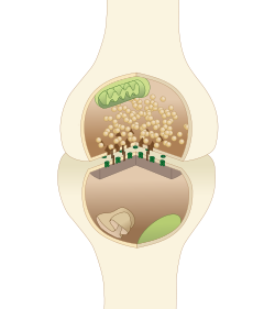
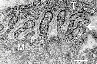
In a neuron, synaptic vesicles store various neurotransmitters that are released at the synapse. The release is regulated by a voltage-dependent calcium channel. Vesicles are essential for propagating nerve impulses between neurons and are constantly recreated by the cell. The area in the axon that holds groups of vesicles is an axon terminal or "terminal bouton". Up to 130 vesicles can be released per bouton over a ten-minute period of stimulation at 0.2 Hz. In the visual cortex of the human brain, synaptic vesicles have an average diameter of 39.5 nanometers (nm) with a standard deviation of 5.1 nm.

<span class="mw-page-title-main">End-plate potential</span>

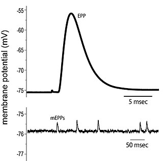
End plate potentials (EPPs) are the voltages which cause depolarization of skeletal muscle fibers caused by neurotransmitters binding to the postsynaptic membrane in the neuromuscular junction. They are called "end plates" because the postsynaptic terminals of muscle fibers have a large, saucer-like appearance. When an action potential reaches the axon terminal of a motor neuron, vesicles carrying neurotransmitters are exocytosed and the contents are released into the neuromuscular junction. These neurotransmitters bind to receptors on the postsynaptic membrane and lead to its depolarization. In the absence of an action potential, acetylcholine vesicles spontaneously leak into the neuromuscular junction and cause very small depolarizations in the postsynaptic membrane. This small response (~0.4mV) is called a miniature end plate potential (MEPP) and is generated by one acetylcholine-containing vesicle. It represents the smallest possible depolarization which can be induced in a muscle.

Molecular neuroscience is a branch of neuroscience that observes concepts in molecular biology applied to the nervous systems of animals. The scope of this subject covers topics such as molecular neuroanatomy, mechanisms of molecular signaling in the nervous system, the effects of genetics and epigenetics on neuronal development, and the molecular basis for neuroplasticity and neurodegenerative diseases. As with molecular biology, molecular neuroscience is a relatively new field that is considerably dynamic.

<span class="mw-page-title-main">Neurotransmission</span> Impulse transmission between neurons

Neurotransmission is the process by which signaling molecules called neurotransmitters are released by the axon terminal of a neuron, and bind to and react with the receptors on the dendrites of another neuron a short distance away. A similar process occurs in retrograde neurotransmission, where the dendrites of the postsynaptic neuron release retrograde neurotransmitters that signal through receptors that are located on the axon terminal of the presynaptic neuron, mainly at GABAergic and glutamatergic synapses.

<span class="mw-page-title-main">Synapse</span> Structure connecting neurons in the nervous system

In the nervous system, a synapse is a structure that permits a neuron to pass an electrical or chemical signal to another neuron or to the target effector cell.

<span class="mw-page-title-main">Synaptic potential</span>

Synaptic potential refers to the potential difference across the postsynaptic membrane that results from the action of neurotransmitters at a neuronal synapse. In other words, it is the “incoming” signal that a neuron receives. There are two forms of synaptic potential: excitatory and inhibitory. The type of potential produced depends on both the postsynaptic receptor, more specifically the changes in conductance of ion channels in the post synaptic membrane, and the nature of the released neurotransmitter. Excitatory post-synaptic potentials (EPSPs) depolarize the membrane and move the potential closer to the threshold for an action potential to be generated. Inhibitory postsynaptic potentials (IPSPs) hyperpolarize the membrane and move the potential farther away from the threshold, decreasing the likelihood of an action potential occurring. The Excitatory Post Synaptic potential is most likely going to be carried out by the neurotransmitters glutamate and acetylcholine, while the Inhibitory post synaptic potential will most likely be carried out by the neurotransmitters gamma-aminobutyric acid (GABA) and glycine. In order to depolarize a neuron enough to cause an action potential, there must be enough EPSPs to both depolarize the postsynaptic membrane from its resting membrane potential to its threshold and counterbalance the concurrent IPSPs that hyperpolarize the membrane. As an example, consider a neuron with a resting membrane potential of -70 mV (millivolts) and a threshold of -50 mV. It will need to be raised 20 mV in order to pass the threshold and fire an action potential. The neuron will account for all the many incoming excitatory and inhibitory signals via summative neural integration, and if the result is an increase of 20 mV or more, an action potential will occur.

<span class="mw-page-title-main">Complexin</span>

Complexin (also known as synaphin) refers to a one of a small set of eukaryotic cytoplasmic neuronal proteins which binds to the SNARE protein complex (SNAREpin) with a high affinity. These are called synaphin 1 and 2. In the presence of Ca2+, the transport vesicle protein synaptotagmin displaces complexin, allowing the SNARE protein complex to bind the transport vesicle to the presynaptic membrane.

<span class="mw-page-title-main">Calyx of Held</span>

The Calyx of Held is a particularly large synapse in the mammalian auditory central nervous system, so named after Hans Held who first described it in his 1893 article Die centrale Gehörleitung because of its resemblance to the calyx of a flower. Globular bushy cells in the anteroventral cochlear nucleus (AVCN) send axons to the contralateral medial nucleus of the trapezoid body (MNTB), where they synapse via these calyces on MNTB principal cells. These principal cells then project to the ipsilateral lateral superior olive (LSO), where they inhibit postsynaptic neurons and provide a basis for interaural level detection (ILD), required for high frequency sound localization. This synapse has been described as the largest in the brain.

Neuromuscular junction disease is a medical condition where the normal conduction through the neuromuscular junction fails to function correctly.

<span class="mw-page-title-main">Axon terminal</span>

Axon terminals are distal terminations of the telodendria (branches) of an axon. An axon, also called a nerve fiber, is a long, slender projection of a nerve cell, or neuron, that conducts electrical impulses called action potentials away from the neuron's cell body, or soma, in order to transmit those impulses to other neurons, muscle cells or glands.

Cellular neuroscience is a branch of neuroscience concerned with the study of neurons at a cellular level. This includes morphology and physiological properties of single neurons. Several techniques such as intracellular recording, patch-clamp, and voltage-clamp technique, pharmacology, confocal imaging, molecular biology, two photon laser scanning microscopy and Ca2+ imaging have been used to study activity at the cellular level. Cellular neuroscience examines the various types of neurons, the functions of different neurons, the influence of neurons upon each other, and how neurons work together.

The ribbon synapse is a type of neuronal synapse characterized by the presence of an electron-dense structure, the synaptic ribbon, that holds vesicles close to the active zone. It is characterized by a tight vesicle-calcium channel coupling that promotes rapid neurotransmitter release and sustained signal transmission. Ribbon synapses undergo a cycle of exocytosis and endocytosis in response to graded changes of membrane potential. It has been proposed that most ribbon synapses undergo a special type of exocytosis based on coordinated multivesicular release. This interpretation has recently been questioned at the inner hair cell ribbon synapse, where it has been instead proposed that exocytosis is described by uniquantal release shaped by a flickering vesicle fusion pore.

Vesicle fusion is the merging of a vesicle with other vesicles or a part of a cell membrane. In the latter case, it is the end stage of secretion from secretory vesicles, where their contents are expelled from the cell through exocytosis. Vesicles can also fuse with other target cell compartments, such as a lysosome. Exocytosis occurs when secretory vesicles transiently dock and fuse at the base of cup-shaped structures at the cell plasma membrane called porosome, the universal secretory machinery in cells. Vesicle fusion may depend on SNARE proteins in the presence of increased intracellular calcium (Ca2+) concentration.

<span class="mw-page-title-main">Thomas C. Südhof</span> German-American biochemist

Thomas Christian Südhof, ForMemRS, is a German-American biochemist known for his study of synaptic transmission. Currently, he is a professor in the School of Medicine in the Department of Molecular and Cellular Physiology, and by courtesy in Neurology, and in Psychiatry and Behavioral Sciences at Stanford University.

<span class="mw-page-title-main">Synaptic fatigue</span>

Synaptic fatigue, or short-term synaptic depression, is an activity-dependent form of short term synaptic plasticity that results in the temporary inability of neurons to fire and therefore transmit an input signal. It is thought to be a form of negative feedback in order to physiologically control particular forms of nervous system activity.

Neurotransmitters are released into a synapse in packaged vesicles called quanta. One quantum generates what is known as a miniature end plate potential (MEPP) which is the smallest amount of stimulation that one neuron can send to another neuron. Quantal release is the mechanism by which most traditional endogenous neurotransmitters are transmitted throughout the body. The aggregate sum of many MEPPs is known as an end plate potential (EPP). A normal end plate potential usually causes the postsynaptic neuron to reach its threshold of excitation and elicit an action potential. Electrical synapses do not use quantal neurotransmitter release and instead use gap junctions between neurons to send current flows between neurons. The goal of any synapse is to produce either an excitatory postsynaptic potential (EPSP) or an inhibitory postsynaptic potential (IPSP), which generate or repress the expression, respectively, of an action potential in the postsynaptic neuron. It is estimated that an action potential will trigger the release of approximately 20% of an axon terminal's neurotransmitter load.

References

  1. Missler M, Südhof TC, Biederer T (2012). "Synaptic cell adhesion". Cold Spring Harb Perspect Biol. 4 (4): a005694. doi:10.1101/cshperspect.a005694. PMC   3312681 . PMID   22278667.
  2. Craig C. Garner and Kang Shen. Structure and Function of Vertebrate and Invertebrate Active Zones. Structure and Functional Organization of the Synapse. Ed: Johannes Hell and Michael Ehlers. Springer, 2008.
  3. Zhai R. Grace; Bellen Hugo J. (2004). "The Architecture of the Active Zone in the Presynaptic Nerve Terminal". Physiology. 19 (5): 262–270. doi:10.1152/physiol.00014.2004. PMID   15381754. S2CID   9609266.
  4. 1 2 Phillips GR; et al. (2001). "The presynaptic particle web: ultrastructure, composition, dissolution, and reconstitution". Neuron. 32 (1): 63–77. doi: 10.1016/s0896-6273(01)00450-0 . PMID   11604139. S2CID   5996555.
  5. Mark L.; et al. "Harlow et al. The architecture of active zone material at the frog's. neuromuscular junction". Nature. 409: 2001.
  6. Siksou; et al. (2007). "Three-Dimensional Architecture of Presynaptic Terminal Cytomatrix". The Journal of Neuroscience. 27 (26): 6868–6877. doi:10.1523/jneurosci.1773-07.2007. PMC   6672225 . PMID   17596435.
  7. Ziv, Garner (2004). "Cellular and molecular mechanisms of presynaptic assembly". Nature Reviews Neuroscience. 5 (5): 385–399. doi:10.1038/nrn1370. PMID   15100721. S2CID   21516580.
  8. Georgiev, Danko D .; James F . Glazebrook (2007). "Subneuronal processing of information by solitary waves and stochastic processes". In Lyshevski, Sergey Edward (ed.). Nano and Molecular Electronics Handbook. Nano and Microengineering Series. CRC Press. pp. 17-1–17-41. doi:10.1201/9781315221670-17. ISBN   978-0-8493-8528-5. S2CID   199021983.
  9. Heidelberger; et al. (1994). "Calcium dependence of the rate of exocytosis in a synaptic terminal". Nature. 371 (6497): 513–515. Bibcode:1994Natur.371..513H. doi:10.1038/371513a0. PMID   7935764. S2CID   4316464.
  10. Stanley EF (1993). "Single calcium channels and acetylcholine release at a presynaptic nerve terminal". Neuron. 11 (6): 1007–1011. doi:10.1016/0896-6273(93)90214-c. PMID   8274272. S2CID   7311805.
  11. Atasoy and Kavalali. Neurotransmitter Release Machinery: Components of the Neuronal SNARE Complex and Their Function. Structural and Functional Organization of the Synapse Hell and Ehlers (eds.) 2008
  12. Pang Z.; Sudhof T. (2010). "Cell biology of Ca2+-triggered exocytosis". Current Opinion in Cell Biology. 22 (4): 496–505. doi:10.1016/j.ceb.2010.05.001. PMC   2963628 . PMID   20561775.
  13. Carr C.; Munson M. (2007). "Tag team action at the synapse". EMBO Reports. 8 (9): 834–838. doi:10.1038/sj.embor.7401051. PMC   1973957 . PMID   17767192.
  14. Jung Nadja; Haucke Volker (2007). "Clathrin-Mediated Endocytosis at Synapses". Traffic. 8 (9): 1129–1136. doi: 10.1111/j.1600-0854.2007.00595.x . PMID   17547698. S2CID   11320827.
  15. Ping Chi; Paul Greengard; Timothy A Ryan (10 April 2003). "Synaptic Vesicle Mobilization Is Regulated by Distinct Synapsin I Phosphorylation Pathways at Different Frequencies". Neuron . 38 (1): 69–78. doi: 10.1016/S0896-6273(03)00151-X . PMID   12691665. S2CID   17405359.
  16. Cesca et al. (2010) The synapsins: Key actors of synapse function and plasticity. Progress in Neurobiology. Vol. 91. 313-348.
  17. Dergai; et al. (2010). "Intersectin 1 forms complexes with SGIP1 and Reps1 in clathrin-coated pits". Biochemical and Biophysical Research Communications. 402 (2): 408–413. doi:10.1016/j.bbrc.2010.10.045. PMID   20946875.
  18. Marie; et al. (2004). "Dap160/Intersectin Scaffolds the Periactive Zone to Achieve High-Fidelity Endocytosis and Normal Synaptic Growth". Neuron. 43 (2): 207–219. doi: 10.1016/j.neuron.2004.07.001 . PMID   15260957. S2CID   16296285.
  19. George Zanazzi & Gary Matthews. The Molecular Architecture of Ribbon Presynaptic Terminals.Mol Neurobiol (2009) 39:130-148
  20. Gersdorff H. and Matthews G. (1994) Dynamics of synaptic vesicle fusion and membrane retrieval in synaptic terminals. Nature. Vol 367. 735-739