Perineurium

Last updated
Perineurium
Gray636.png
Transverse section of human tibial nerve (perineurium labeled at upper right)
Illu nerve structure.jpg
Nerve structure
Identifiers
TA98 A14.2.00.015
TA2 6156
FMA 52585
Anatomical terminology

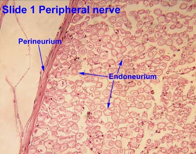
The perineurium is a protective sheath that surrounds a nerve fascicle. [1] This bundles together axons targeting the same anatomical location. [1] The perineurium is composed from fibroblasts. [2]

Contents

In the peripheral nervous system, the myelin sheath of each axon in a nerve is wrapped in a delicate protective sheath known as the endoneurium. Fascicles, bundles of neurons, are surrounded by the perineurium. Several fascicles may be in turn bundled together with a blood supply and fatty tissue within yet another sheath, the epineurium. This grouping structure is analogous to the muscular organization system of epimysium, perimysium and endomysium.

Structure

The perineurium is composed of connective tissue, which has a distinctly lamellar arrangement consisting of one to several concentric layers. The perineurium is composed of perineurial cells, which are epithelioid myofibroblasts. Perineurial cells are sometimes referred to as myoepithelioid due to their epithelioid and myofibroblastoid properties including tight junctions, gap junctions, external laminae and contractility. The tight junctions provide selective barrier to chemical substances.

The perineurium is a smooth, transparent tubular membrane which may be easily separated from the fibers it encloses. In contrast, the epineurium is a tough and mechanically resistant tissue which is not easily penetrated by a needle.

Clinical importance

The perineurium, as the epineurium, has a clinical importance following a trauma, like a fracture. A sort of lesion called axonotmesis [3] can happen, where the axon of the nerve is damaged while the integrity of the perineurium and epineurium is preserved. In that case, they will be a loss of neural transmission which will be causing a diminished response in the distal part of the nerve. The axon will be able to regenerate itself at a rate of 3 cm per month, generally indicating a return to a physiological state in roughly three months.

See also

Related Research Articles

<span class="mw-page-title-main">Central nervous system</span> Brain and spinal cord

The central nervous system (CNS) is the part of the nervous system consisting primarily of the brain and spinal cord. The CNS is so named because the brain integrates the received information and coordinates and influences the activity of all parts of the bodies of bilaterally symmetric and triploblastic animals—that is, all multicellular animals except sponges and diploblasts. It is a structure composed of nervous tissue positioned along the rostral to caudal axis of the body and may have an enlarged section at the rostral end which is a brain. Only arthropods, cephalopods and vertebrates have a true brain.

<span class="mw-page-title-main">Nerve</span> Enclosed, cable-like bundle of axons in the peripheral nervous system

A nerve is an enclosed, cable-like bundle of nerve fibers in the peripheral nervous system.

<span class="mw-page-title-main">Schwann cell</span> Glial cell type

Schwann cells or neurolemmocytes are the principal glia of the peripheral nervous system (PNS). Glial cells function to support neurons and in the PNS, also include satellite cells, olfactory ensheathing cells, enteric glia and glia that reside at sensory nerve endings, such as the Pacinian corpuscle. The two types of Schwann cells are myelinating and nonmyelinating. Myelinating Schwann cells wrap around axons of motor and sensory neurons to form the myelin sheath. The Schwann cell promoter is present in the downstream region of the human dystrophin gene that gives shortened transcript that are again synthesized in a tissue-specific manner.

<span class="mw-page-title-main">Neurilemma</span> Layer present on Schwann cells of PNS neurons

Neurilemma is the outermost nucleated cytoplasmic layer of Schwann cells that surrounds the axon of the neuron. It forms the outermost layer of the nerve fiber in the peripheral nervous system.

<span class="mw-page-title-main">Nervous tissue</span> Main component of the nervous system

Nervous tissue, also called neural tissue, is the main tissue component of the nervous system. The nervous system regulates and controls body functions and activity. It consists of two parts: the central nervous system (CNS) comprising the brain and spinal cord, and the peripheral nervous system (PNS) comprising the branching peripheral nerves. It is composed of neurons, also known as nerve cells, which receive and transmit impulses, and neuroglia, also known as glial cells or glia, which assist the propagation of the nerve impulse as well as provide nutrients to the neurons.

<span class="mw-page-title-main">Motor nerve</span> Nerve located in the central nervous system

A motor nerve is a nerve located in the central nervous system (CNS), usually the spinal cord, that sends motor signals from the CNS to the muscles of the body. This is different from the motor neuron, which includes a cell body and branching of dendrites, while the nerve is made up of a bundle of axons. Motor nerves act as efferent nerves which carry information out from the CNS to muscles, as opposed to afferent nerves, which send signals from sensory receptors in the periphery to the CNS. Efferent nerves can also connect to glands or other organs/issues instead of muscles. In addition, there are nerves that serve as both sensory and motor nerves called mixed nerves.

<span class="mw-page-title-main">Node of Ranvier</span> Gaps between myelin sheaths on the axon of a neuron

In neuroscience and anatomy, Nodes of Ranvier, also known as myelin-sheath gaps, occur along a myelinated axon where the axolemma is exposed to the extracellular space. Nodes of Ranvier are uninsulated and highly enriched in ion channels, allowing them to participate in the exchange of ions required to regenerate the action potential. Nerve conduction in myelinated axons is referred to as saltatory conduction due to the manner in which the action potential seems to "jump" from one node to the next along the axon. This results in faster conduction of the action potential.

Axonotmesis is an injury to the peripheral nerve of one of the extremities of the body. The axons and their myelin sheath are damaged in this kind of injury, but the endoneurium, perineurium and epineurium remain intact. Motor and sensory functions distal to the point of injury are completely lost over time leading to Wallerian degeneration due to ischemia, or loss of blood supply. Axonotmesis is usually the result of a more severe crush or contusion than neurapraxia.

<span class="mw-page-title-main">Epineurium</span> Outermost layer of a nerve

The epineurium is the outermost layer of dense irregular connective tissue surrounding a peripheral nerve. It usually surrounds multiple nerve fascicles as well as blood vessels which supply the nerve. Smaller branches of these blood vessels penetrate into the perineurium. In addition to blood vessels which supply the nerve, lymphocytes and fibroblasts are also present and contribute to the production of collagen fibers that form the backbone of the epineurium. In addition to providing structural support, lymphocytes and fibroblasts also play a vital role in maintenance and repair of the surrounding tissues.

<span class="mw-page-title-main">Funiculus (neuroanatomy)</span>

A funiculus or column is a small bundle of axons, enclosed by the perineurium. A small nerve may consist of a single funiculus, but a larger nerve will have several funiculi collected together into larger bundles known as fascicles. Fascicles are bound together in a common membrane, the epineurium.

<span class="mw-page-title-main">Myelin incisure</span>

Myelin incisures, are small pockets of cytoplasm left behind during the Schwann cell myelination process.

<span class="mw-page-title-main">Endoneurium</span> Connective tissue layer around myelinated nerve fibers in peripheral nervous system

The endoneurium is a layer of delicate connective tissue around the myelin sheath of each myelinated nerve fiber in the peripheral nervous system. Its component cells are called endoneurial cells. The endoneuria with their enclosed nerve fibers are bundled into groups called nerve fascicles, each fascicle within its own protective sheath called a perineurium. In sufficiently large nerves multiple fascicles, each with its blood supply and fatty tissue, may be bundled within yet another sheath, the epineurium.

<span class="mw-page-title-main">Nerve fascicle</span> Bundle of funiculi (small bundles of axons) in the peripheral nervous system

A nerve fascicle, is a bundle of nerve fibers belonging to a nerve in the peripheral nervous system. A nerve fascicle is also called a fasciculus. A nerve fascicle is enclosed by perineurium, a layer of fascial connective tissue. Each enclosed nerve fiber in the fascicle is enclosed by a connective tissue layer of endoneurium. Bundles of nerve fascicles are called fasciculi and are constituents of a nerve trunk. A main nerve trunk may contain a great many fascicles enclosing many thousands of axons.

<span class="mw-page-title-main">Satellite glial cell</span>

Satellite glial cells, formerly called amphicytes, are glial cells that cover the surface of neuron cell bodies in ganglia of the peripheral nervous system. Thus, they are found in sensory, sympathetic, and parasympathetic ganglia. Both satellite glial cells (SGCs) and Schwann cells are derived from the neural crest of the embryo during development. SGCs have been found to play a variety of roles, including control over the microenvironment of sympathetic ganglia. They are thought to have a similar role to astrocytes in the central nervous system (CNS). They supply nutrients to the surrounding neurons and also have some structural function. Satellite cells also act as protective, cushioning cells. Additionally, they express a variety of receptors that allow for a range of interactions with neuroactive chemicals. Many of these receptors and other ion channels have recently been implicated in health issues including chronic pain and herpes simplex. There is much more to be learned about these cells, and research surrounding additional properties and roles of the SGCs is ongoing.

Neuroregeneration refers to the regrowth or repair of nervous tissues, cells or cell products. Such mechanisms may include generation of new neurons, glia, axons, myelin, or synapses. Neuroregeneration differs between the peripheral nervous system (PNS) and the central nervous system (CNS) by the functional mechanisms involved, especially in the extent and speed of repair. When an axon is damaged, the distal segment undergoes Wallerian degeneration, losing its myelin sheath. The proximal segment can either die by apoptosis or undergo the chromatolytic reaction, which is an attempt at repair. In the CNS, synaptic stripping occurs as glial foot processes invade the dead synapse.

In neurobiology, a mesaxon is a pair of parallel plasma membranes of a Schwann cell. It marks the point of edge-to-edge contact by the Schwann cell encircling the axon. A single Schwann cell of the peripheral nervous system will wrap around and support only one individual axon, while the oligodendrocytes found in the central nervous system can wrap around and support 5-8 axons. Thin unmyelinated axons are often bundled, with several unmyelinated axons to a single mesaxon.

<span class="mw-page-title-main">Peripheral nerve injury classification</span>

Classification of peripheral nerve injury assists in prognosis and determination of treatment strategy. Classification of nerve injury was described by Seddon in 1943 and by Sunderland in 1951. The lowest degree of nerve injury in which the nerve remains intact but signaling ability is damaged is called neurapraxia. The second degree in which the axon is damaged but the surrounding connecting tissue remains intact is called axonotmesis. The last degree in which both the axon and connective tissue are damaged is called neurotmesis.

<span class="mw-page-title-main">Epineurial repair</span>

Epineurial repair is a common surgical procedure to repair a nerve laceration via the epineurium, the connective tissue surrounding nerve fibers originating from the spinal cord. It is intended to allow the restoration of sensory function. When a nerve is lacerated or cut, repair is done by sewing the cut ends together through the epineurium to increase the potential of the proximal part growing correctly along the route the degrading distal part leaves behind. Usual sensation and mobility will not be an immediate result because nerves grow at a rate of approximately 1 millimeter per day, so it will take a few months to notice the final outcome. Research in use of nerve grafts and nerve growth factors is being done to speed recovery time.

Nerve allotransplantation is the transplantation of a nerve to a receiver from a donor of the same species. For example, nerve tissue is transplanted from one person to another. Allotransplantation is a commonly used type of transplantation of which nerve repair is one specific aspect.

Cryoneurolysis, also referred to as cryoanalgesia, is a medical procedure that temporarily blocks nerve conduction along peripheral nerve pathways. The procedure, which inserts a small probe to freeze the target nerve, can facilitate complete regeneration of the structure and function of the affected nerve. Cryoneurolysis has been used to treat a variety of painful conditions.

References

  1. 1 2 McCracken, Thomas (1999). New Atlas of Human Anatomy. China: Metro Books. pp. 96–97. ISBN   1-5866-3097-0.
  2. Weerasuriya, ANANDA (2005-01-01), Dyck, Peter J.; Thomas, P. K. (eds.), "Chapter 29 - Blood-Nerve Interface and Endoneurial Homeostasis", Peripheral Neuropathy (Fourth Edition), Philadelphia: W.B. Saunders, pp. 651–665, doi:10.1016/b978-0-7216-9491-7.50032-6, ISBN   978-0-7216-9491-7 , retrieved 2020-11-18
  3. Netter, Frank. Netter's Orthopaedics.