Pacinian corpuscle

Last updated
Pacinian corpuscle
Gray935.png
Pacinian corpuscle, with its system of capsules and central cavity.
a. Arterial twig, ending in capillaries, which form loops in some of the intercapsular spaces, and one penetrates to the central capsule.
b. The fibrous tissue of the stalk.
n. Nerve tube advancing to the central capsule, there losing its white matter and stretching along the axis to the opposite end, where it ends by a tuberculated enlargement.
Skin.png
Pacinian corpuscle labeled at bottom
Details
Location Skin
Identifiers
Latin corpusculum Pacinian
MeSH D010141
TH H3.11.06.0.00009
FMA 83604
Anatomical terms of microanatomy

The Pacinian corpuscle, lamellar corpuscle or Vater-Pacini corpuscle [1] is one of the four major types of mechanoreceptors (specialized nerve ending) for mechanical sensation) found in mammalian skin. This type of mechanoreceptor is found in both hairy, and hairless skin, viscera, joints, and attached to the periosteum of bone, primarily responsible for sensitivity to vibration. [2] A few are also sensitive to quasi-static or low frequency pressure stimuli.[ citation needed ] Most of them respond only to sudden disturbances and are especially sensitive to vibration of a few hundreds hertz. [3] The vibrational role may be used for detecting surface texture, such as rough and smooth. Most of the Pacinian corpuscles act as rapidly adapting mechanoreceptors. Groups of corpuscles respond to pressure changes, such as on grasping or releasing an object.

Contents

Structure

Pacinian corpuscles are larger and fewer in number than Meissner's corpuscle, Merkel cells and Ruffini's corpuscles. [4]

The Pacinian corpuscle is approximately oval-cylindrical-shaped and 1 mm in length. The entire corpuscle is wrapped by a layer of connective tissue. Its capsule consists of 20 to 60 concentric lamellae (hence the alternative lamellar corpuscle) including fibroblasts and fibrous connective tissue (mainly Type IV and Type II collagen network), separated by gelatinous material, more than 92% of which is water. [5] It presents a whorled pattern on micrographs.

Function

Pacinian corpuscles are rapidly adapting (phasic) receptors that detect gross pressure changes and vibrations in the skin. [6] Any deformation in the corpuscle leads to opening of pressure-sensitive or stretch-activated ion channels or mechanosensitive channels present in the axon membrane or axolemma of the neurite inside the core of the corpuscles or end-organ.[ citation needed ] This initiates generation of the receptor potential inside the corpuscles which is also secondarily supported by the voltage-activated ion channels present in the core of the corpuscles. Finally the receptor potential is modulated to neural spikes or action potential with the help of opening of sodium ion channels present at the first Ranveir's Node of the axon. [3]

These corpuscles are especially sensitive to vibrations, which they can sense even centimeters away. [4] Their optimal sensitivity is 250 Hz, and this is the frequency range generated upon fingertips by textures made of features smaller than 1  µm. [7] [8] Pacinian corpuscles respond when the skin is rapidly indented but not when the pressure is steady, due to the layers of connective tissue that cover the nerve ending. [4] It is thought that they respond to high-velocity changes in joint position. They have also been implicated in detecting the location of touch sensations on handheld tools. [9]

Pacinian corpuscles have a large receptive field on the skin's surface with an especially sensitive center. [4]

Mechanism

Pacinian corpuscles sense stimuli due to the deformation of their lamellae, which press on the membrane of the sensory neuron and causes it to bend or stretch. [10] When the lamellae are deformed, due to either application or release of pressure, a generator or receptor potential is created as it physically deforms the plasma membrane of the receptive area of the neuron, making it "leak" different cations through Mechanosensitive channels which initiates the receptor potential. This mechanotransduction process is also supported by distributed voltage sensitive ion channels in the inner-core and neurite of the corpuscles. [3] Due to generation of receptor potential in the receptive area of the neurite (especially near the heminode or half-node of the axon) the potential at the first Ranvier's node can reach certain threshold, triggering nerve impulses or action potentials at the first node of Ranvier. The first Ranvier's node of the myelinated section of the neurite is often found inside the capsule.[ citation needed ] This impulse is then transferred along the axon from node to node with the use of sodium channels and sodium/potassium pumps in the axon membrane.

Once the receptive area of the neurite is depolarized, it will depolarize the first node of Ranvier; however, as it is a rapidly adapting fibre, this does not carry on indefinitely, and the signal propagation ceases. This is a graded response, meaning that the greater the deformation, the greater the generator potential. This information is encoded in the frequency of impulses, since a bigger or faster deformation induces a higher impulse frequency. Action potentials are formed when the skin is rapidly distorted but not when pressure is continuous because of the mechanical filtering of the stimulus in the lamellar structure. The frequencies of the impulses decrease quickly and soon stop due to the relaxation of the inner layers of connective tissue that cover the nerve ending.

Discovery

Pacinian corpuscles were the first cellular sensory receptor ever observed. They were first reported by German anatomist and botanist Abraham Vater and his student Johannes Gottlieb Lehmann in 1741, but ultimately named after Italian anatomist Filippo Pacini, who rediscovered them in 1835. [11] [12] John Shekleton, a curator of the Royal College of Surgeons in Ireland, also discovered them before Pacini, but his results were published later. [11] Similar to Pacinian corpuscles, Herbst corpuscles and Grandry corpuscles are found in bird species.[ citation needed ]

Additional images

See also

Related Research Articles

<span class="mw-page-title-main">Axon</span> Long projection on a neuron that conducts signals to other neurons

An axon or nerve fiber is a long, slender projection of a nerve cell, or neuron, in vertebrates, that typically conducts electrical impulses known as action potentials away from the nerve cell body. The function of the axon is to transmit information to different neurons, muscles, and glands. In certain sensory neurons, such as those for touch and warmth, the axons are called afferent nerve fibers and the electrical impulse travels along these from the periphery to the cell body and from the cell body to the spinal cord along another branch of the same axon. Axon dysfunction can be the cause of many inherited and acquired neurological disorders that affect both the peripheral and central neurons. Nerve fibers are classed into three types – group A nerve fibers, group B nerve fibers, and group C nerve fibers. Groups A and B are myelinated, and group C are unmyelinated. These groups include both sensory fibers and motor fibers. Another classification groups only the sensory fibers as Type I, Type II, Type III, and Type IV.

<span class="mw-page-title-main">Sensory nervous system</span> Part of the nervous system

The sensory nervous system is a part of the nervous system responsible for processing sensory information. A sensory system consists of sensory neurons, neural pathways, and parts of the brain involved in sensory perception and interoception. Commonly recognized sensory systems are those for vision, hearing, touch, taste, smell, balance and visceral sensation. Sense organs are transducers that convert data from the outer physical world to the realm of the mind where people interpret the information, creating their perception of the world around them.

Stimulus modality, also called sensory modality, is one aspect of a stimulus or what is perceived after a stimulus. For example, the temperature modality is registered after heat or cold stimulate a receptor. Some sensory modalities include: light, sound, temperature, taste, pressure, and smell. The type and location of the sensory receptor activated by the stimulus plays the primary role in coding the sensation. All sensory modalities work together to heighten stimuli sensation when necessary.

A cutaneous receptor is the type of sensory receptor found in the skin. They are a part of the somatosensory system. Cutaneous receptors include mechanoreceptors, nociceptors (pain), and thermoreceptors (temperature).

A mechanoreceptor, also called mechanoceptor, is a sensory receptor that responds to mechanical pressure or distortion. Mechanoreceptors are innervated by sensory neurons that convert mechanical pressure into electrical signals that, in animals, are sent to the central nervous system.

<span class="mw-page-title-main">Dorsal column–medial lemniscus pathway</span> Sensory spinal pathway

The dorsal column–medial lemniscus pathway (DCML) is a sensory pathway of the central nervous system that conveys sensations of fine touch, vibration, two-point discrimination, and proprioception from the skin and joints. It transmits information from the body to the primary somatosensory cortex in the postcentral gyrus of the parietal lobe of the brain. The pathway receives information from sensory receptors throughout the body, and carries this in nerve tracts in the white matter of the dorsal column of the spinal cord to the medulla, where it is continued in the medial lemniscus, on to the thalamus and relayed from there through the internal capsule and transmitted to the somatosensory cortex. The name dorsal-column medial lemniscus comes from the two structures that carry the sensory information: the dorsal columns of the spinal cord, and the medial lemniscus in the brainstem.

<span class="mw-page-title-main">Tactile corpuscle</span> Type of mechanoreceptor that detects light touch

Tactile corpuscles or Meissner's corpuscles are a type of mechanoreceptor discovered by anatomist Georg Meissner (1829–1905) and Rudolf Wagner. This corpuscle is a type of nerve ending in the skin that is responsible for sensitivity to pressure. In particular, they have their highest sensitivity when sensing vibrations between 10 and 50 hertz. They are rapidly adaptive receptors. They are most concentrated in thick hairless skin, especially at the finger pads.

<span class="mw-page-title-main">Sensory neuron</span> Nerve cell that converts environmental stimuli into corresponding internal stimuli

Sensory neurons, also known as afferent neurons, are neurons in the nervous system, that convert a specific type of stimulus, via their receptors, into action potentials or graded receptor potentials. This process is called sensory transduction. The cell bodies of the sensory neurons are located in the dorsal ganglia of the spinal cord.

<span class="mw-page-title-main">Node of Ranvier</span> Gaps between myelin sheaths on the axon of a neuron

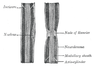
In neuroscience and anatomy, nodes of Ranvier, also known as myelin-sheath gaps, occur along a myelinated axon where the axolemma is exposed to the extracellular space. Nodes of Ranvier are uninsulated and highly enriched in ion channels, allowing them to participate in the exchange of ions required to regenerate the action potential. Nerve conduction in myelinated axons is referred to as saltatory conduction due to the manner in which the action potential seems to "jump" from one node to the next along the axon. This results in faster conduction of the action potential.

<span class="mw-page-title-main">Dorsal root ganglion</span> Cluster of neurons in a dorsal root of a spinal nerve

A dorsal root ganglion is a cluster of neurons in a dorsal root of a spinal nerve. The cell bodies of sensory neurons known as first-order neurons are located in the dorsal root ganglia.

<span class="mw-page-title-main">Star-nosed mole</span> Species of Mole

The star-nosed mole is a small semiaquatic mole found in moist, low elevation areas in the northern parts of North America. It is the only extant member of the tribe Condylurini and genus Condylura, and it has more than 25,000 minute sensory receptors in touch organs, known as Eimer's organs, with which this hamster-sized mole feels its way around. With the help of its Eimer's organs, it may be perfectly poised to detect seismic wave vibrations.

<span class="mw-page-title-main">Merkel cell</span> Receptors in the skin of vertebrates

Merkel cells, also known as Merkel-Ranvier cells or tactile epithelial cells, are oval-shaped mechanoreceptors essential for light touch sensation and found in the skin of vertebrates. They are abundant in highly sensitive skin like that of the fingertips in humans, and make synaptic contacts with somatosensory afferent nerve fibers. It has been reported that Merkel cells are derived from neural crest cells, though more recent experiments in mammals have indicated that they are epithelial in origin.

Merkel nerve endings are mechanoreceptors, a type of sensory receptor, that are found in the basal epidermis and hair follicles. They are nerve endings and provide information on mechanical pressure, position, and deep static touch features, such as shapes and edges.

<span class="mw-page-title-main">Tactile corpuscles of Grandry</span>

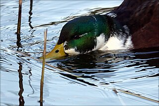
The tactile corpuscles of Grandry or Grandry corpuscles are mechanoreceptors found in the beak skin and oral mucosa of aquatic birds. They were first described by Grandry in 1869 in the bill skin of ducks and geese. Their general structure includes the flattened endings of an afferent nerve fiber sandwiched between two or more somewhat flattened sensory cells called Grandry cells, all surrounded by a layer of satellite cells and a partial capsule of collagen protein. Electrophysiological studies have shown that Grandry corpuscles function as rapidly adapting velocity detectors. In birds, Grandry and Merkel corpuscles share many morphological similarities, which has led to some confusion in the literature over their classification.

<span class="mw-page-title-main">Microneurography</span>

Microneurography is a neurophysiological method employed to visualize and record the traffic of nerve impulses that are conducted in peripheral nerves of waking human subjects. It can also be used in animal recordings. The method has been successfully employed to reveal functional properties of a number of neural systems, e.g. sensory systems related to touch, pain, and muscle sense as well as sympathetic activity controlling the constriction state of blood vessels. To study nerve impulses of an identified nerve, a fine tungsten needle microelectrode is inserted into the nerve and connected to a high input impedance differential amplifier. The exact position of the electrode tip within the nerve is then adjusted in minute steps until the electrode discriminates nerve impulses of interest. A unique feature and a significant strength of the microneurography method is that subjects are fully awake and able to cooperate in tests requiring mental attention, while impulses in a representative nerve fibre or set of nerve fibres are recorded, e.g. when cutaneous sense organs are stimulated or subjects perform voluntary precision movements.

<span class="mw-page-title-main">Abraham Vater</span> German anatomist

Abraham Vater was a German anatomist from Wittenberg.

Eimer's organs are sensory organs in which the epidermis is modified to form bulbous papillae. First isolated by Theodor Eimer from the European mole in 1871, these organs are present in many moles, and are particularly common in the star-nosed mole, which bears 25,000 of them on its unique tentacled snout. The organs are formed from a stack of epidermal cells, which is innervated by nerve processes from myelinated fibers in the dermis, which form terminal swellings just below the outer keratinized layer of epidermis. They contain a Merkel cell-neurite complex in the epidermis and a lamellated corpuscle in the dermal connective tissue.

Pallesthesia, or vibratory sensation, is the ability to perceive vibration. This sensation, often conducted through skin and bone, is usually generated by mechanoreceptors such as Pacinian corpuscles, Merkel disk receptors, and tactile corpuscles. All of these receptors stimulate an action potential in afferent nerves found in various layers of the skin and body. The afferent neuron travels to the spinal column and then to the brain where the information is processed. Damage to the peripheral nervous system or central nervous system can result in a decline or loss of pallesthesia.

<span class="mw-page-title-main">Somatosensory system</span> Nerve system for sensing touch, temperature, body position, and pain

Touch is perceiving the environment using skin. Specialized receptors in the skin send signals to the brain indicating light and soft pressure, hot and cold, body position and pain. It is a subset of the sensory nervous system, which also includes the visual, auditory, olfactory, gustatory and vestibular senses.

A sense is a biological system used by an organism for sensation, the process of gathering information about the surroundings through the detection of stimuli. Although, in some cultures, five human senses were traditionally identified as such, many more are now recognized. Senses used by non-human organisms are even greater in variety and number. During sensation, sense organs collect various stimuli for transduction, meaning transformation into a form that can be understood by the brain. Sensation and perception are fundamental to nearly every aspect of an organism's cognition, behavior and thought.

References

  1. Germann, C.; Sutter, R.; Nanz, D. (June 2021). "Novel observations of Pacinian corpuscle distribution in the hands and feet based on high-resolution 7-T MRI in healthy volunteers". Skeletal Radiology. 50 (6): 1249–1255. doi:10.1007/s00256-020-03667-7. PMC   8035111 . PMID   33156397.
  2. Biswas, Abhijit; Manivannan, M.; Srinivasan, Mandyam A. (2015). "Multiscale layered biomechanical model of the Pacinian corpuscle". IEEE Transactions on Haptics. 8 (1): 31–42. doi:10.1109/TOH.2014.2369416. PMID   25398182. S2CID   24658742.
  3. 1 2 3 Biswas, Abhijit; Manivannan, M.; Srinivasan, Mandyam A. (2015). "Vibrotactile sensitivity threshold: Nonlinear stochastic mechanotransduction model of the Pacinian corpuscle". IEEE Transactions on Haptics. 8 (1): 102–113. doi:10.1109/TOH.2014.2369422. PMID   25398183. S2CID   15326972.
  4. 1 2 3 4 Kandel, Eric R.; Schwartz, James H.; Jessell, Thomas M., eds. (2000). Principles of Neural Science . New York, NY: McGraw-Hill, Health Professions Division. ISBN   0-8385-7701-6.
  5. Cherepnov, V.L.; Chadaeva, N.I. (1981). "Some characteristics of soluble proteins of Pacinian corpuscles". Bulletin of Experimental Biology and Medicine. 91 (3): 346–348. doi:10.1007/BF00839370. PMID   7248510. S2CID   26734354.
  6. Purves, Dale; Augustine, George J.; Fitzpatrick, David; Katz, Lawrence C.; LaMantia, Anthony-Samuel; McNamara, James O.; Williams, S. Mark (2001). "Cutaneous and Subcutaneous Somatic Sensory Receptors". Neuroscience. 2nd edition. Sinauer Associates. Retrieved 31 July 2023.
  7. Scheibert, J; Leurent, S; Prevost, A; Debrégeas, G (2009). "The role of fingerprints in the coding of tactile information probed with a biomimetic sensor". Science. 323 (5920): 1503–6. arXiv: 0911.4885 . Bibcode:2009Sci...323.1503S. doi:10.1126/science.1166467. PMID   19179493. S2CID   14459552.
  8. Skedung, Lisa, Martin Arvidsson, Jun Young Chung, Christopher M. Stafford, Birgitta Berglund, and Mark W. Rutland. 2013. "Feeling Small: Exploring the Tactile Perception Limits." Sci. Rep. 3 (September 12). doi : 10.1038/srep02617.
  9. Sima, Richard (23 December 2019). "The Brain Senses Touch beyond the Body". Scientific American. Retrieved 17 February 2020.
  10. Klein, Stephen B.; Michael Thorne, B. (2006-10-03). Biological Psychology. Macmillan. ISBN   9780716799221.
  11. 1 2 Bentivoglio, Marina; Pacini, Paolo (1995). "Filippo Pacini: a determined observer". Brain Research Bulletin. 38 (2): 161–165. doi:10.1016/0361-9230(95)00083-Q. PMID   7583342. S2CID   6094598.
  12. Cauna, N.; Mannan, G. (1958). "The structure of human digital pacinian corpuscles (corpus cula lamellosa) and its functional significance". Journal of Anatomy. 92 (1): 1–20. ISSN   0021-8782. PMC   1244958 . PMID   13513492.