Sensory neuron

Last updated
Four types of sensory neuron Structure of sensory system (4 models) E.PNG
Four types of sensory neuron

Sensory neurons, also known as afferent neurons, are neurons in the nervous system, that convert a specific type of stimulus, via their receptors, into action potentials or graded receptor potentials. [1] This process is called sensory transduction. The cell bodies of the sensory neurons are located in the dorsal ganglia of the spinal cord. [2]

Contents

The sensory information travels on the afferent nerve fibers in a sensory nerve, to the brain via the spinal cord. Spinal nerves transmit external sensations via sensory nerves to the brain through the spinal cord. [3] The stimulus can come from exteroreceptors outside the body, for example those that detect light and sound, or from interoreceptors inside the body, for example those that are responsive to blood pressure or the sense of body position.

Types and function

Sensory neurons in vertebrates are predominantly pseudounipolar or bipolar, and different types of sensory neurons have different sensory receptors that respond to different kinds of stimuli. There are at least six external and two internal sensory receptors:

External receptors

External receptors that respond to stimuli from outside the body are called exteroreceptors. [4] Exteroreceptors include chemoreceptors such as olfactory receptors (smell) and taste receptors, photoreceptors (vision), thermoreceptors (temperature), nociceptors (pain), hair cells (hearing and balance), and a number of other different mechanoreceptors for touch and proprioception (stretch, distortion and stress).

Smell

The sensory neurons involved in smell are called olfactory sensory neurons. These neurons contain receptors, called olfactory receptors, that are activated by odor molecules in the air. The molecules in the air are detected by enlarged cilia and microvilli. [5] These sensory neurons produce action potentials. Their axons form the olfactory nerve, and they synapse directly onto neurons in the cerebral cortex (olfactory bulb). They do not use the same route as other sensory systems, bypassing the brain stem and the thalamus. The neurons in the olfactory bulb that receive direct sensory nerve input, have connections to other parts of the olfactory system and many parts of the limbic system. 9.

Taste

Taste sensation is facilitated by specialized sensory neurons located in the taste buds of the tongue and other parts of the mouth and throat. These sensory neurons are responsible for detecting different taste qualities, such as sweet, sour, salty, bitter, and savory. When you eat or drink something, chemicals in the food or liquid interact with receptors on these sensory neurons, triggering signals that are sent to the brain. The brain then processes these signals and interprets them as specific taste sensations, allowing you to perceive and enjoy the flavors of the foods you consume. [6] When taste receptor cells are stimulated by the binding of these chemical compounds (tastants), it can lead to changes in the flow of ions, such as sodium (Na+), calcium (Ca2+), and potassium (K+), across the cell membrane. [7] In response to tastant binding, ion channels on the taste receptor cell membrane can open or close. This can lead to depolarization of the cell membrane, creating an electrical signal.

Similar to olfactory receptors, taste receptors (gustatory receptors) in taste buds interact with chemicals in food to produce an action potential.

Vision

Photoreceptor cells are capable of phototransduction, a process which converts light (electromagnetic radiation) into electrical signals. These signals are refined and controlled by the interactions with other types of neurons in the retina. The five basic classes of neurons within the retina are photoreceptor cells, bipolar cells, ganglion cells, horizontal cells, and amacrine cells. The basic circuitry of the retina incorporates a three-neuron chain consisting of the photoreceptor (either a rod or cone), bipolar cell, and the ganglion cell. The first action potential occurs in the retinal ganglion cell. This pathway is the most direct way for transmitting visual information to the brain. There are three primary types of photoreceptors: Cones are photoreceptors that respond significantly to color. In humans the three different types of cones correspond with a primary response to short wavelength (blue), medium wavelength (green), and long wavelength (yellow/red). [8] Rods are photoreceptors that are very sensitive to the intensity of light, allowing for vision in dim lighting. The concentrations and ratio of rods to cones is strongly correlated with whether an animal is diurnal or nocturnal. In humans, rods outnumber cones by approximately 20:1, while in nocturnal animals, such as the tawny owl, the ratio is closer to 1000:1. [8] Retinal ganglion cells are involved in the sympathetic response. Of the ~1.3 million ganglion cells present in the retina, 1-2% are believed to be photosensitive. [9]

Issues and decay of sensory neurons associated with vision lead to disorders such as:

  1. Macular degeneration – degeneration of the central visual field due to either cellular debris or blood vessels accumulating between the retina and the choroid, thereby disturbing and/or destroying the complex interplay of neurons that are present there. [10]
  2. Glaucoma – loss of retinal ganglion cells which causes some loss of vision to blindness. [11]
  3. Diabetic retinopathy – poor blood sugar control due to diabetes damages the tiny blood vessels in the retina. [12]

Auditory

The auditory system is responsible for converting pressure waves generated by vibrating air molecules or sound into signals that can be interpreted by the brain.

This mechanoelectrical transduction is mediated with hair cells within the ear. Depending on the movement, the hair cell can either hyperpolarize or depolarize. When the movement is towards the tallest stereocilia, the Na+ cation channels open allowing Na+ to flow into cell and the resulting depolarization causes the Ca++ channels to open, thus releasing its neurotransmitter into the afferent auditory nerve. There are two types of hair cells: inner and outer. The inner hair cells are the sensory receptors . [13]

Problems with sensory neurons associated with the auditory system leads to disorders such as:

  1. Auditory processing disorder – Auditory information in the brain is processed in an abnormal way. Patients with auditory processing disorder can usually gain the information normally, but their brain cannot process it properly, leading to hearing disability. [14]
  2. Auditory verbal agnosia – Comprehension of speech is lost but hearing, speaking, reading, and writing ability is retained. This is caused by damage to the posterior superior temporal lobes, again not allowing the brain to process auditory input correctly. [15]

Temperature

Thermoreceptors are sensory receptors, which respond to varying temperatures. While the mechanisms through which these receptors operate is unclear, recent discoveries have shown that mammals have at least two distinct types of thermoreceptors. [16] The bulboid corpuscle, is a cutaneous receptor a cold-sensitive receptor, that detects cold temperatures. The other type is a warmth-sensitive receptor.

Mechanoreceptors

Mechanoreceptors are sensory receptors which respond to mechanical forces, such as pressure or distortion. [17]

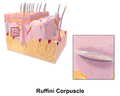
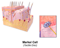
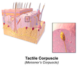
Specialized sensory receptor cells called mechanoreceptors often encapsulate afferent fibers to help tune the afferent fibers to the different types of somatic stimulation. Mechanoreceptors also help lower thresholds for action potential generation in afferent fibers and thus make them more likely to fire in the presence of sensory stimulation. [18]

Some types of mechanoreceptors fire action potentials when their membranes are physically stretched.

Proprioceptors are another type of mechanoreceptors which literally means "receptors for self". These receptors provide spatial information about limbs and other body parts. [19]

Nociceptors are responsible for processing pain and temperature changes. The burning pain and irritation experienced after eating a chili pepper (due to its main ingredient, capsaicin), the cold sensation experienced after ingesting a chemical such as menthol or icillin, as well as the common sensation of pain are all a result of neurons with these receptors. [20]

Problems with mechanoreceptors lead to disorders such as:

  1. Neuropathic pain - a severe pain condition resulting from a damaged sensory nerve [20]
  2. Hyperalgesia - an increased sensitivity to pain caused by sensory ion channel, TRPM8, which is typically responds to temperatures between 23 and 26 degrees, and provides the cooling sensation associated with menthol and icillin [20]
  3. Phantom limb syndrome - a sensory system disorder where pain or movement is experienced in a limb that does not exist [21]

Internal receptors

Internal receptors that respond to changes inside the body are known as interoceptors. [4]

Blood

The aortic bodies and carotid bodies contain clusters of glomus cellsperipheral chemoreceptors that detect changes in chemical properties in the blood such as oxygen concentration. [22] These receptors are polymodal responding to a number of different stimuli.

Nociceptors

Nociceptors respond to potentially damaging stimuli by sending signals to the spinal cord and brain. This process, called nociception, usually causes the perception of pain. [23] [24] They are found in internal organs as well as on the surface of the body to "detect and protect". [24] Nociceptors detect different kinds of noxious stimuli indicating potential for damage, then initiate neural responses to withdraw from the stimulus. [24]

  1. Thermal nociceptors are activated by noxious heat or cold at various temperatures. [24]
  2. Mechanical nociceptors respond to excess pressure or mechanical deformation, such as a pinch. [24]
  3. Chemical nociceptors respond to a wide variety of chemicals, some of which signal a response. They are involved in the detection of some spices in food, such as the pungent ingredients in Brassica and Allium plants, which target the sensory neural receptor to produce acute pain and subsequent pain hypersensitivity. [25]

Connection with the central nervous system

Information coming from the sensory neurons in the head enters the central nervous system (CNS) through cranial nerves. Information from the sensory neurons below the head enters the spinal cord and passes towards the brain through the 31 spinal nerves. [26] The sensory information traveling through the spinal cord follows well-defined pathways. The nervous system codes the differences among the sensations in terms of which cells are active.

Classification

Adequate stimulus

A sensory receptor's adequate stimulus is the stimulus modality for which it possesses the adequate sensory transduction apparatus. Adequate stimulus can be used to classify sensory receptors:

  1. Baroreceptors respond to pressure in blood vessels
  2. Chemoreceptors respond to chemical stimuli
  3. Electromagnetic radiation receptors respond to electromagnetic radiation [27]
    1. Infrared receptors respond to infrared radiation
    2. Photoreceptors respond to visible light
    3. Ultraviolet receptors respond to ultraviolet radiation [ citation needed ]
  4. Electroreceptors respond to electric fields
    1. Ampullae of Lorenzini respond to electric fields, salinity, and to temperature, but function primarily as electroreceptors
  5. Hydroreceptors respond to changes in humidity
  6. Magnetoreceptors respond to magnetic fields
  7. Mechanoreceptors respond to mechanical stress or mechanical strain
  8. Nociceptors respond to damage, or threat of damage, to body tissues, leading (often but not always) to pain perception
  9. Osmoreceptors respond to the osmolarity of fluids (such as in the hypothalamus)
  10. Proprioceptors provide the sense of position
  11. Thermoreceptors respond to temperature, either heat, cold or both

Location

Sensory receptors can be classified by location:

  1. Cutaneous receptors are sensory receptors found in the dermis or epidermis. [28]
  2. Muscle spindles contain mechanoreceptors that detect stretch in muscles.

Morphology

Somatic sensory receptors near the surface of the skin can usually be divided into two groups based on morphology:

  1. Free nerve endings characterize the nociceptors and thermoreceptors and are called thus because the terminal branches of the neuron are unmyelinated and spread throughout the dermis and epidermis.
  2. Encapsulated receptors consist of the remaining types of cutaneous receptors. Encapsulation exists for specialized functioning.

Rate of adaptation

  1. A tonic receptor is a sensory receptor that adapts slowly to a stimulus [29] and continues to produce action potentials over the duration of the stimulus. [30] In this way it conveys information about the duration of the stimulus. Some tonic receptors are permanently active and indicate a background level. Examples of such tonic receptors are pain receptors, joint capsule, and muscle spindle. [31]
  2. A phasic receptor is a sensory receptor that adapts rapidly to a stimulus. The response of the cell diminishes very quickly and then stops. [32] It does not provide information on the duration of the stimulus; [30] instead some of them convey information on rapid changes in stimulus intensity and rate. [31] An example of a phasic receptor is the Pacinian corpuscle.

Drugs

There are many drugs currently on the market that are used to manipulate or treat sensory system disorders. For instance, gabapentin is a drug that is used to treat neuropathic pain by interacting with one of the voltage-dependent calcium channels present on non-receptive neurons. [20] Some drugs may be used to combat other health problems, but can have unintended side effects on the sensory system. Dysfunction in the hair cell mechanotransduction complex, along with the potential loss of specialized ribbon synapses, can lead to hair cell death, often caused by ototoxic drugs like aminoglycoside antibiotics poisoning the cochlea. [33] Through the use of these toxins, the K+ pumping hair cells cease their function. Thus, the energy generated by the endocochlear potential which drives the auditory signal transduction process is lost, leading to hearing loss. [34]

Neuroplasticity

Ever since scientists observed cortical remapping in the brain of Taub's Silver Spring monkeys, there has been a large amount of research into sensory system plasticity. Huge strides have been made in treating disorders of the sensory system. Techniques such as constraint-induced movement therapy developed by Taub have helped patients with paralyzed limbs regain use of their limbs by forcing the sensory system to grow new neural pathways. [35] Phantom limb syndrome is a sensory system disorder in which amputees perceive that their amputated limb still exists and they may still be experiencing pain in it. The mirror box developed by V.S. Ramachandran, has enabled patients with phantom limb syndrome to relieve the perception of paralyzed or painful phantom limbs. It is a simple device which uses a mirror in a box to create an illusion in which the sensory system perceives that it is seeing two hands instead of one, therefore allowing the sensory system to control the "phantom limb". By doing this, the sensory system can gradually get acclimated to the amputated limb, and thus alleviate this syndrome. [36]

Other animals

Hydrodynamic reception is a form of mechanoreception used in a range of animal species.

Additional images

See also

Related Research Articles

In physiology, nociception, also nocioception; from Latin nocere 'to harm/hurt') is the sensory nervous system's process of encoding noxious stimuli. It deals with a series of events and processes required for an organism to receive a painful stimulus, convert it to a molecular signal, and recognize and characterize the signal to trigger an appropriate defensive response.

<span class="mw-page-title-main">Trigeminal nerve</span> Cranial nerve responsible for the faces senses and motor functions

In neuroanatomy, the trigeminal nerve (lit. triplet nerve), also known as the fifth cranial nerve, cranial nerve V, or simply CN V, is a cranial nerve responsible for sensation in the face and motor functions such as biting and chewing; it is the most complex of the cranial nerves. Its name (trigeminal, from Latin tri- 'three', and -geminus 'twin') derives from each of the two nerves (one on each side of the pons) having three major branches: the ophthalmic nerve (V1), the maxillary nerve (V2), and the mandibular nerve (V3). The ophthalmic and maxillary nerves are purely sensory, whereas the mandibular nerve supplies motor as well as sensory (or "cutaneous") functions. Adding to the complexity of this nerve is that autonomic nerve fibers as well as special sensory fibers (taste) are contained within it.

<span class="mw-page-title-main">Sensory nervous system</span> Part of the nervous system

The sensory nervous system is a part of the nervous system responsible for processing sensory information. A sensory system consists of sensory neurons, neural pathways, and parts of the brain involved in sensory perception and interoception. Commonly recognized sensory systems are those for vision, hearing, touch, taste, smell, balance and visceral sensation. Sense organs are transducers that convert data from the outer physical world to the realm of the mind where people interpret the information, creating their perception of the world around them.

<span class="mw-page-title-main">Afferent nerve fiber</span> Axonal projections that arrive at a particular brain region

Afferent nerve fibers are axons of sensory neurons that carry sensory information from sensory receptors to the central nervous system. Many afferent projections arrive at a particular brain region.

<span class="mw-page-title-main">Stimulus (physiology)</span> Detectable change in the internal or external surroundings

In physiology, a stimulus is a detectable change in the physical or chemical structure of an organism's internal or external environment. The ability of an organism or organ to detect external stimuli, so that an appropriate reaction can be made, is called sensitivity (excitability). Sensory receptors can receive information from outside the body, as in touch receptors found in the skin or light receptors in the eye, as well as from inside the body, as in chemoreceptors and mechanoreceptors. When a stimulus is detected by a sensory receptor, it can elicit a reflex via stimulus transduction. An internal stimulus is often the first component of a homeostatic control system. External stimuli are capable of producing systemic responses throughout the body, as in the fight-or-flight response. In order for a stimulus to be detected with high probability, its level of strength must exceed the absolute threshold; if a signal does reach threshold, the information is transmitted to the central nervous system (CNS), where it is integrated and a decision on how to react is made. Although stimuli commonly cause the body to respond, it is the CNS that finally determines whether a signal causes a reaction or not.

The adequate stimulus is a property of a sensory receptor that determines the type of energy to which a sensory receptor responds with the initiation of sensory transduction. Sensory receptor are specialized to respond to certain types of stimuli. The adequate stimulus is the amount and type of energy required to stimulate a specific sensory organ.

Stimulus modality, also called sensory modality, is one aspect of a stimulus or what is perceived after a stimulus. For example, the temperature modality is registered after heat or cold stimulate a receptor. Some sensory modalities include: light, sound, temperature, taste, pressure, and smell. The type and location of the sensory receptor activated by the stimulus plays the primary role in coding the sensation. All sensory modalities work together to heighten stimuli sensation when necessary.

A cutaneous receptor is the type of sensory receptor found in the skin. They are a part of the somatosensory system. Cutaneous receptors include mechanoreceptors, nociceptors (pain), and thermoreceptors (temperature).

A mechanoreceptor, also called mechanoceptor, is a sensory receptor that responds to mechanical pressure or distortion. Mechanoreceptors are innervated by sensory neurons that convert mechanical pressure into electrical signals that, in animals, are sent to the central nervous system.

<span class="mw-page-title-main">Thermoreceptor</span> Receptive portion of a sensory neuron

A thermoreceptor is a non-specialised sense receptor, or more accurately the receptive portion of a sensory neuron, that codes absolute and relative changes in temperature, primarily within the innocuous range. In the mammalian peripheral nervous system, warmth receptors are thought to be unmyelinated C-fibres, while those responding to cold have both C-fibers and thinly myelinated A delta fibers. The adequate stimulus for a warm receptor is warming, which results in an increase in their action potential discharge rate. Cooling results in a decrease in warm receptor discharge rate. For cold receptors their firing rate increases during cooling and decreases during warming. Some cold receptors also respond with a brief action potential discharge to high temperatures, i.e. typically above 45 °C, and this is known as a paradoxical response to heat. The mechanism responsible for this behavior has not been determined.

<span class="mw-page-title-main">Nociceptor</span> Sensory neuron that detects pain

A nociceptor is a sensory neuron that responds to damaging or potentially damaging stimuli by sending "possible threat" signals to the spinal cord and the brain. The brain creates the sensation of pain to direct attention to the body part, so the threat can be mitigated; this process is called nociception.

<span class="mw-page-title-main">Dorsal column–medial lemniscus pathway</span> Sensory spinal pathway

The dorsal column–medial lemniscus pathway (DCML) is a sensory pathway of the central nervous system that conveys sensations of fine touch, vibration, two-point discrimination, and proprioception from the skin and joints. It transmits information from the body to the primary somatosensory cortex in the postcentral gyrus of the parietal lobe of the brain. The pathway receives information from sensory receptors throughout the body, and carries this in nerve tracts in the white matter of the dorsal column of the spinal cord to the medulla, where it is continued in the medial lemniscus, on to the thalamus and relayed from there through the internal capsule and transmitted to the somatosensory cortex. The name dorsal-column medial lemniscus comes from the two structures that carry the sensory information: the dorsal columns of the spinal cord, and the medial lemniscus in the brainstem.

<span class="mw-page-title-main">Dorsal root ganglion</span> Cluster of neurons in a dorsal root of a spinal nerve

A dorsal root ganglion is a cluster of neurons in a dorsal root of a spinal nerve. The cell bodies of sensory neurons known as first-order neurons are located in the dorsal root ganglia.

In medicine and anatomy, the special senses are the senses that have specialized organs devoted to them:

A topographic map is the ordered projection of a sensory surface, like the retina or the skin, or an effector system, like the musculature, to one or more structures of the central nervous system. Topographic maps can be found in all sensory systems and in many motor systems.

<span class="mw-page-title-main">Group C nerve fiber</span> One of three classes of nerve fiber in the central nervous system and peripheral nervous system

Group C nerve fibers are one of three classes of nerve fiber in the central nervous system (CNS) and peripheral nervous system (PNS). The C group fibers are unmyelinated and have a small diameter and low conduction velocity, whereas Groups A and B are myelinated. Group C fibers include postganglionic fibers in the autonomic nervous system (ANS), and nerve fibers at the dorsal roots. These fibers carry sensory information.

Pallesthesia, or vibratory sensation, is the ability to perceive vibration. This sensation, often conducted through skin and bone, is usually generated by mechanoreceptors such as Pacinian corpuscles, Merkel disk receptors, and tactile corpuscles. All of these receptors stimulate an action potential in afferent nerves found in various layers of the skin and body. The afferent neuron travels to the spinal column and then to the brain where the information is processed. Damage to the peripheral nervous system or central nervous system can result in a decline or loss of pallesthesia.

A sense is a biological system used by an organism for sensation, the process of gathering information about the surroundings through the detection of stimuli. Although, in some cultures, five human senses were traditionally identified as such, many more are now recognized. Senses used by non-human organisms are even greater in variety and number. During sensation, sense organs collect various stimuli for transduction, meaning transformation into a form that can be understood by the brain. Sensation and perception are fundamental to nearly every aspect of an organism's cognition, behavior and thought.

Many types of sense loss occur due to a dysfunctional sensation process, whether it be ineffective receptors, nerve damage, or cerebral impairment. Unlike agnosia, these impairments are due to damages prior to the perception process.

Group A nerve fibers are one of the three classes of nerve fiber as generally classified by Erlanger and Gasser. The other two classes are the group B nerve fibers, and the group C nerve fibers. Group A are heavily myelinated, group B are moderately myelinated, and group C are unmyelinated.

References

  1. Parsons, Richard (2018). CGP: A-Level Biology Complete Revision & Practice. Newcastle Upon Thynde: Coordination Group Publishing Ltd. p. 138. ISBN   9781789080261.
  2. Purves, Dale; Augustine, George; Fitzpatrick, David; Hall, William; LaMantia, Anthony-Samuel; McNamara, James; White, Leonard (2008). Neuroscience (4 ed.). Sinauer Associates, Inc. pp.  207. ISBN   978-0878936977.
  3. Koop LK, Tadi P. Neuroanatomy, Sensory Nerves. 2022 Jul 25. In: StatPearls [Internet]. Treasure Island (FL): StatPearls Publishing; 2023 Jan–. PMID: 30969668.
  4. 1 2 Campbell, Neil (1996). Biology (4th ed.). Benjamin/Cummings Pub. Co. p.  1028. ISBN   0805319409.
  5. Breed, Michael D., and Moore, Janice. Encyclopedia of Animal Behavior . London: Elsevier, 2010. Print.
  6. Vincis R, Fontanini A. Central taste anatomy and physiology. Handb Clin Neurol. 2019;164:187-204. doi: 10.1016/B978-0-444-63855-7.00012-5. PMID: 31604547; PMCID: PMC6989094.
  7. Taruno A, Nomura K, Kusakizako T, Ma Z, Nureki O, Foskett JK. Taste transduction and channel synapses in taste buds. Pflugers Arch. 2021 Jan;473(1):3-13. doi: 10.1007/s00424-020-02464-4. Epub 2020 Sep 16. PMID: 32936320; PMCID: PMC9386877.
  8. 1 2 "eye, human." Encyclopædia Britannica. Encyclopædia Britannica Ultimate Reference Suite. Chicago: Encyclopædia Britannica, 2010.
  9. Foster, R. G.; Provencio, I.; Hudson, D.; Fiske, S.; Grip, W.; Menaker, M. (1991). "Circadian photoreception in the retinally degenerate mouse (rd/rd)". Journal of Comparative Physiology A 169. doi : 10.1007/BF00198171
  10. de Jong, Paulus T.V.M. (2006-10-05). "Age-Related Macular Degeneration". New England Journal of Medicine. 355 (14): 1474–1485. doi:10.1056/NEJMra062326. ISSN   0028-4793. PMID   17021323.
  11. Alguire, Patrick; Dallas, Wilbur; Willis, John; Kenneth, Henry (1990). "Chapter 118 Tonometry". Clinical methods : the history, physical, and laboratory examinations (3 ed.). Butterworths. ISBN   978-0409900774. OCLC   15695765.
  12. "NIHSeniorHealth: Diabetic Retinopathy - Causes and Risk Factors". nihseniorhealth.gov. Archived from the original on 2017-01-14. Retrieved 2016-12-19.
  13. Purves, Dale; Augustine, George; Fitzpatrick, David; Hall, William; LaMantia, Anthony-Samuel; McNamara, James; White, Leonard (2008). Neuroscience (4 ed.). Sinauer Associates, Inc. pp.  327–330. ISBN   978-0878936977.
  14. "Auditory Processing Disorder (APD)" (PDF). British Society of Audiology APD Special Interest Group MRC Institute of Hearing Research. Archived from the original (PDF) on 2016-04-02. Retrieved 2016-12-19.
  15. Stefanatos, Gerry A.; Gershkoff, Arthur; Madigan, Sean (2005-07-01). "On pure word deafness, temporal processing, and the left hemisphere". Journal of the International Neuropsychological Society. 11 (4): 456–470, discussion 455. doi:10.1017/S1355617705050538. ISSN   1355-6177. PMID   16209426. S2CID   25584363.
  16. Krantz, John. Experiencing Sensation and Perception Archived 2017-11-17 at the Wayback Machine . Pearson Education, Limited, 2009. p. 12.3
  17. Winter, R., Harrar, V., Gozdzik, M., & Harris, L. R. (2008). The relative timing of active and passive touch. [Proceedings Paper]. Brain Research, 1242, 54-58. doi : 10.1016/j.brainres.2008.06.090
  18. Purves, Dale; Augustine, George; Fitzpatrick, David; Hall, William; LaMantia, Anthony-Samuel; McNamara, James; White, Leonard (2008). Neuroscience (4 ed.). Sinauer Associates, Inc. pp.  209. ISBN   978-0878936977.
  19. Purves, Dale; Augustine, George; Fitzpatrick, David; Hall, William; LaMantia, Anthony-Samuel; McNamara, James; White, Leonard (2008). Neuroscience (4 ed.). Sinauer Associates, Inc. pp.  215–216. ISBN   978-0878936977.
  20. 1 2 3 4 Lee, Y; Lee, C; Oh, U (2005). "Painful channels in sensory neurons". Molecules and Cells. 20 (3): 315–324. PMID   16404144.
  21. Halligan, Peter W; Zeman, Adam; Berger, Abi (1999-09-04). "Phantoms in the brain". BMJ: British Medical Journal. 319 (7210): 587–588. doi:10.1136/bmj.319.7210.587. ISSN   0959-8138. PMC   1116476 . PMID   10473458.
  22. Satir, P. & Christensen, S.T. (2008) Structure and function of mammalian cilia. in Histochemistry and Cell Biology, Vol 129:6
  23. Sherrington C. The Integrative Action of the Nervous System. Oxford: Oxford University Press; 1906.
  24. 1 2 3 4 5 St. John Smith, Ewan (2017-10-14). "Advances in understanding nociception and neuropathic pain". Journal of Neurology. 265 (2): 231–238. doi:10.1007/s00415-017-8641-6. ISSN   0340-5354. PMC   5808094 . PMID   29032407.
  25. Zhao, Jianhua; Lin King, John V.; Paulsen, Candice E.; Cheng, Yifan; Julius, David (2020-07-08). "Irritant-evoked activation and calcium modulation of the TRPA1 receptor". Nature. 585 (7823): 141–145. Bibcode:2020Natur.585..141Z. doi:10.1038/s41586-020-2480-9. ISSN   1476-4687. PMC   7483980 . PMID   32641835.
  26. Kalat, James W. (2013). Biological Psychology (11th ed.). Wadsworth Publishing. ISBN   978-1111831004.
  27. Michael J. Gregory. "Sensory Systems". Clinton Community College. Archived from the original on 2013-06-25. Retrieved 2013-06-06.
  28. "Cutaneous receptor".
  29. Binder, Marc D.; Hirokawa, Nobutaka; Windhorst, Uwe (2009). Encyclopedia of neuroscience ([Online-Ausg.]. ed.). Berlin: Springer. ISBN   978-3-540-29678-2.
  30. 1 2 mentor.lscf.ucsb.edu/course/fall/eemb157/lecture/Lectures%2016,%2017%2018.ppt [ dead link ]
  31. 1 2 "Sensory Receptor Function". frank.mtsu.edu. Archived from the original on August 3, 2008.
  32. Sherwood, Lauralee; Klandorf, Hillar; Yancey, Paul (2012). Animal Physiology: From Genes to Organisms. Cengage Learning. ISBN   978-0840068651 . Retrieved 13 December 2017.
  33. Wagner EL, Shin JB. Mechanisms of Hair Cell Damage and Repair. Trends Neurosci. 2019 Jun;42(6):414-424. doi: 10.1016/j.tins.2019.03.006. Epub 2019 Apr 13. PMID: 30992136; PMCID: PMC6556399.
  34. Priuska, E.M.; Schacht, J. (1997). "Mechanism and prevention of aminoglycoside ototoxicity: Outer hair cells as targets and tools". Ear, Nose, & Throat Journal. 76 (3): 164–171. doi:10.1177/014556139707600310. PMID   9086645. S2CID   8216716.
  35. Schwartz and Begley 2002, p. 160; "Constraint-Induced Movement Therapy", excerpted from "A Rehab Revolution," Stroke Connection Magazine, September/October 2004. Print.
  36. Blakeslee, Sandra; Ramachandran, V. S. (1998). Phantoms in the brain : probing the mysteries of the human mind . William Morrow & Company, Inc. ISBN   978-0688152475. OCLC   43344396.