
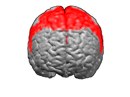
The cerebral cortex, also known as the cerebral mantle, is the outer layer of neural tissue of the cerebrum of the brain in humans and other mammals. It is the largest site of neural integration in the central nervous system, and plays a key role in attention, perception, awareness, thought, memory, language, and consciousness. The cerebral cortex is the part of the brain responsible for cognition.

In neuroanatomy, the optic chiasm, or optic chiasma, is the part of the brain where the optic nerves cross. It is located at the bottom of the brain immediately inferior to the hypothalamus. The optic chiasm is found in all vertebrates, although in cyclostomes, it is located within the brain.

The corpus callosum, also callosal commissure, is a wide, thick nerve tract, consisting of a flat bundle of commissural fibers, beneath the cerebral cortex in the brain. The corpus callosum is only found in placental mammals. It spans part of the longitudinal fissure, connecting the left and right cerebral hemispheres, enabling communication between them. It is the largest white matter structure in the human brain, about 10 cm (3.9 in) in length and consisting of 200–300 million axonal projections.

The cerebrum, telencephalon or endbrain is the largest part of the brain, containing the cerebral cortex as well as several subcortical structures, including the hippocampus, basal ganglia, and olfactory bulb. In the human brain, the cerebrum is the uppermost region of the central nervous system. The cerebrum develops prenatally from the forebrain (prosencephalon). In mammals, the dorsal telencephalon, or pallium, develops into the cerebral cortex, and the ventral telencephalon, or subpallium, becomes the basal ganglia. The cerebrum is also divided into approximately symmetric left and right cerebral hemispheres.
Brodmann area 6 (BA6) is part of the frontal cortex in the human brain. Situated just anterior to the primary motor cortex (BA4), it is composed of the premotor cortex and, medially, the supplementary motor area (SMA). This large area of the frontal cortex is believed to play a role in planning complex, coordinated movements.

The internal capsule is a paired white matter structure, as a two-way tract, carrying ascending and descending fibers, to and from the cerebral cortex. The internal capsule is situated in the inferomedial part of each cerebral hemisphere of the brain. It carries information past the subcortical basal ganglia. As it courses it separates the caudate nucleus and the thalamus from the putamen and the globus pallidus. It also separates the caudate nucleus and the putamen in the dorsal striatum, a brain region involved in motor and reward pathways.

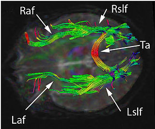
In neuroanatomy, a neural pathway is the connection formed by axons that project from neurons to make synapses onto neurons in another location, to enable neurotransmission. Neurons are connected by a single axon, or by a bundle of axons known as a nerve tract, or fasciculus. Shorter neural pathways are found within grey matter in the brain, whereas longer projections, made up of myelinated axons, constitute white matter.

The spinothalamic tract is a nerve tract in the anterolateral system in the spinal cord. This tract is an ascending sensory pathway to the thalamus. From the ventral posterolateral nucleus in the thalamus, sensory information is relayed upward to the somatosensory cortex of the postcentral gyrus.

The dorsal column–medial lemniscus pathway (DCML) (also known as the posterior column-medial lemniscus pathway is the major sensory pathway of the central nervous system that conveys sensations of fine touch, vibration, two-point discrimination, and proprioception from the skin and joints. It transmits this information to the somatosensory cortex of the postcentral gyrus in the parietal lobe of the brain. The pathway receives information from sensory receptors throughout the body, and carries this in the gracile fasciculus and the cuneate fasciculus, tracts that make up the white matter dorsal columns of the spinal cord. At the level of the medulla oblongata, the fibers of the tracts decussate and are continued in the medial lemniscus, on to the thalamus and relayed from there through the internal capsule and transmitted to the somatosensory cortex. The name dorsal-column medial lemniscus comes from the two structures that carry the sensory information: the dorsal columns of the spinal cord, and the medial lemniscus in the brainstem.

The precentral gyrus is a prominent gyrus on the surface of the posterior frontal lobe of the brain. It is the site of the primary motor cortex that in humans is cytoarchitecturally defined as Brodmann area 4.

The spinocerebellar tracts are nerve tracts originating in the spinal cord and terminating in the same side (ipsilateral) of the cerebellum. The two main tracts are the dorsal spinocerebellar tract, and the ventral spinocerebellar tract. Both of these tracts are located in the peripheral region of the lateral funiculi. Other tracts are the rostral spinocerebellar tract, and the cuneocerebellar tract.

The posterior cerebral artery (PCA) is one of a pair of cerebral arteries that supply oxygenated blood to the occipital lobe, as well as the medial and inferior aspects of the temporal lobe of the human brain. The two arteries originate from the distal end of the basilar artery, where it bifurcates into the left and right posterior cerebral arteries. These anastomose with the middle cerebral arteries and internal carotid arteries via the posterior communicating arteries.

In neuroanatomy, a sulcus is a shallow depression or groove in the cerebral cortex. One or more sulci surround a gyrus, a ridge on the surface of the cortex, creating the characteristic folded appearance of the brain in humans and most other mammals. The larger sulci are also called fissures. The cortex develops in the fetal stage of corticogenesis, preceding the cortical folding stage known as gyrification. The large fissures and main sulci are the first to develop.
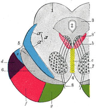
Projection fibers consist of efferent and afferent fibers uniting the cortex with the lower parts of the brain and with the spinal cord. In human neuroanatomy, bundles of axons called nerve tracts, within the brain, can be categorized by their function into association tracts, projection tracts, and commissural tracts.

The commissural fibers or transverse fibers are axons that connect the two hemispheres of the brain. Huge numbers of commissural fibers make up the commissural tracts in the brain, the largest of which is the corpus callosum.

The salivatory nuclei are two general visceral efferent nuclei located in the caudal pons, dorsal and lateral to the facial nucleus. Their neurons give rise to preganglionic parasympathetic nerve fibers in the control of salivation. The superior salivatory nucleus supplies fibers to the intermediate nerve (part of the facial nerve. The inferior salivatory nucleus supplies fibers to the glossopharyngeal nerve. The nuclei may also be involved in parasympathetic control of head vasculature.
Corticopontine fibers are projections from layer V of the cerebral cortex to the pontine nuclei of the ventral pons. They represent the first link in a cortico-cerebello-cortical pathway mediating neocerebellar control of the motor cortex. The pathway is especially important for voluntary movements.

A nerve tract is a bundle of nerve fibers (axons) connecting nuclei of the central nervous system. In the peripheral nervous system, this is known as a nerve fascicle, and has associated connective tissue. The main nerve tracts in the central nervous system are of three types: association fibers, commissural fibers, and projection fibers. A nerve tract may also be referred to as a commissure, decussation, or neural pathway. A commissure connects the two cerebral hemispheres at the same levels, while a decussation connects at different levels.
Disconnection syndrome is a general term for a collection of neurological symptoms caused – via lesions to associational or commissural nerve fibres – by damage to the white matter axons of communication pathways in the cerebrum, independent of any lesions to the cortex. The behavioral effects of such disconnections are relatively predictable in adults. Disconnection syndromes usually reflect circumstances where regions A and B still have their functional specializations except in domains that depend on the interconnections between the two regions.

