 Animation. Inferior parietal lobule is shown in red.
 Lateral view of a human brain, main gyri labeled.
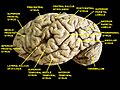 Cerebrum. Lateral view. Deep dissection.
 Cerebrum. Lateral view. Deep dissection.
 Cerebrum. Lateral view. Deep dissection.
 Inferior parietal lobule, right hemisphere view.
 Inferior parietal lobule highlighted in green on coronal T1 MRI images
 Inferior parietal lobule highlighted in green on sagittal T1 MRI images
 Inferior parietal lobule highlighted in green on transversal T1 MRI images
| Inferior parietal lobule | |
|---|---|
 Lateral surface of left cerebral hemisphere, viewed from the side. (Inferior parietal lobule is shown in orange.) | |
 Superficial anatomy of the inferior parietal lobule. LS: Lateral sulcus (Sylvian fissure), CS: Central sulcus, IPS: Intraparietal sulcus, STS:Superior temporal sulcus, PN: Preoccipital notch. | |
| Details | |
| Part of | Parietal lobe |
| Identifiers | |
| Latin | Lobulus parietalis inferior |
| NeuroNames | 107 |
| NeuroLex ID | birnlex_1194 |
| TA98 | A14.1.09.125 |
| TA2 | 5471 |
| FMA | 77536 |
| Anatomical terms of neuroanatomy | |
The inferior parietal lobule (subparietal district) lies below the horizontal portion of the intraparietal sulcus, and behind the lower part of the postcentral sulcus. Also known as Geschwind's territory after Norman Geschwind, an American neurologist, who in the early 1960s recognised its importance. [1] It is a part of the parietal lobe.


