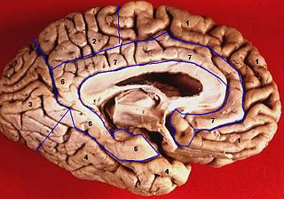
In neuroanatomy, the precuneus is the portion of the superior parietal lobule on the medial surface of each brain hemisphere. It is located in front of the cuneus. The precuneus is bounded in front by the marginal branch of the cingulate sulcus, at the rear by the parieto-occipital sulcus, and underneath by the subparietal sulcus. It is involved with episodic memory, visuospatial processing, reflections upon self, and aspects of consciousness.

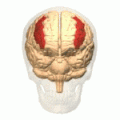
The frontal lobe is the largest of the four major lobes of the brain in mammals, and is located at the front of each cerebral hemisphere. It is parted from the parietal lobe by a groove between tissues called the central sulcus and from the temporal lobe by a deeper groove called the lateral sulcus. The most anterior rounded part of the frontal lobe is known as the frontal pole, one of the three poles of the cerebrum.

Brodmann area 44, or BA44, is part of the frontal cortex in the human brain. Situated just anterior to premotor cortex (BA6) and on the lateral surface, inferior to BA9.

The fusiform gyrus, also known as the lateral occipitotemporal gyrus,is part of the temporal lobe and occipital lobe in Brodmann area 37. The fusiform gyrus is located between the lingual gyrus and parahippocampal gyrus above, and the inferior temporal gyrus below. Though the functionality of the fusiform gyrus is not fully understood, it has been linked with various neural pathways related to recognition. Additionally, it has been linked to various neurological phenomena such as synesthesia, dyslexia, and prosopagnosia.
The limbic lobe is an arc-shaped cortical region of the limbic system, on the medial surface of each cerebral hemisphere of the mammalian brain, consisting of parts of the frontal, parietal and temporal lobes. The term is ambiguous, with some authors including the paraterminal gyrus, the subcallosal area, the cingulate gyrus, the parahippocampal gyrus, the dentate gyrus, the hippocampus and the subiculum; while the Terminologia Anatomica includes the cingulate sulcus, the cingulate gyrus, the isthmus of cingulate gyrus, the fasciolar gyrus, the parahippocampal gyrus, the parahippocampal sulcus, the dentate gyrus, the fimbrodentate sulcus, the fimbria of hippocampus, the collateral sulcus, and the rhinal sulcus, and omits the hippocampus.

In neuroanatomy, the postcentral gyrus is a prominent gyrus in the lateral parietal lobe of the human brain. It is the location of the primary somatosensory cortex, the main sensory receptive area for the sense of touch. Like other sensory areas, there is a map of sensory space in this location, called the sensory homunculus.

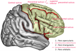
The inferior frontal gyrus (IFG),, is the lowest positioned gyrus of the frontal gyri, of the frontal lobe, and is part of the prefrontal cortex.
In neuroanatomy, the superior frontal gyrus is a gyrus – a ridge on the brain's cerebral cortex – which makes up about one third of the frontal lobe. It is bounded laterally by the superior frontal sulcus.

The postcentral sulcus of the parietal lobe lies parallel to, and behind, the central sulcus in the human brain.

The superior temporal gyrus (STG) is one of three gyri in the temporal lobe of the human brain, which is located laterally to the head, situated somewhat above the external ear.

The angular gyrus is a region of the brain lying mainly in the posteroinferior region of the parietal lobe, occupying the posterior part of the inferior parietal lobule. It represents the Brodmann area 39.
The precentral gyrus is a prominent gyrus on the surface of the posterior frontal lobe of the brain. It is the site of the primary motor cortex that in humans is cytoarchitecturally defined as Brodmann area 4.

The middle cerebral artery (MCA) is one of the three major paired cerebral arteries that supply blood to the cerebrum. The MCA arises from the internal carotid artery and continues into the lateral sulcus where it then branches and projects to many parts of the lateral cerebral cortex. It also supplies blood to the anterior temporal lobes and the insular cortices.

The lobes of the brain are the major identifiable zones of the human cerebral cortex, and they comprise the surface of each hemisphere of the cerebrum. The two hemispheres are roughly symmetrical in structure, and are connected by the corpus callosum. They traditionally have been divided into four lobes, but are today considered as having six lobes each. The lobes are large areas that are anatomically distinguishable, and are also functionally distinct to some degree. Each lobe of the brain has numerous ridges, or gyri, and furrows, the sulci that constitute further subzones of the cortex. The expression "lobes of the brain" usually refers only to those of the cerebrum, not to the distinct areas of the cerebellum.

The cuneus is a smaller lobe in the occipital lobe of the brain. The cuneus is bounded anteriorly by the parieto-occipital sulcus and inferiorly by the calcarine sulcus.

Middle temporal gyrus is a gyrus in the brain on the temporal lobe. It is located between the superior temporal gyrus and inferior temporal gyrus. It corresponds largely to Brodmann area 21.

The inferior temporal gyrus is one of three gyri of the temporal lobe and is located below the middle temporal gyrus, connected behind with the inferior occipital gyrus; it also extends around the infero-lateral border on to the inferior surface of the temporal lobe, where it is limited by the inferior sulcus. This region is one of the higher levels of the ventral stream of visual processing, associated with the representation of objects, places, faces, and colors. It may also be involved in face perception, and in the recognition of numbers and words.

The superior parietal lobule is bounded in front by the upper part of the postcentral sulcus, but is usually connected with the postcentral gyrus above the end of the sulcus. The superior parietal lobule contains Brodmann's areas 5 and 7.

The inferior parietal lobule lies below the horizontal portion of the intraparietal sulcus, and behind the lower part of the postcentral sulcus. Also known as Geschwind's territory after Norman Geschwind, an American neurologist, who in the early 1960s recognised its importance. It is a part of the parietal lobe.

The superior longitudinal fasciculus (SLF) is an association tract in the brain that is composed of three separate components. It is present in both hemispheres and can be found lateral to the centrum semiovale and connects the frontal, occipital, parietal, and temporal lobes. This bundle of tracts (fasciculus) passes from the frontal lobe through the operculum to the posterior end of the lateral sulcus where they either radiate to and synapse on neurons in the occipital lobe, or turn downward and forward around the putamen and then radiate to and synapse on neurons in anterior portions of the temporal lobe.