Parahippocampal gyrus

Last updated
Parahippocampal gyrus
Sobo 1909 630 - Parahippocampal gyrus.png
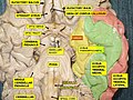
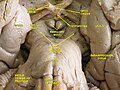
Human brain seen from below. Parahippocampal gyrus shown in blue
Cerebral Gyri - Medial Surface2.png
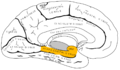
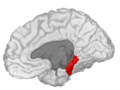
Medial view of left cerebral hemisphere. Parahippocampal gyrus shown in orange.
Details
Identifiers
Latin gyrus parahippocampalis
MeSH D020534
NeuroNames 164
NeuroLex ID birnlex_807
TA98 A14.1.09.234
TA2 5515
FMA 61918
Anatomical terminology

The parahippocampal gyrus (or hippocampal gyrus [1] ) is a grey matter cortical region of the brain that surrounds the hippocampus and is part of the limbic system. The region plays an important role in memory encoding and retrieval. It has been involved in some cases of hippocampal sclerosis. [2] Asymmetry has been observed in schizophrenia. [3]

Contents

Structure

The anterior part of the gyrus includes the perirhinal and entorhinal cortices[ citation needed ].

The term parahippocampal cortex is used to refer to an area that encompasses both the posterior parahippocampal gyrus and the medial portion of the fusiform gyrus [ citation needed ].

Function

Scene recognition

The parahippocampal place area (PPA) is a sub-region of the parahippocampal cortex that lies medially in the inferior temporo-occipital cortex. PPA plays an important role in the encoding and recognition of environmental scenes (rather than faces). fMRI studies indicate that this region of the brain becomes highly active when human subjects view topographical scene stimuli such as images of landscapes, cityscapes, or rooms (i.e. images of "places"). Furthermore, according to work by Pierre Mégevand et al. in 2014, stimulation of the region via intracranial electrodes yields intense topographical visual hallucinations of places and situations. [4] The region was first described by Russell Epstein and Nancy Kanwisher in 1998 at MIT, [5] see also other similar reports by Geoffrey Aguirre [6] [7] and Alumit Ishai. [8]

Damage to the PPA (for example, due to stroke) often leads to a syndrome in which patients cannot visually recognize scenes even though they can recognize the individual objects in the scenes (such as people, furniture, etc.). The PPA is often considered the complement of the fusiform face area (FFA), a nearby cortical region that responds strongly whenever faces are viewed, and that is believed to be important for face recognition.

Social context

Additional research has suggested that the right parahippocampal gyrus in particular has functions beyond the contextualizing of visual background. Tests by a California-based group led by Katherine P. Rankin indicate that the lobe may play a crucial role in identifying social context as well, including paralinguistic elements of verbal communication. [9] For example, Rankin's research suggests that the right parahippocampal gyrus enables people to detect sarcasm.

Additional images

Related Research Articles

<span class="mw-page-title-main">Entorhinal cortex</span> Area of the temporal lobe of the brain

The entorhinal cortex (EC) is an area of the brain's allocortex, located in the medial temporal lobe, whose functions include being a widespread network hub for memory, navigation, and the perception of time. The EC is the main interface between the hippocampus and neocortex. The EC-hippocampus system plays an important role in declarative (autobiographical/episodic/semantic) memories and in particular spatial memories including memory formation, memory consolidation, and memory optimization in sleep. The EC is also responsible for the pre-processing (familiarity) of the input signals in the reflex nictitating membrane response of classical trace conditioning; the association of impulses from the eye and the ear occurs in the entorhinal cortex.

<span class="mw-page-title-main">Brodmann area</span> Region of the brain

A Brodmann area is a region of the cerebral cortex, in the human or other primate brain, defined by its cytoarchitecture, or histological structure and organization of cells.

<span class="mw-page-title-main">Precuneus</span> Region of the parietal lobe of the brain

In neuroanatomy, the precuneus is the portion of the superior parietal lobule on the medial surface of each brain hemisphere. It is located in front of the cuneus. The precuneus is bounded in front by the marginal branch of the cingulate sulcus, at the rear by the parieto-occipital sulcus, and underneath by the subparietal sulcus. It is involved with episodic memory, visuospatial processing, reflections upon self, and aspects of consciousness.

<span class="mw-page-title-main">Temporal lobe</span> One of the four lobes of the mammalian brain

The temporal lobe is one of the four major lobes of the cerebral cortex in the brain of mammals. The temporal lobe is located beneath the lateral fissure on both cerebral hemispheres of the mammalian brain.

A mental image is an experience that, on most occasions, significantly resembles the experience of 'perceiving' some object, event, or scene, but occurs when the relevant object, event, or scene is not actually present to the senses. There are sometimes episodes, particularly on falling asleep and waking up, when the mental imagery may be dynamic, phantasmagoric and involuntary in character, repeatedly presenting identifiable objects or actions, spilling over from waking events, or defying perception, presenting a kaleidoscopic field, in which no distinct object can be discerned. Mental imagery can sometimes produce the same effects as would be produced by the behavior or experience imagined.

<span class="mw-page-title-main">Fusiform gyrus</span> Gyrus of the temporal and occipital lobes of the brain

The fusiform gyrus, also known as the lateral occipitotemporal gyrus,is part of the temporal lobe and occipital lobe in Brodmann area 37. The fusiform gyrus is located between the lingual gyrus and parahippocampal gyrus above, and the inferior temporal gyrus below. Though the functionality of the fusiform gyrus is not fully understood, it has been linked with various neural pathways related to recognition. Additionally, it has been linked to various neurological phenomena such as synesthesia, dyslexia, and prosopagnosia.

<span class="mw-page-title-main">Limbic lobe</span> Region of a cerebral cortex

The limbic lobe is an arc-shaped cortical region of the limbic system, on the medial surface of each cerebral hemisphere of the mammalian brain, consisting of parts of the frontal, parietal and temporal lobes. The term is ambiguous, with some authors including the paraterminal gyrus, the subcallosal area, the cingulate gyrus, the parahippocampal gyrus, the dentate gyrus, the hippocampus and the subiculum; while the Terminologia Anatomica includes the cingulate sulcus, the cingulate gyrus, the isthmus of cingulate gyrus, the fasciolar gyrus, the parahippocampal gyrus, the parahippocampal sulcus, the dentate gyrus, the fimbrodentate sulcus, the fimbria of hippocampus, the collateral sulcus, and the rhinal sulcus, and omits the hippocampus.

<span class="mw-page-title-main">Superior frontal gyrus</span> Region of the frontal lobe of the brain

In neuroanatomy, the superior frontal gyrus is a gyrus – a ridge on the brain's cerebral cortex – which makes up about one third of the frontal lobe. It is bounded laterally by the superior frontal sulcus.

Visual processing is a term that is used to refer to the brain's ability to use and interpret visual information from the world around us. The process of converting light energy into a meaningful image is a complex process that is facilitated by numerous brain structures and higher level cognitive processes. On an anatomical level, light energy first enters the eye through the cornea, where the light is bent. After passing through the cornea, light passes through the pupil and then lens of the eye, where it is bent to a greater degree and focused upon the retina. The retina is where a group of light-sensing cells, called photoreceptors are located. There are two types of photoreceptors: rods and cones. Rods are sensitive to dim light and cones are better able to transduce bright light. Photoreceptors connect to bipolar cells, which induce action potentials in retinal ganglion cells. These retinal ganglion cells form a bundle at the optic disc, which is a part of the optic nerve. The two optic nerves from each eye meet at the optic chiasm, where nerve fibers from each nasal retina cross which results in the right half of each eye's visual field being represented in the left hemisphere and the left half of each eye's visual fields being represented in the right hemisphere. The optic tract then diverges into two visual pathways, the geniculostriate pathway and the tectopulvinar pathway, which send visual information to the visual cortex of the occipital lobe for higher level processing.

<span class="mw-page-title-main">Brodmann areas 35 and 36</span>

Brodmann area 35, together with Brodmann area 36, comprise the perirhinal cortex. They are cytoarchitecturally defined temporal regions of the cerebral cortex.

<span class="mw-page-title-main">Inferior temporal gyrus</span> One of three gyri of the temporal lobe of the brain

The inferior temporal gyrus is one of three gyri of the temporal lobe and is located below the middle temporal gyrus, connected behind with the inferior occipital gyrus; it also extends around the infero-lateral border on to the inferior surface of the temporal lobe, where it is limited by the inferior sulcus. This region is one of the higher levels of the ventral stream of visual processing, associated with the representation of objects, places, faces, and colors. It may also be involved in face perception, and in the recognition of numbers and words.

<span class="mw-page-title-main">Lingual gyrus</span> Gyrus of the occipital lobe of the brain

The lingual gyrus, also known as the medialoccipitotemporal gyrus, is a brain structure that is linked to processing vision, especially related to letters. It is thought to also play a role in analysis of logical conditions and encoding visual memories. It is named after its shape, which is somewhat similar to a tongue. Contrary to the name, the region has little to do with speech.

<span class="mw-page-title-main">Uncus</span>

The uncus is an anterior extremity of the parahippocampal gyrus. It is separated from the apex of the temporal lobe by a slight fissure called the incisura temporalis.

<span class="mw-page-title-main">Colour centre</span> Brain region responsible for colour processing

The colour centre is a region in the brain primarily responsible for visual perception and cortical processing of colour signals received by the eye, which ultimately results in colour vision. The colour centre in humans is thought to be located in the ventral occipital lobe as part of the visual system, in addition to other areas responsible for recognizing and processing specific visual stimuli, such as faces, words, and objects. Many functional magnetic resonance imaging (fMRI) studies in both humans and macaque monkeys have shown colour stimuli to activate multiple areas in the brain, including the fusiform gyrus and the lingual gyrus. These areas, as well as others identified as having a role in colour vision processing, are collectively labelled visual area 4 (V4). The exact mechanisms, location, and function of V4 are still being investigated.

<span class="mw-page-title-main">Fusiform face area</span> Part of the human visual system that is specialized for facial recognition

The fusiform face area is a part of the human visual system that is specialized for facial recognition. It is located in the inferior temporal cortex (IT), in the fusiform gyrus.

The trisynaptic circuit, or trisynaptic loop is a relay of synaptic transmission in the hippocampus. The circuit was initially described by the neuroanatomist Santiago Ramon y Cajal, in the early twentieth century, using the Golgi staining method. After the discovery of the trisynaptic circuit, a series of research has been conducted to determine the mechanisms driving this circuit. Today, research is focused on how this loop interacts with other parts of the brain, and how it influences human physiology and behaviour. For example, it has been shown that disruptions within the trisynaptic circuit leads to behavioural changes in rodent and feline models.

Discrete categories of objects such as faces, body parts, tools, animals and buildings have been associated with preferential activation in specialised areas of the cerebral cortex, leading to the suggestion that they may be produced separately in discrete neural regions.

Visual object recognition refers to the ability to identify the objects in view based on visual input. One important signature of visual object recognition is "object invariance", or the ability to identify objects across changes in the detailed context in which objects are viewed, including changes in illumination, object pose, and background context.

<span class="mw-page-title-main">Functional specialization (brain)</span> Neuroscientific theory that different regions of the brain are specialized for different functions

In neuroscience, functional specialization is a theory which suggests that different areas in the brain are specialized for different functions.

The occipital face area (OFA) is a region of the human cerebral cortex which is specialised for face perception. The OFA is located on the lateral surface of the occipital lobe adjacent to the inferior occipital gyrus. The OFA comprises a network of brain regions including the fusiform face area (FFA) and posterior superior temporal sulcus (STS) which support facial processing.

References

  1. Reuter P.: Der Grobe Reuter Springer Universalworterbuch Medizin, Pharmakologie Und Zahnmedizin: Englisch-deutsch (Band 2), Birkhäuser, 2005, ISBN   3-540-25102-2, p. 648 here online
  2. Ferreira NF, de Oliveira V, Amaral L, Mendonça R, Lima SS (September 2003). "Analysis of parahippocampal gyrus in 115 patients with hippocampal sclerosis". Arq Neuropsiquiatr. 61 (3B): 707–11. doi: 10.1590/s0004-282x2003000500001 . PMID   14595469.
  3. McDonald B, Highley JR, Walker MA, et al. (January 2000). "Anomalous asymmetry of fusiform and parahippocampal gyrus gray matter in schizophrenia: A postmortem study". Am J Psychiatry. 157 (1): 40–7. doi:10.1176/ajp.157.1.40. PMID   10618011. S2CID   5915984.
  4. Mégevand P, Groppe DM, Goldfinger MS, et al. (2014). "Seeing Scenes: Topographic Visual Hallucinations Evoked by Direct Electrical Stimulation of the Parahippocampal Place Area". Journal of Neuroscience. 34 (16): 5399–5405. doi:10.1523/jneurosci.5202-13.2014. PMC   6608225 . PMID   24741031.
  5. Epstein R (1998). "A cortical representation of the local visual environment". Nature. 392 (6676): 598–601. Bibcode:1998Natur.392..598E. doi:10.1038/33402. PMID   9560155. S2CID   920141.
  6. Aguirre; et al. (1996). "The Parahippocampus Subserves Topographical Learning in Man". Cerebral Cortex. 6 (6): 823–9. doi: 10.1093/cercor/6.6.823 . PMID   8922339.
  7. "Neuron - An Area within Human Ventral Cortex Sensitive to "Building" Stimuli". Archived from the original on 2013-01-13. Retrieved 2009-11-03.
  8. Ishai A (1999). "Distributed representation of objects in the human ventral visual pathway — PNAS". Proceedings of the National Academy of Sciences. 96 (16): 9379–9384. Bibcode:1999PNAS...96.9379I. doi: 10.1073/pnas.96.16.9379 . PMC   17791 . PMID   10430951.
  9. Hurley, Dan (2008-06-03). "Katherine P. Rankin, a Neuropsychologist, Studies Sarcasm - NYTimes.com". The New York Times. Retrieved 2009-11-03.