Articles related to anatomy include:

Brodmann area 40 (BA40) is part of the parietal cortex in the human brain. The inferior part of BA40 is in the area of the supramarginal gyrus, which lies at the posterior end of the lateral fissure, in the inferior lateral part of the parietal lobe.

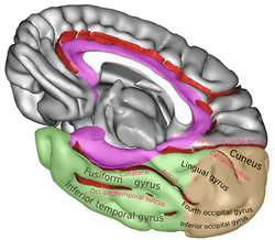
The fusiform gyrus, also known as the lateral occipitotemporal gyrus,is part of the temporal lobe and occipital lobe in Brodmann area 37. The fusiform gyrus is located between the lingual gyrus and parahippocampal gyrus above, and the inferior temporal gyrus below. Though the functionality of the fusiform gyrus is not fully understood, it has been linked with various neural pathways related to recognition. Additionally, it has been linked to various neurological phenomena such as synesthesia, dyslexia, and prosopagnosia.

In neuroanatomy, the postcentral gyrus is a prominent gyrus in the lateral parietal lobe of the human brain. It is the location of the primary somatosensory cortex, the main sensory receptive area for the sense of touch. Like other sensory areas, there is a map of sensory space in this location, called the sensory homunculus.

The inferior frontal gyrus is the lowest positioned gyrus of the frontal gyri, of the frontal lobe, and is part of the prefrontal cortex.
The precentral gyrus is a prominent gyrus on the surface of the posterior frontal lobe of the brain. It is the site of the primary motor cortex that in humans is cytoarchitecturally defined as Brodmann area 4.
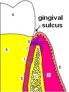
In biological morphology and anatomy, a sulcus is a furrow or fissure. It may be a groove, natural division, deep furrow, elongated cleft, or tear in the surface of a limb or an organ, most notably on the surface of the brain, but also in the lungs, certain muscles, as well as in bones, and elsewhere. Many sulci are the product of a surface fold or junction, such as in the gums, where they fold around the neck of the tooth.

The middle cerebral artery (MCA) is one of the three major paired cerebral arteries that supply blood to the cerebrum. The MCA arises from the internal carotid artery and continues into the lateral sulcus where it then branches and projects to many parts of the lateral cerebral cortex. It also supplies blood to the anterior temporal lobes and the insular cortices.

The lobes of the brain are the four major identifiable regions of the human cerebral cortex, and they comprise the surface of each hemisphere of the cerebrum. The two hemispheres are roughly symmetrical in structure, and are connected by the corpus callosum. Some sources include the insula and limbic lobe but the limbic lobe incorporates parts of the other lobes. The lobes are large areas that are anatomically distinguishable, and are also functionally distinct. Each lobe of the brain has numerous ridges, or gyri, and furrows, sulci that constitute further subzones of the cortex. The expression "lobes of the brain" usually refers only to those of the cerebrum, not to the distinct areas of the cerebellum.

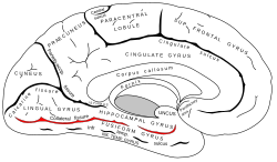
In neuroanatomy, the parieto-occipital sulcus is a deep sulcus in the cerebral cortex that marks the boundary between the cuneus and precuneus, and also between the parietal and occipital lobes. Only a small part can be seen on the lateral surface of the hemisphere, its chief part being on the medial surface.

The calcarine sulcus is an anatomical landmark located at the caudal end of the medial surface of the brain of humans and other primates. Its name comes from the Latin "calcar" meaning "spur". It is very deep, and known as a complete sulcus.

The inferior parietal lobule lies below the horizontal portion of the intraparietal sulcus, and behind the lower part of the postcentral sulcus. Also known as Geschwind's territory after Norman Geschwind, an American neurologist, who in the early 1960s recognised its importance. It is a part of the parietal lobe.

The lingual gyrus, also known as the medialoccipitotemporal gyrus, is a brain structure that is linked to processing vision, especially related to letters. It is thought to also play a role in analysis of logical conditions and encoding visual memories. It is named after its shape, which is somewhat similar to a tongue. Contrary to the name, the region has little to do with speech.

In neuroanatomy, a sulcus is a shallow depression or groove in the cerebral cortex. One or more sulci surround a gyrus, a ridge on the surface of the cortex, creating the characteristic folded appearance of the brain in humans and most other mammals. The larger sulci are also called fissures. The cortex develops in the fetal stage of corticogenesis, preceding the cortical folding stage known as gyrification. The large fissures and main sulci are the first to develop.

In the human brain, the entorhinal cortex appears as a longitudinal elevation anterior to the parahippocampal gyrus, with a corresponding internal furrow, the external rhinal sulcus. The rhinal sulcus separates the parahippocampal uncus from the rest of the temporal lobe in the neocortex. The rhinal sulcus and the hippocampal sulcus were both present in early mammals.
The sensory cortex can refer sometimes to the primary somatosensory cortex, or it can be used as a term for the primary and secondary cortices of the different senses : the visual cortex on the occipital lobes, the auditory cortex on the temporal lobes, the primary olfactory cortex on the uncus of the piriform region of the temporal lobes, the gustatory cortex on the insular lobe, and the primary somatosensory cortex on the anterior parietal lobes. Just posterior to the primary somatosensory cortex lies the somatosensory association cortex or area, which integrates sensory information from the primary somatosensory cortex to construct an understanding of the object being felt. Inferior to the frontal lobes are found the olfactory bulbs, which receive sensory input from the olfactory nerves and route those signals throughout the brain. Not all olfactory information is routed to the olfactory cortex: some neural fibers are routed to the supraorbital region of the frontal lobe, while others are routed directly to limbic structures. The direct limbic connection makes the olfactory sense unique.
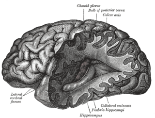
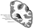
The collateral eminence is an elongated swelling lying lateral parallel with the hippocampus. It corresponds with the medial part of the collateral fissure, and its size depends on the depth and direction of this fissure. It is continuous behind with a flattened triangular area, the trigone of the lateral ventricle, situated between the posterior and inferior horn. It is not always present.

The hippocampal sulcus, also known as the hippocampal fissure, is a sulcus that separates the dentate gyrus from the subiculum and the CA1 field in the hippocampus.

The inferior surface of the temporal lobe is concave, and is continuous posteriorly with the tentorial surface of the occipital lobe. It is traversed by the occipitotemporal sulcus, also known as the lateral occipitotemporal sulcus which extends from near the occipital pole behind, to within a short distance of the temporal pole in front, but is frequently subdivided by bridging gyri.

The occipital gyri (OcG) are three gyri in parallel, along the lateral portion of the occipital lobe, also referred to as a composite structure in the brain. The gyri are the superior occipital gyrus, the middle occipital gyrus, and the inferior occipital gyrus, and these are also known as the occipital face area. The superior and inferior occipital sulci separates the three occipital gyri.