The location of the precuneus makes it difficult to study. Furthermore, it is rarely subject to isolated injury due to strokes, or trauma such as gunshot wounds. This has resulted in it being "one of the less accurately mapped areas of the whole cortical surface".[1] While originally described as homogeneous by Korbinian Brodmann, it is now appreciated to contain three subdivisions.[2]
It is also known after Achille-Louis Foville as the quadrate lobule of Foville.[1][3][4] The Latin form of praecuneus was first used in 1868 and the English precuneus in 1879.[5]
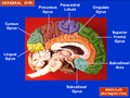
Structure
Subdivisions of precuneus and posterior cingulate in the human based upon resting state functional connectivity.
Blue: Sensorimotor Anterior Region and its connections
Axon tracing research on macaque monkeys has established that it consists of three subdivisions which now have been confirmed by fMRI upon resting-state functional connectivity to also exist in humans (parallel fMRI research has also been done upon monkeys).[2]
The mental imagery concerning the self has been located in the forward part of the precuneus with posterior areas being involved with episodic memory.[6] Another area has been linked to visuospatial imagery. (It is not though clear how these—and the functions noted below—link with the above three subdivisions.)
The precuneus plays a role in itch sensations (there are many different types of itch) and their brain processing [7] "'We can't [yet] pinpoint what the precuneus does in itch, but it's uniquely activated with itch and not pain.'" [8]
Self
Functional imaging has linked the precuneus to the processes involved in self-consciousness, such as reflective self-awareness, that involve rating one's own personality traits compared to those judged of other people.[9][10]
The precuneus is involved in memory tasks, such as when people look at images and try to respond based on what they have remembered in regard to verbal questions about their spatial details.[12] It is involved with the left prefrontal cortex in the recall of episodic memories[13][14] including past episodes related to the self.[10] The precuneus is also involved in source memory (in which the "source" circumstances of a memory are recalled) with the left inferior prefrontal cortex: here its role is postulated to be providing rich episodic contextual associations used by the prefrontal cortex to select the correct past memory.[15] In the recollection of memories, it has been postulated that the precuneus discerns whether contextual information exists that can be useful for involving the aid of the hippocampus.[citation needed] Alternatively it has a different involvement when judging the familiarity as it decides whether the processing of perceptual features would be more useful.[16] In this way the precuneus gets involved in diverse processes such as attention, episodic memory retrieval, working memory and conscious perception.[16]
Visuospatial
The precuneus has been suggested to be involved in directing attention in space both when an individual makes movements and when imaging or preparing them.[1][17] It is involved in motor imagery and shifting attention between motor targets.[1] It is also involved in motor coordination that requires shifting attention to different spatial locations.[18] It is also together with the dorsal premotor cortex involved in visuospatial mental operations (such as in a modified form of the game of Amidakuji). It is suggested that while the premotor area engages in the mental operation, the precuneus aids monitoring the success of that operation in terms of internally represented visual images.[19]
The precuneus' role in mental imagery has been suggested to extend to that of modeling other people's views. It is activated when a person takes a third-person versus first-person visual point of view.[20] Together with the superior frontal gyrus and orbitofrontal cortex, the precuneus is activated when people make judgments that require understanding whether to act out of empathy and forgiveness.[21]
Executive functions
Precuneus is thought to be related to response inhibition.[22]
Consciousness
It has been suggested that together with the posterior cingulate, the precuneus is "pivotal for conscious information processing".[23] The evidence for this link with consciousness comes from the effects of its disruption in epilepsy, brain lesions and vegetative state.[3][23] Also, cerebral glucose metabolism is at its highest in these two areas during wakefulness but is most reduced in them during anesthesia.[3][23] In addition, it is one of the areas of the brain most deactivated during slow-wave sleep and rapid eye movement sleep.[3]
Together with the prefrontal cortex, the precuneus, is more activated upon the learning of words briefly flashed when they are supraliminal (and so enter consciousness) than subliminal (and so do not enter consciousness).[24]
Default network
It has been suggested to be the 'core node' or 'hub' of the default mode network that is activated during "resting consciousness" in which people do not engage intentionally in sensory or motor activity.[3] This involvement in the default network is suggested to underlie its role in self-consciousness. However its involvement in the default network has been questioned.[2][25] Though one of the authors raising these doubts noted "our findings in this regard should be treated as preliminary."[2] A later study in 2012 showed that only ventral precuneus is involved in the default network.[26]
Parietal prefrontal central hub
Olaf Sporns and Ed Bullmore have proposed that its functions link to its role as a central and well connected "small-world network" hub between parietal and prefrontal regions.
These clusters or modules are interlinked by specialized hub regions, ensuring that overall path lengths across the network are short. Most studies identified [such] hubs among parietal and prefrontal regions, providing a potential explanation for their well-documented activation by many cognitive functions. Particularly notable is the prominent structural role of the precuneus, a region that is homologous to the highly connected posteromedial cortex in the macaque. The precuneus is involved in self-referential processing, imagery and memory, and its deactivation is associated with anaesthetic-induced loss of consciousness. An intriguing hypothesis suggests that these functional aspects can be explained on the basis of its high centrality in the cortical network.[27]
Correlation of grey matter volume and subjective happiness score
A positive relationship has been found between the volume of grey matter in the right precuneus and the subject's subjective happiness score.[28]
Impact of mindfulness
A 6-week mindfulness based intervention was found to correlate with a significant grey matter increase within the precuneus.[29]
Other animals
The precuneus seems to be a recently expanded part of the brain, as in less developed primates such as New world monkeys "the superior parietal and precuneate regions are poorly developed".[1] It has been noted that "the precuneus is more highly developed (i.e. comprises a larger portion of the brain volume) in human beings than in non-human primates or other animals, has the most complex columnar cortical organization and is among the last regions to myelinate".[1]
Additional images
Precuneus of left cerebral hemisphere (shown in red).
Medial surface of left cerebral hemisphere. (Precuneus visible at top left.)
Medial surface of left cerebral hemisphere. (Precuneus colored in red.)
Boundaries of precuneus are defined by the three sulci. (shown in red)
↑ Foville AL. (1844). Traité complêt de l'anatomie, de la physiologie et de la pathologie du système nerveux cérébro-spinal. Paris, France: Fortin, Masson
↑ Sadigh-Eteghad S, Majdi A, Farhoudi M, Talebi M, Mahmoudi J (2014). "Different patterns of brain activation in normal aging and Alzheimer's disease from cognitional sight: meta analysis using activation likelihood estimation". Journal of the Neurological Sciences. 343 (1): 159–66. doi:10.1016/j.jns.2014.05.066. PMID24950901. S2CID24359894.
↑ Kawashima R, Roland PE, O'Sullivan BT (1995). "Functional anatomy of reaching and visuomotor learning: a positron emission tomography study". Cereb Cortex. 5 (2): 111–22. doi:10.1093/cercor/5.2.111. PMID7620288.
↑ Wenderoth N, Debaere F, Sunaert S, Swinnen SP (2005). "The role of anterior cingulate cortex and precuneus in the coordination of motor behaviour". Eur J Neurosci. 22 (1): 235–46. doi:10.1111/j.1460-9568.2005.04176.x. PMID16029213. S2CID25754084.
↑ Oshio R, Tanaka S, Sadato N, Sokabe M, Hanakawa T, Honda M (2010). "Differential effect of double-pulse TMS applied to dorsal premotor cortex and precuneus during internal operation of visuospatial information". NeuroImage. 49 (1): 1108–15. doi:10.1016/j.neuroimage.2009.07.034. PMID19632337. S2CID17788847.
↑ Vogeley K, May M, Ritzl A, Falkai P, Zilles K, Fink GR (2004). "Neural correlates of first-person perspective as one constituent of human self-consciousness". J Cogn Neurosci. 16 (5): 817–27. doi:10.1162/089892904970799. PMID15200709. S2CID17578549.
↑ Farrow TF, Zheng Y, Wilkinson ID, Spence SA, Deakin JF, Tarrier N, Griffiths PD, Woodruff PW (2001). "Investigating the functional anatomy of empathy and forgiveness". NeuroReport. 12 (11): 2433–8. doi:10.1097/00001756-200108080-00029. PMID11496124. S2CID34437619.
1 2 3 Vogt BA, Laureys S (2005). "Posterior cingulate, precuneal and retrosplenial cortices: cytology and components of the neural network correlates of consciousness". The Boundaries of Consciousness: Neurobiology and Neuropathology. Progress in Brain Research. Vol.150. pp.205–17. doi:10.1016/S0079-6123(05)50015-3. ISBN978-0-444-51851-4. PMC2679949. PMID16186025.
↑ Kjaer TW, Nowak M, Kjaer KW, Lou AR, Lou HC (2001). "Precuneus-prefrontal activity during awareness of visual verbal stimuli". Conscious. Cogn. 10 (3): 356–65. doi:10.1006/ccog.2001.0509. PMID11697869. S2CID39449352.
This page is based on this Wikipedia article Text is available under the CC BY-SA 4.0 license; additional terms may apply. Images, videos and audio are available under their respective licenses.