Pulvinar nuclei

Last updated
Pulvinar nuclei
Gray719.png
Hind- and mid-brains; postero-lateral view. (Pulvinar visible near top.)
ThalamicNuclei.svg
Details
Part of thalamus
Identifiers
Latin nuclei pulvinares (the nuclei plurally); pulvinar thalami (the set of nuclei singularly)
MeSH D020649
NeuroNames 328
NeuroLex ID birnlex_824
TA98 A14.1.08.104
A14.1.08.610
TA2 5665, 5698
FMA 62178
Anatomical terms of neuroanatomy

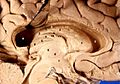
The pulvinar nuclei or nuclei of the pulvinar (nuclei pulvinares) are the nuclei (cell bodies of neurons) located in the thalamus (a part of the vertebrate brain). [1] As a group they make up the collection called the pulvinar of the thalamus (pulvinar thalami), usually just called the pulvinar.

Contents

The pulvinar is usually grouped as one of the lateral thalamic nuclei in rodents and carnivores, and stands as an independent complex in primates.

Structure

By convention, the pulvinar is divided into four nuclei:

TA alphanumeric identifierTA nameEnglish translation
A14.1.08.611nucleus pulvinaris anterior anterior pulvinar nucleus
A14.1.08.612nucleus pulvinaris inferior inferior pulvinar nucleus
A14.1.08.613nucleus pulvinaris lateralis lateral pulvinar nucleus
A14.1.08.614nucleus pulvinaris medialis medial pulvinar nucleus

Their connectomic details are as follows:

Clinical significance

Lesions of the pulvinar can result in neglect syndromes and attentional deficits. [7] In addition, lesions in early life can impact normal visuomotor behaviors such as reaching and grasping. [8] Furthermore, the pulvinar was demonstrated to be instrumental in the preservation of vision afforded to a boy who lost his primary visual cortex bilaterally at birth [9] as well as other forms of blindsight in monkeys [10] [11] and humans. [12] Strokes affecting the pulvinar have also been implicated in the development of chronic pain. [13] In a case study of photophobia caused by blue light, pulvinar nuclei associated with the melanopsin containing ipRGCs visual pathway where bilaterally activated. [14]

Other animals

The pulvinar varies in importance in different animals: it is virtually nonexistent in the rat, and grouped as the lateral posterior-pulvinar complex with the lateral posterior thalamic nucleus due to its small size in cats. In humans it makes up roughly 40% of the thalamus making it the largest of its nuclei. [15] Significant research has been undertaken in the marmoset examining the role of the retinorecipient region of the inferior pulvinar (medial subdivision), which projects to visual cortical area MT, in the early development of MT and the dorsal stream, as well as following early-life lesions of the primary visual cortex (V1). [16] [17] [18]

Etymology

The word pulvinar ( /pʌlˈvnər/ ) in Latin broadly means an armchair lined with numerous pillows. It was first neuroanatomically named by Karl Friedrich Burdach in 1817: [19] "The cushion (pulvinar), a swelling at the posterior end of the inner edge of the upper quadrigemina like a pillow over seats", English translation [20] (original German: "Das Polster (pulvinar), eine Anschwellung am hintern Ende des inner Randes der obern Vierhügel wie ein Kissen herüber legt" [19] ). In Latin pulvinus could refer to "a sofa, cushioned seat, seat of honor, easy couch; of the couch or marriage-bed ", or more specifically, "a couch made of cushions, and spread over with a splendid covering, for the gods and persons who received divine honors; a couch or cushioned seat of the gods" [21] In the religion of ancient Rome, a pulvinar was an hetoimasia or empty throne, cushioned for occupation by a deity. [22] While anatomically, neuroanatomically there was no Roman deity between its arms, there was the pineal gland, that had in the 17th century, been identified by the French philosopher René Descartes as the seat of intellect and soul, and it has been suggested this link contributed to the first naming of this part of the brain by Karl Friedrich Burdach. [20]

Related Research Articles

<span class="mw-page-title-main">Thalamus</span> Structure within the brain

The thalamus is a large mass of gray matter located in the dorsal part of the diencephalon. Nerve fibers project out of the thalamus to the cerebral cortex in all directions, known as the thalamocortical radiations, allowing hub-like exchanges of information. It has several functions, such as the relaying of sensory signals, including motor signals to the cerebral cortex and the regulation of consciousness, sleep, and alertness.

<span class="mw-page-title-main">Basal ganglia</span> Group of subcortical nuclei involved in the motor and reward systems

The basal ganglia (BG), or basal nuclei, are a group of subcortical nuclei found in the brains of vertebrates. In humans and some primates, differences exist, primarily in the division of the globus pallidus into external and internal regions, and in the division of the striatum. Positioned at the base of the forebrain and the top of the midbrain, they have strong connections with the cerebral cortex, thalamus, brainstem and other brain areas. The basal ganglia are associated with a variety of functions, including regulating voluntary motor movements, procedural learning, habit formation, conditional learning, eye movements, cognition, and emotion.

Blindsight is the ability of people who are cortically blind to respond to visual stimuli that they do not consciously see due to lesions in the primary visual cortex, also known as the striate cortex or Brodmann Area 17. The term was coined by Lawrence Weiskrantz and his colleagues in a paper published in a 1974 issue of Brain. A previous paper studying the discriminatory capacity of a cortically blind patient was published in Nature in 1973. The assumed existence of blindsight is controversial, with some arguing that it is merely degraded conscious vision.

<span class="mw-page-title-main">Lateral geniculate nucleus</span> Component of the visual system in the brains thalamus

In neuroanatomy, the lateral geniculate nucleus is a structure in the thalamus and a key component of the mammalian visual pathway. It is a small, ovoid, ventral projection of the thalamus where the thalamus connects with the optic nerve. There are two LGNs, one on the left and another on the right side of the thalamus. In humans, both LGNs have six layers of neurons alternating with optic fibers.

<span class="mw-page-title-main">Cingulate cortex</span> Part of the brain within the cerebral cortex

The cingulate cortex is a part of the brain situated in the medial aspect of the cerebral cortex. The cingulate cortex includes the entire cingulate gyrus, which lies immediately above the corpus callosum, and the continuation of this in the cingulate sulcus. The cingulate cortex is usually considered part of the limbic lobe.

<span class="mw-page-title-main">Parietal lobe</span> Part of the brain responsible for sensory input and some language processing

The parietal lobe is one of the four major lobes of the cerebral cortex in the brain of mammals. The parietal lobe is positioned above the temporal lobe and behind the frontal lobe and central sulcus.

<span class="mw-page-title-main">Mammillary body</span> Part of the limbic system

The mammillary bodies are a pair of small round bodies, located on the undersurface of the brain that, as part of the diencephalon, form part of the limbic system. They are located at the ends of the anterior arches of the fornix. They consist of two groups of nuclei, the medial mammillary nuclei and the lateral mammillary nuclei.

<span class="mw-page-title-main">Internal capsule</span> White matter structure situated in the inferomedial part of each cerebral hemisphere of the brain

The internal capsule is a white matter structure situated in the inferomedial part of each cerebral hemisphere of the brain. It carries information past the basal ganglia, separating the caudate nucleus and the thalamus from the putamen and the globus pallidus. The internal capsule contains both ascending and descending axons, going to and coming from the cerebral cortex. It also separates the caudate nucleus and the putamen in the dorsal striatum, a brain region involved in motor and reward pathways.

<span class="mw-page-title-main">Superior colliculus</span> Structure in the midbrain

In neuroanatomy, the superior colliculus is a structure lying on the roof of the mammalian midbrain. In non-mammalian vertebrates, the homologous structure is known as the optic tectum, or optic lobe. The adjective form tectal is commonly used for both structures.

<span class="mw-page-title-main">Pretectal area</span> Structure in the midbrain which mediates responses to ambient light

In neuroanatomy, the pretectal area, or pretectum, is a midbrain structure composed of seven nuclei and comprises part of the subcortical visual system. Through reciprocal bilateral projections from the retina, it is involved primarily in mediating behavioral responses to acute changes in ambient light such as the pupillary light reflex, the optokinetic reflex, and temporary changes to the circadian rhythm. In addition to the pretectum's role in the visual system, the anterior pretectal nucleus has been found to mediate somatosensory and nociceptive information.

<span class="mw-page-title-main">Reticular formation</span> Spinal trigeminal nucleus

The reticular formation is a set of interconnected nuclei that are located throughout the brainstem. It is not anatomically well defined, because it includes neurons located in different parts of the brain. The neurons of the reticular formation make up a complex set of networks in the core of the brainstem that extend from the upper part of the midbrain to the lower part of the medulla oblongata. The reticular formation includes ascending pathways to the cortex in the ascending reticular activating system (ARAS) and descending pathways to the spinal cord via the reticulospinal tracts.

<span class="mw-page-title-main">Thalamocortical radiations</span> Neural pathways between the thalamus and cerebral cortex

In neuroanatomy, thalamocortical radiations also known as thalamocortical fibres, are the efferent fibres that project from the thalamus to distinct areas of the cerebral cortex. They form fibre bundles that emerge from the lateral surface of the thalamus.

The zona incerta (ZI) is a horizontally elongated region of gray matter in the subthalamus below the thalamus. Its connections project extensively over the brain from the cerebral cortex down into the spinal cord.

<span class="mw-page-title-main">Medial dorsal nucleus</span>

The medial dorsal nucleus is a large nucleus in the thalamus.

The isothalamus is a division used by some researchers in describing the thalamus.

<span class="mw-page-title-main">Inferior pulvinar nucleus</span>

Inferior pulvinar nucleus is one of four traditionally anatomically distinguished nuclei of the pulvinar of the thalamus. The other three nuclei of the pulvinar are called lateral, anterior and medial pulvinar nuclei.

<span class="mw-page-title-main">Lateral pulvinar nucleus</span>

Lateral pulvinar nucleus is one of four traditionally anatomically distinguished nuclei of the pulvinar of the thalamus. The other three nuclei of the pulvinar are called anterior, inferior and medial pulvinar nuclei.

<span class="mw-page-title-main">Medial pulvinar nucleus</span>

Medial pulvinar nucleus is one of four traditionally anatomically distinguished nuclei of the pulvinar of the thalamus. The other three nuclei of the pulvinar are called lateral, inferior and anterior pulvinar nuclei.

The tectopulvinar pathway and the geniculostriate pathway are the two visual pathways that travel from the retina to the early visual cortical areas. From the optic tract, the tectopulvinar pathway sends neuronal radiations to the superior colliculus in the tectum, then to the lateral posterior-pulvinar thalamic complex. Approximately 10% of retinal ganglion cells project onto the tectopulvinar pathway.

References

  1. Baud, RH; et al., "Latin index of TA98, Terminologia Anatomica version 1998", Federative International Programme on Anatomical Terminologies (FIPAT), International Federation of Associations of Anatomists (IFAA), hosted by the University of Fribourg (Switzerland)
  2. Cappe, Céline; Morel, Anne; Barone, Pascal; Rouiller, Eric M. (September 2009). "The Thalamocortical Projection Systems in Primate: An Anatomical Support for Multisensory and Sensorimotor Interplay". Cerebral Cortex. 19 (9): 2025–2037. doi:10.1093/cercor/bhn228. PMC   2722423 . PMID   19150924.
  3. Berman, R. A.; Wurtz, R. H. (12 January 2011). "Signals Conveyed in the Pulvinar Pathway from Superior Colliculus to Cortical Area MT". Journal of Neuroscience. 31 (2): 373–384. doi: 10.1523/JNEUROSCI.4738-10.2011 . PMC   6623455 . PMID   21228149.
  4. Robinson, David Lee; Petersen, Steven E. (July 1985). "Responses of pulvinar neurons to real and self-induced stimulus movement". Brain Research. 338 (2): 392–394. doi:10.1016/0006-8993(85)90176-3. PMID   4027606. S2CID   7547426.
  5. Petersen, Steven E.; Robinson, David Lee; Morris, J.David (January 1987). "Contributions of the pulvinar to visual spatial attention". Neuropsychologia. 25 (1): 97–105. doi:10.1016/0028-3932(87)90046-7. PMID   3574654. S2CID   23143322.
  6. Chalupa, L. (1991). Visual function of the pulvinar. The Neural Basis of Visual Function. CRC Press, Boca Raton, Florida, pp. 140-159.
  7. Arend, I.; Rafal, R.; Ward, R. (10 January 2008). "Spatial and temporal deficits are regionally dissociable in patients with pulvinar lesions". Brain. 131 (8): 2140–2152. doi: 10.1093/brain/awn135 . PMID   18669494.
  8. Mundinano, Inaki (2018). "Transient visual pathway critical for normal development of primate grasping behavior". Proceedings of the National Academy of Sciences. 115 (6): 1364–1369. doi: 10.1073/pnas.1717016115 . PMC   5819431 . PMID   29298912.
  9. Mundinano, IC; Chen, J; de Souza, M; Sarossy, MG; Joanisse, MF; Goodale, MA; Bourne, JA (2017). "More than blindsight: Case report of a child with extraordinary visual capacity following perinatal bilateral occipital lobe injury". Neuropsychologia. 128: 178–186. doi:10.1016/j.neuropsychologia.2017.11.017. PMID   29146465. S2CID   207242249.
  10. Kinoshita, Masaharu; Kato, Rikako; Isa, Kaoru; Kobayashi, Kenta; Kobayashi, Kazuto; Onoe, Hirotaka; Isa, Tadashi (December 2019). "Dissecting the circuit for blindsight to reveal the critical role of pulvinar and superior colliculus". Nature Communications. 10 (1): 135. Bibcode:2019NatCo..10..135K. doi:10.1038/s41467-018-08058-0. PMC   6329824 . PMID   30635570.
  11. Takakuwa, Norihiro; Isa, Kaoru; Onoe, Hirotaka; Takahashi, Jun; Isa, Tadashi (24 February 2021). "Contribution of the Pulvinar and Lateral Geniculate Nucleus to the Control of Visually Guided Saccades in Blindsight Monkeys". The Journal of Neuroscience. 41 (8): 1755–1768. doi:10.1523/JNEUROSCI.2293-20.2020. PMC   8115889 . PMID   33443074.
  12. Kletenik, Isaiah; Ferguson, Michael A.; Bateman, James R.; Cohen, Alexander L.; Lin, Christopher; Tetreault, Aaron; Pelak, Victoria S.; Anderson, Clark Alan; Prasad, Sashank; Darby, Richard Ryan; Fox, Michael D. (February 2022). "Network Localization of Unconscious Visual Perception in Blindsight". Annals of Neurology. 91 (2): 217–224. doi:10.1002/ana.26292. PMC   10013845 . PMID   34961965. S2CID   245553461.
  13. Vartiainen, Nuutti; Perchet, Caroline; Magnin, Michel; Creac’h, Christelle; Convers, Philippe; Nighoghossian, Norbert; Mauguière, François; Peyron, Roland; Garcia-Larrea, Luis (March 2016). "Thalamic pain: anatomical and physiological indices of prediction". Brain. 139 (3): 708–722. doi: 10.1093/brain/awv389 . PMID   26912644.
  14. Panorgias, Athanasios; Lee, Danielle; Silva, Katie E.; Borsook, David; Moulton, Eric A. (2019). "Blue light activates pulvinar nuclei in longstanding idiopathic photophobia: A case report". NeuroImage: Clinical. 24: 102096. doi:10.1016/j.nicl.2019.102096. PMC   6879998 . PMID   31795037.
  15. LaBerge, D. (1999). Attention pp. 44-98. In Cognitive science (Handbook of Perception and Cognition, Second Edition), Bly BM, Rumelhart DE. (edits). Academic Press ISBN   978-0-12-601730-4 p. 73
  16. Warner, Claire E.; Kwan, William C.; Bourne, James A. (28 November 2012). "The Early Maturation of Visual Cortical Area MT is Dependent on Input from the Retinorecipient Medial Portion of the Inferior Pulvinar". Journal of Neuroscience. 32 (48): 17073–17085. doi: 10.1523/JNEUROSCI.3269-12.2012 . PMC   6621860 . PMID   23197701.
  17. Warner, Claire; Goldshmit, Yona; Bourne, James (2010). "Retinal afferents synapse with relay cells targeting the middle temporal area in the pulvinar and lateral geniculate nuclei". Frontiers in Neuroanatomy. 4: 8. doi: 10.3389/neuro.05.008.2010 . PMC   2826187 . PMID   20179789.
  18. Warner, Claire E.; Kwan, William C.; Wright, David; Johnston, Leigh A.; Egan, Gary F.; Bourne, James A. (16 February 2015). "Preservation of Vision by the Pulvinar following Early-Life Primary Visual Cortex Lesions". Current Biology. 25 (4): 424–434. doi: 10.1016/j.cub.2014.12.028 . PMID   25601551.
  19. 1 2 Burdach, K.F. (1817). Ueber die Aufgabe der Morphologie (in German). In der Dyk'schen Buchhandlung. Retrieved 2023-08-29.
  20. 1 2 Turliuc, Dana; Turliuc, Şerban; Cucu, Andrei; Dumitrescu, Gabriela Florenţa; Cărăuleanu, Alexandru; Buzdugă, Cătălin; Tamaş, Camelia; Sava, Anca; Costea, Claudia Florida (2016). "A review of analogies between some neuroanatomical terms and roman household objects". Annals of Anatomy. 204: 127–133. doi:10.1016/j.aanat.2015.07.001. ISSN   0940-9602.
  21. "Charlton T. Lewis, Charles Short, A Latin Dictionary, pulvīnar". Perseus Digital Library. Retrieved 2023-08-29.
  22. Vranesevic, Branka; Spehar, Olga (2021). "Enthronement of the invisible. Understanding the origin and evolution of the iconography of empty thrones and hetoimasia in the late antique period". Zograf (45): 1–14. doi: 10.2298/zog2145001v . ISSN   0350-1361.

Additional images