Subthalamic nucleus

Last updated
Subthalamic nucleus
Basal-ganglia-coronal-sections-large.png
Coronal slices of human brain showing the basal ganglia (external globus pallidus (GPe) and internal globus pallidus (GPi)), subthalamic nucleus (STN) and substantia nigra (SN).
DA-loops in PD.svg
DA-loops in Parkinson's disease
Details
Part of Subthalamus (physically); basal ganglia (functionally)
Identifiers
Latin nucleus subthalamicus
Acronym(s)STN
MeSH D020531
NeuroNames 435
NeuroLex ID nlx_anat_1010002
TA98 A14.1.08.702
TA2 5709
FMA 62035
Anatomical terms of neuroanatomy

The subthalamic nucleus (STN) is a small lens-shaped nucleus in the brain where it is, from a functional point of view, part of the basal ganglia system. In terms of anatomy, it is the major part of the subthalamus. As suggested by its name, the subthalamic nucleus is located ventral to the thalamus. It is also dorsal to the substantia nigra and medial to the internal capsule. It was first described by Jules Bernard Luys in 1865, [1] and the term corpus Luysi or Luys' body is still sometimes used.

Contents

Anatomy

Structural connectivity of the human subthalamic nucleus as visualized through diffusion-weighted MRI.

Structure

The principal type of neuron found in the subthalamic nucleus has rather long, sparsely spiny dendrites. [2] [3] In the more centrally located neurons, the dendritic arbors have a more ellipsoidal shape. [4] The dimensions of these arbors (1200 μm, 600 μm, and 300 μm) are similar across many species—including rat, cat, monkey and human—which is unusual. However, the number of neurons increases with brain size as well as the external dimensions of the nucleus. The principal neurons are glutamatergic, which give them a particular functional position in the basal ganglia system. In humans there are also a small number (about 7.5%) of GABAergic interneurons that participate in the local circuitry; however, the dendritic arbors of subthalamic neurons shy away from the border and primarily interact with one another. [5]

The structure of the subthalamic nucleus has not yet been fully explored and understood, but it is likely composed of several internal domains. The primate subthalamic nucleus is often divided in three internal anatomical-functional domains. However, this so-called tripartite model has been debated because it does not fully explain the complexity of the subthalamic nucleus in brain function. [6] [7]

Afferent axons

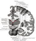
The subthalamic nucleus receives its main input from the external globus pallidus (GPe), [8] not so much through the ansa lenticularis as often said but by radiating fibers crossing the medial pallidum first and the internal capsule (see figure). These afferents are GABAergic, inhibiting neurons in the subthalamic nucleus. Excitatory, glutamatergic inputs come from the cerebral cortex (particularly the motor cortex), and from the pars parafascicularis of the central complex. The subthalamic nucleus also receives neuromodulatory inputs, notably dopaminergic axons from the substantia nigra pars compacta. [9] It also receives inputs from the pedunculopontine nucleus.

Efferent targets

The axons of subthalamic nucleus neurons leave the nucleus dorsally. The efferent axons are glutamatergic (excitatory). Except for the connection to the striatum (17.3% in macaques), most of the subthalamic principal neurons are multitargets and directed to the other elements of the core of the basal ganglia. [10] Some send axons to the substantia nigra medially and to the medial and lateral nuclei of the pallidum laterally (3-target, 21.3%). Some are 2-target with the lateral pallidum and the substantia nigra (2.7%) or the lateral pallidum and the medial (48%). Less are single target for the lateral pallidum. In the pallidum, subthalamic terminals end in bands parallel to the pallidal border. [10] [11] When all axons reaching this target are added, the main efference of the subthalamic nucleus is, in 82.7% of the cases, clearly the internal globus pallidus (GPi).

Some researchers have reported internal axon collaterals. [12] However, there is little functional evidence for this.

Physiology

Anatomical overview of the main circuits of the basal ganglia. Subthalamic nucleus is shown in red. Picture shows 2 coronal slices that have been superimposed to include the involved basal ganglia structures. + and - signs at the point of the arrows indicate respectively whether the pathway is excitatory or inhibitory in effect. Green arrows refer to excitatory glutamatergic pathways, red arrows refer to inhibitory GABAergic pathways and turquoise arrows refer to dopaminergic pathways that are excitatory on the direct pathway and inhibitory on the indirect pathway. Basal ganglia circuits.svg
Anatomical overview of the main circuits of the basal ganglia. Subthalamic nucleus is shown in red. Picture shows 2 coronal slices that have been superimposed to include the involved basal ganglia structures. + and - signs at the point of the arrows indicate respectively whether the pathway is excitatory or inhibitory in effect. Green arrows refer to excitatory glutamatergic pathways, red arrows refer to inhibitory GABAergic pathways and turquoise arrows refer to dopaminergic pathways that are excitatory on the direct pathway and inhibitory on the indirect pathway.

Subthalamic nucleus

The first intracellular electrical recordings of subthalamic neurons were performed using sharp electrodes in a rat slice preparation.[ citation needed ] In these recordings three key observations were made, all three of which have dominated subsequent reports of subthalamic firing properties. The first observation was that, in the absence of current injection or synaptic stimulation, the majority of cells were spontaneously firing. The second observation is that these cells are capable of transiently firing at very high frequencies. The third observation concerns non-linear behaviors when cells are transiently depolarized after being hyperpolarized below –65mV. They are then able to engage voltage-gated calcium and sodium currents to fire bursts of action potentials.

Several recent studies have focused on the autonomous pacemaking ability of subthalamic neurons. These cells are often referred to as "fast-spiking pacemakers", [13] since they can generate spontaneous action potentials at rates of 80 to 90 Hz in primates.

Oscillatory and synchronous activity [14] [15] is likely to be a typical pattern of discharge in subthalamic neurons recorded from patients and animal models characterized by the loss of dopaminergic cells in the substantia nigra pars compacta, which is the principal pathology that underlies Parkinson's disease.

Lateropallido-subthalamic system

Strong reciprocal connections link the subthalamic nucleus and the external segment of the globus pallidus. Both are fast-spiking pacemakers. Together, they are thought to constitute the "central pacemaker of the basal ganglia" [16] with synchronous bursts.

The connection of the lateral pallidum with the subthalamic nucleus is also the one in the basal ganglia system where the reduction between emitter/receiving elements is likely the strongest. In terms of volume, in humans, the lateral pallidum measures 808 mm³, the subthalamic nucleus only 158 mm³. [17] This translated in numbers of neurons represents a strong compression with loss of map precision.

Some axons from the lateral pallidum go to the striatum. [18] The activity of the medial pallidum is influenced by afferences from the lateral pallidum and from the subthalamic nucleus. [19] The same for the substantia nigra pars reticulata. [11] The subthalamic nucleus sends axons to another regulator: the pedunculo-pontine complex (id).

The lateropallido-subthalamic system is thought to play a key role in the generation of the patterns of activity seen in Parkinson's disease. [20]

Pathophysiology

Chronic stimulation of the STN, called deep brain stimulation (DBS), is used to treat patients with Parkinson disease. The first to be stimulated are the terminal arborisations of afferent axons, which modify the activity of subthalamic neurons. However, it has been shown in thalamic slices from mice, [21] that the stimulus also causes nearby astrocytes to release adenosine triphosphate (ATP), a precursor to adenosine (through a catabolic process). In turn, adenosine A1 receptor activation depresses excitatory transmission in the thalamus, thus mimicking ablation of the subthalamic nucleus.

Unilateral destruction or disruption of the subthalamic nucleus — which can commonly occur via a small vessel stroke in patients with diabetes, hypertension, or a history of smoking – produces hemiballismus.

As one of the STN's suspected functions is in impulse control, dysfunction in this region has been implicated in obsessive–compulsive disorder. [22] Artificially stimulating the STN has shown some promise in correcting severe impulsive behavior and may later be used as an alternative treatment for the disorder. [23]

Function

The function of the STN is unknown, but current theories place it as a component of the basal ganglia control system that may perform action selection. It is thought to implement the so-called "hyperdirect pathway" of motor control, contrasting with the direct and indirect pathways implemented elsewhere in the basal ganglia. STN dysfunction has also been shown to increase impulsivity in individuals presented with two equally rewarding stimuli. [24]

Research has suggested that the subthalamus is an extrapyramidal center. It holds muscular responses in check, and damage may result in hemiballism (a violent flinging of the arm and leg on one side of the body). [25]

The physiological role of the STN has been for long hidden by its pathological role. But lately, the research on the physiology of the STN lead to the discovery that the STN is required to achieve intended movement, including locomotion, balance and motor coordination. It is indeed involved in stopping or interrupting on-going motor tasks. Moreover, STN excitation was generally correlated with significant reduction in locomotor activity, while in contrast, STN inhibition enhanced locomotion. [26] [27] [28]

Additional images

See also

Related Research Articles

<span class="mw-page-title-main">Putamen</span> Round structure at the base of the forebrain

The putamen is a round structure located at the base of the forebrain (telencephalon). The putamen and caudate nucleus together form the dorsal striatum. It is also one of the structures that compose the basal nuclei. Through various pathways, the putamen is connected to the substantia nigra, the globus pallidus, the claustrum, and the thalamus, in addition to many regions of the cerebral cortex. A primary function of the putamen is to regulate movements at various stages and influence various types of learning. It employs GABA, acetylcholine, and enkephalin to perform its functions. The putamen also plays a role in degenerative neurological disorders, such as Parkinson's disease.

<span class="mw-page-title-main">Striatum</span> Nucleus in the basal ganglia of the brain

The striatum, or corpus striatum, is a nucleus in the subcortical basal ganglia of the forebrain. The striatum is a critical component of the motor and reward systems; receives glutamatergic and dopaminergic inputs from different sources; and serves as the primary input to the rest of the basal ganglia.

<span class="mw-page-title-main">Substantia nigra</span> Structure in the basal ganglia of the brain

The substantia nigra (SN) is a basal ganglia structure located in the midbrain that plays an important role in reward and movement. Substantia nigra is Latin for "black substance", reflecting the fact that parts of the substantia nigra appear darker than neighboring areas due to high levels of neuromelanin in dopaminergic neurons. Parkinson's disease is characterized by the loss of dopaminergic neurons in the substantia nigra pars compacta.

<span class="mw-page-title-main">Basal ganglia</span> Group of subcortical nuclei involved in the motor and reward systems

The basal ganglia (BG), or basal nuclei, are a group of subcortical nuclei found in the brains of vertebrates. In humans and some primates, differences exist, primarily in the division of the globus pallidus into external and internal regions, and in the division of the striatum. Positioned at the base of the forebrain and the top of the midbrain, they have strong connections with the cerebral cortex, thalamus, brainstem and other brain areas. The basal ganglia are associated with a variety of functions, including regulating voluntary motor movements, procedural learning, habit formation, conditional learning, eye movements, cognition, and emotion.

<span class="mw-page-title-main">Globus pallidus</span> Structure of the basal ganglia of the brain

The globus pallidus (GP), also known as paleostriatum or dorsal pallidum, is a subcortical structure of the brain. It consists of two adjacent segments, one external, known in rodents simply as the globus pallidus, and one internal, known in rodents as the entopeduncular nucleus. It is part of the telencephalon, but retains close functional ties with the subthalamus in the diencephalon – both of which are part of the extrapyramidal motor system. The globus pallidus is a major component of the basal ganglia, with principal inputs from the striatum, and principal direct outputs to the thalamus and the substantia nigra. The latter is made up of similar neuronal elements, has similar afferents from the striatum, similar projections to the thalamus, and has a similar synaptology. Neither receives direct cortical afferents, and both receive substantial additional inputs from the intralaminar thalamus.

<span class="mw-page-title-main">Nigrostriatal pathway</span>

The nigrostriatal pathway is a bilateral dopaminergic pathway in the brain that connects the substantia nigra pars compacta (SNc) in the midbrain with the dorsal striatum in the forebrain. It is one of the four major dopamine pathways in the brain, and is critical in the production of movement as part of a system called the basal ganglia motor loop. Dopaminergic neurons of this pathway release dopamine from axon terminals that synapse onto GABAergic medium spiny neurons (MSNs), also known as spiny projection neurons (SPNs), located in the striatum.

<span class="mw-page-title-main">Indirect pathway</span> Neuronal circuit that suppresses unwanted movements

The indirect pathway, sometimes known as the indirect pathway of movement, is a neuronal circuit through the basal ganglia and several associated nuclei within the central nervous system (CNS) which helps to prevent unwanted muscle contractions from competing with voluntary movements. It operates in conjunction with the direct pathway.

Hypokinesia is one of the classifications of movement disorders, and refers to decreased bodily movement. Hypokinesia is characterized by a partial or complete loss of muscle movement due to a disruption in the basal ganglia. Hypokinesia is a symptom of Parkinson's disease shown as muscle rigidity and an inability to produce movement. It is also associated with mental health disorders and prolonged inactivity due to illness, amongst other diseases.

The pars reticulata (SNpr) is a portion of the substantia nigra and is located lateral to the pars compacta. Most of the neurons that project out of the pars reticulata are inhibitory GABAergic neurons.

The pedunculopontine nucleus (PPN) or pedunculopontine tegmental nucleus is a collection of neurons located in the upper pons in the brainstem. It lies caudal to the substantia nigra and adjacent to the superior cerebellar peduncle. It has two divisions of subnuclei; the pars compacta containing mainly cholinergic neurons, and the pars dissipata containing mainly glutamatergic neurons and some non-cholinergic neurons. The pedunculopontine nucleus is one of the main components of the reticular activating system. It was first described in 1909 by Louis Jacobsohn-Lask, a German neuroanatomist.

<span class="mw-page-title-main">Habenula</span>

The habenula is a small brain structure present in virtually all vertebrate species. Located within the epithalamus, alongside the pineal gland, it borders the third ventricle and is closely situated to the midline of the brain. It has been described as having "one of the most conserved circuits in the brain of vertebrates".

The zona incerta (ZI) is a horizontally elongated region of gray matter in the subthalamus below the thalamus. Its connections project extensively over the brain from the cerebral cortex down into the spinal cord.

<span class="mw-page-title-main">Primate basal ganglia</span>

The basal ganglia form a major brain system in all species of vertebrates, but in primates there are special features that justify a separate consideration. As in other vertebrates, the primate basal ganglia can be divided into striatal, pallidal, nigral, and subthalamic components. In primates, however, there are two pallidal subdivisions called the external globus pallidus (GPe) and internal globus pallidus (GPi). Also in primates, the dorsal striatum is divided by a large tract called the internal capsule into two masses named the caudate nucleus and the putamen—in most other species no such division exists, and only the striatum as a whole is recognized. Beyond this, there is a complex circuitry of connections between the striatum and cortex that is specific to primates. This complexity reflects the difference in functioning of different cortical areas in the primate brain.

The pars compacta (SNpc) is one of two subdivisions of the substantia nigra of the midbrain ; it is situated medial to the pars reticulata. It is formed by dopaminergic neurons. It projects to the striatum and portions of the cerebral cortex. It is functionally involved in fine motor control.

<span class="mw-page-title-main">Medium spiny neuron</span> Type of GABAergic neuron in the striatum

Medium spiny neurons (MSNs), also known as spiny projection neurons (SPNs), are a special type of GABAergic inhibitory cell representing 95% of neurons within the human striatum, a basal ganglia structure. Medium spiny neurons have two primary phenotypes : D1-type MSNs of the direct pathway and D2-type MSNs of the indirect pathway. Most striatal MSNs contain only D1-type or D2-type dopamine receptors, but a subpopulation of MSNs exhibit both phenotypes.

<span class="mw-page-title-main">External globus pallidus</span> Part of the globus pallidus

The external globus pallidus combines with the internal globus pallidus (GPi) to form the globus pallidus, an anatomical subset of the basal ganglia. Globus pallidus means "pale globe" in Latin, indicating its appearance. The external globus pallidus is the segment of the globus pallidus that is relatively further (lateral) from the midline of the brain.

<span class="mw-page-title-main">Internal globus pallidus</span>

The internal globus pallidus and the external globus pallidus (GPe) make up the globus pallidus. The GPi is one of the output nuclei of the basal ganglia. The GABAergic neurons of the GPi send their axons to the ventral anterior nucleus (VA) and the ventral lateral nucleus (VL) in the dorsal thalamus, to the centromedian complex, and to the pedunculopontine complex.

<span class="mw-page-title-main">Basal ganglia disease</span> Group of physical problems resulting from basal ganglia dysfunction

Basal ganglia disease is a group of physical problems that occur when the group of nuclei in the brain known as the basal ganglia fail to properly suppress unwanted movements or to properly prime upper motor neuron circuits to initiate motor function. Research indicates that increased output of the basal ganglia inhibits thalamocortical projection neurons. Proper activation or deactivation of these neurons is an integral component for proper movement. If something causes too much basal ganglia output, then the ventral anterior (VA) and ventral lateral (VL) thalamocortical projection neurons become too inhibited, and one cannot initiate voluntary movement. These disorders are known as hypokinetic disorders. However, a disorder leading to abnormally low output of the basal ganglia leads to reduced inhibition, and thus excitation, of the thalamocortical projection neurons which synapse onto the cortex. This situation leads to an inability to suppress unwanted movements. These disorders are known as hyperkinetic disorders.

<span class="mw-page-title-main">Blocq's disease</span> Loss of memory of specialized movements causing the inability to maintain an upright posture

Blocq's disease was first considered by Paul Blocq (1860–1896), who described this phenomenon as the loss of memory of specialized movements causing the inability to maintain an upright posture, despite normal function of the legs in the bed. The patient is able to stand up, but as soon as the feet are on the ground, the patient cannot hold himself upright nor walk; however when lying down, the subject conserved the integrity of muscular force and the precision of movements of the lower limbs. The motivation of this study came when a fellow student Georges Marinesco (1864) and Paul published a case of parkinsonian tremor (1893) due to a tumor located in the substantia nigra.

The ventral pallidum (VP) is a structure within the basal ganglia of the brain. It is an output nucleus whose fibres project to thalamic nuclei, such as the ventral anterior nucleus, the ventral lateral nucleus, and the medial dorsal nucleus. The VP is a core component of the reward system which forms part of the limbic loop of the basal ganglia, a pathway involved in the regulation of motivational salience, behavior, and emotions. It is involved in addiction.

References

  1. Luys JB (1865). Recherches sur le système cérébro-spinal, sa structure, ses fonctions et ses maladies (in French). Paris: Baillière.
  2. Afsharpour S (June 1985). "Light microscopic analysis of Golgi-impregnated rat subthalamic neurons". The Journal of Comparative Neurology. 236 (1): 1–13. doi:10.1002/cne.902360102. PMID   4056088. S2CID   12482772.
  3. Rafols JA, Fox CA (July 1976). "The neurons in the primate subthalamic nucleus: a Golgi and electron microscopic study". The Journal of Comparative Neurology. 168 (1): 75–111. doi:10.1002/cne.901680105. PMID   819471. S2CID   11962279.
  4. Yelnik J, Percheron G (1979). "Subthalamic neurons in primates: a quantitative and comparative analysis". Neuroscience. 4 (11): 1717–43. doi:10.1016/0306-4522(79)90030-7. PMID   117397. S2CID   40909863.
  5. Lévesque JC, Parent A (May 2005). "GABAergic interneurons in human subthalamic nucleus". Movement Disorders. 20 (5): 574–84. doi:10.1002/mds.20374. PMID   15645534. S2CID   9551517.
  6. Alkemade, Anneke; Forstmann, Birte U. (July 2014). "Do we need to revise the tripartite subdivision hypothesis of the human subthalamic nucleus (STN)?". NeuroImage. 95: 326–329. doi:10.1016/j.neuroimage.2014.03.010. PMID   24642281. S2CID   11010595.
  7. Lambert, Christian; Zrinzo, Ludvic; Nagy, Zoltan; Lutti, Antoine; Hariz, Marwan; Foltynie, Thomas; Draganski, Bogdan; Ashburner, John; Frackowiak, Richard (March 2012). "Confirmation of functional zones within the human subthalamic nucleus: Patterns of connectivity and sub-parcellation using diffusion weighted imaging". NeuroImage. 60 (1): 83–94. doi:10.1016/j.neuroimage.2011.11.082. PMC   3315017 . PMID   22173294.
  8. Canteras NS, Shammah-Lagnado SJ, Silva BA, Ricardo JA (April 1990). "Afferent connections of the subthalamic nucleus: a combined retrograde and anterograde horseradish peroxidase study in the rat". Brain Research. 513 (1): 43–59. doi:10.1016/0006-8993(90)91087-W. PMID   2350684. S2CID   22996045.
  9. Cragg SJ, Baufreton J, Xue Y, Bolam JP, Bevan MD (October 2004). "Synaptic release of dopamine in the subthalamic nucleus". The European Journal of Neuroscience. 20 (7): 1788–802. doi: 10.1111/j.1460-9568.2004.03629.x . PMID   15380000. S2CID   14698708.
  10. 1 2 Nauta HJ, Cole M (July 1978). "Efferent projections of the subthalamic nucleus: an autoradiographic study in monkey and cat". The Journal of Comparative Neurology. 180 (1): 1–16. doi:10.1002/cne.901800102. PMID   418083. S2CID   43046462.
  11. 1 2 Smith Y, Hazrati LN, Parent A (April 1990). "Efferent projections of the subthalamic nucleus in the squirrel monkey as studied by the PHA-L anterograde tracing method". The Journal of Comparative Neurology. 294 (2): 306–23. doi:10.1002/cne.902940213. PMID   2332533. S2CID   9667393.
  12. Kita H, Chang HT, Kitai ST (April 1983). "The morphology of intracellularly labeled rat subthalamic neurons: a light microscopic analysis". The Journal of Comparative Neurology. 215 (3): 245–57. doi:10.1002/cne.902150302. PMID   6304154. S2CID   32152785.
  13. Surmeier DJ, Mercer JN, Chan CS (June 2005). "Autonomous pacemakers in the basal ganglia: who needs excitatory synapses anyway?". Current Opinion in Neurobiology. 15 (3): 312–8. doi:10.1016/j.conb.2005.05.007. PMID   15916893. S2CID   42900941.
  14. Levy R, Hutchison WD, Lozano AM, Dostrovsky JO (October 2000). "High-frequency synchronization of neuronal activity in the subthalamic nucleus of parkinsonian patients with limb tremor". The Journal of Neuroscience. 20 (20): 7766–75. doi:10.1523/JNEUROSCI.20-20-07766.2000. PMC   6772896 . PMID   11027240.
  15. Lintas A, Silkis IG, Albéri L, Villa AE (January 2012). "Dopamine deficiency increases synchronized activity in the rat subthalamic nucleus" (PDF). Brain Research. 1434 (3): 142–51. doi:10.1016/j.brainres.2011.09.005. PMID   21959175. S2CID   14636489.
  16. Plenz D, Kital ST (August 1999). "A basal ganglia pacemaker formed by the subthalamic nucleus and external globus pallidus". Nature. 400 (6745): 677–82. Bibcode:1999Natur.400..677P. doi:10.1038/23281. PMID   10458164. S2CID   4356230.
  17. Yelnik J (2002). "Functional anatomy of the basal ganglia". Movement Disorders. 17 (Suppl. 3): S15-21. doi:10.1002/mds.10138. PMID   11948751. S2CID   40925638.
  18. Sato F, Lavallée P, Lévesque M, Parent A (January 2000). "Single-axon tracing study of neurons of the external segment of the globus pallidus in primate". The Journal of Comparative Neurology. 417 (1): 17–31. doi:10.1002/(SICI)1096-9861(20000131)417:1<17::AID-CNE2>3.0.CO;2-I. PMID   10660885. S2CID   84665164.
  19. Smith Y, Wichmann T, DeLong MR (May 1994). "Synaptic innervation of neurones in the internal pallidal segment by the subthalamic nucleus and the external pallidum in monkeys". The Journal of Comparative Neurology. 343 (2): 297–318. doi:10.1002/cne.903430209. PMID   8027445. S2CID   24968074.
  20. Bevan MD, Magill PJ, Terman D, Bolam JP, Wilson CJ (October 2002). "Move to the rhythm: oscillations in the subthalamic nucleus-external globus pallidus network". Trends in Neurosciences. 25 (10): 525–31. doi:10.1016/S0166-2236(02)02235-X. PMID   12220881. S2CID   8127062.
  21. Bekar L, Libionka W, Tian GF, Xu Q, Torres A, Wang X, et al. (January 2008). "Adenosine is crucial for deep brain stimulation-mediated attenuation of tremor". Nature Medicine. 14 (1): 75–80. doi:10.1038/nm1693. PMID   18157140. S2CID   7107064.
  22. Carter R. The Human Brain Book. pp. 58, 233.
  23. Mallet L, Polosan M, Jaafari N, Baup N, Welter ML, Fontaine D, et al. (November 2008). "Subthalamic nucleus stimulation in severe obsessive-compulsive disorder". The New England Journal of Medicine. 359 (20): 2121–34. doi: 10.1056/NEJMoa0708514 . PMID   19005196.
  24. Frank MJ, Samanta J, Moustafa AA, Sherman SJ (November 2007). "Hold your horses: impulsivity, deep brain stimulation, and medication in parkinsonism". Science. 318 (5854): 1309–12. Bibcode:2007Sci...318.1309F. doi: 10.1126/science.1146157 . PMID   17962524. S2CID   2718110.
  25. Bruce H. Robinson (2007). Biomedicine - A textbook for Practitioners of Acupuncture & Oriental Medicine. Blue Poppy Press. p. 126. ISBN   978-1-891845-38-3. LCCN   2006940894.
  26. Aron AR, Behrens TE, Smith S, Frank MJ, Poldrack RA (April 2007). "Triangulating a cognitive control network using diffusion-weighted magnetic resonance imaging (MRI) and functional MRI". The Journal of Neuroscience. 27 (14): 3743–52. doi:10.1523/JNEUROSCI.0519-07.2007. PMC   6672420 . PMID   17409238.
  27. Fife KH, Gutierrez-Reed NA, Zell V, Bailly J, Lewis CM, Aron AR, Hnasko TS (July 2017). Uchida N (ed.). "Causal role for the subthalamic nucleus in interrupting behavior". eLife. 6: e27689. doi: 10.7554/eLife.27689 . PMC   5526663 . PMID   28742497.
  28. Guillaumin A, Serra GP, Georges F, Wallén-Mackenzie Å (March 2021). "Experimental investigation into the role of the subthalamic nucleus (STN) in motor control using optogenetics in mice". Brain Research. 1755: 147226. doi: 10.1016/j.brainres.2020.147226 . PMID   33358727.