Anterior corticospinal tract

Last updated
Anterior corticospinal tract
Spinal cord tracts - English.svg
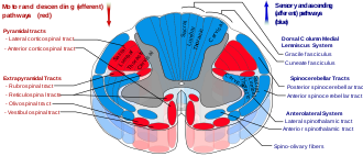
Anterior corticospinal tract seen in red at bottom center in figure (text tag found at upper-left).
Gray680.png
Decussation of pyramids. Scheme showing passage of various fasciculi from medulla spinalis to medulla oblongata. a. Pons. b. Medulla oblongata. c. Decussation of the pyramids. d. Section of cervical part of medulla spinalis. 1. Anterior cerebrospinal fasciculus (in red). 2. Lateral cerebrospinal fasciculus (in red). 3. Dorsal column tracts (gracile fasciculus and cuneate fasciculus) (in blue). 3’. Gracile and cuneate nuclei. 4. Anterolateral corticospinal tract (in dotted line). 5. Pyramid. 6. Lemniscus. 7. Medial longitudinal fasciculus. 8. Ventral spinocerebellar fasciculus (in blue). 9. Dorsal spinocerebellar fasciculus (in yellow).
Details
Identifiers
Latin tractus corticospinalis anterior, fasciculus cerebrospinalis anterior
NeuroLex ID birnlex_1618
TA98 A14.1.02.205
TA2 6112
FMA 72636
Anatomical terminology

The anterior corticospinal tract (also called the ventral corticospinal tract, "Bundle of Turck", medial corticospinal tract, direct pyramidal tract, or anterior cerebrospinal fasciculus) is a small bundle of descending fibers that connect the cerebral cortex to the spinal cord. Descending tracts are pathways by which motor signals are sent from upper motor neurons in the brain to lower motor neurons which then directly innervate muscle to produce movement. The anterior corticospinal tract is usually small, varying inversely in size with the lateral corticospinal tract, which is the main part of the corticospinal tract.

Contents

It lies close to the anterior median fissure, and is present only in the upper part of the spinal cord; gradually diminishing in size as it descends, it ends about the middle of the thoracic region.

It consists of descending fibers that arise from cells in the motor area of the ipsilateral cerebral hemisphere. The impulse travels from these upper motor neurons (located in the pre-central gyrus of the brain) through the anterior column. In contrast to the fibers for the lateral corticospinal tract, the fibers for the anterior corticospinal tract do not decussate at the level of the medulla oblongata, although they do cross over in the spinal level they innervate. [1] They then synapse at the anterior horn with the lower motor neuron which then synapses with the target muscle at the motor end plate. In contrast to the lateral corticospinal tract which controls the movement of the limbs, the anterior corticospinal tract controls the movements of axial muscles (of the trunk).

A few of its fibers pass to the lateral column of the same side and to the gray matter at the base of the posterior grey column.[ citation needed ]

Additional images

Related Research Articles

<span class="mw-page-title-main">Motor neuron</span> Nerve cell sending impulse to muscle

A motor neuron is a neuron whose cell body is located in the motor cortex, brainstem or the spinal cord, and whose axon (fiber) projects to the spinal cord or outside of the spinal cord to directly or indirectly control effector organs, mainly muscles and glands. There are two types of motor neuron – upper motor neurons and lower motor neurons. Axons from upper motor neurons synapse onto interneurons in the spinal cord and occasionally directly onto lower motor neurons. The axons from the lower motor neurons are efferent nerve fibers that carry signals from the spinal cord to the effectors. Types of lower motor neurons are alpha motor neurons, beta motor neurons, and gamma motor neurons.

<span class="mw-page-title-main">Medulla oblongata</span> Structure of the brain stem

The medulla oblongata or simply medulla is a long stem-like structure which makes up the lower part of the brainstem. It is anterior and partially inferior to the cerebellum. It is a cone-shaped neuronal mass responsible for autonomic (involuntary) functions, ranging from vomiting to sneezing. The medulla contains the cardiac, respiratory, vomiting and vasomotor centers, and therefore deals with the autonomic functions of breathing, heart rate and blood pressure as well as the sleep–wake cycle.

Pronator quadratus is a square-shaped muscle on the distal forearm that acts to pronate the hand.

<span class="mw-page-title-main">Brainstem</span> Posterior part of the brain, adjoining and structurally continuous

The brainstem is the stalk-like part of the brain that interconnects the cerebrum and diencephalon with the spinal cord. In the human brain, the brainstem is composed of the midbrain, the pons, and the medulla oblongata. The midbrain is continuous with the thalamus of the diencephalon through the tentorial notch.

<span class="mw-page-title-main">Extrapyramidal system</span> Connection between brain and spinal cord

In anatomy, the extrapyramidal system is a part of the motor system network causing involuntary actions. The system is called extrapyramidal to distinguish it from the tracts of the motor cortex that reach their targets by traveling through the pyramids of the medulla. The pyramidal tracts may directly innervate motor neurons of the spinal cord or brainstem, whereas the extrapyramidal system centers on the modulation and regulation of anterior (ventral) horn cells.

<span class="mw-page-title-main">Internal capsule</span> White matter structure situated in the inferomedial part of each cerebral hemisphere of the brain

The internal capsule is a white matter structure situated in the inferomedial part of each cerebral hemisphere of the brain. It carries information past the basal ganglia, separating the caudate nucleus and the thalamus from the putamen and the globus pallidus. The internal capsule contains both ascending and descending axons, going to and coming from the cerebral cortex. It also separates the caudate nucleus and the putamen in the dorsal striatum, a brain region involved in motor and reward pathways.

<span class="mw-page-title-main">Neural pathway</span> Connection formed between neurons that allows neurotransmission

In neuroanatomy, a neural pathway is the connection formed by axons that project from neurons to make synapses onto neurons in another location, to enable neurotransmission. Neurons are connected by a single axon, or by a bundle of axons known as a nerve tract, or fasciculus. Shorter neural pathways are found within grey matter in the brain, whereas longer projections, made up of myelinated axons, constitute white matter.

<span class="mw-page-title-main">Grey column</span>

The grey column refers to a somewhat ridge-shaped mass of grey matter in the spinal cord. This presents as three columns: the anterior grey column, the posterior grey column, and the lateral grey column, all of which are visible in cross-section of the spinal cord.

<span class="mw-page-title-main">Pyramidal tracts</span> The corticobulbar tract and the corticospinal tract

The pyramidal tracts include both the corticobulbar tract and the corticospinal tract. These are aggregations of efferent nerve fibers from the upper motor neurons that travel from the cerebral cortex and terminate either in the brainstem (corticobulbar) or spinal cord (corticospinal) and are involved in the control of motor functions of the body.

<span class="mw-page-title-main">Spinothalamic tract</span> Sensory pathway from the skin to the thalamus

The spinothalamic tract is a part of the anterolateral system or the ventrolateral system, a sensory pathway to the thalamus. From the ventral posterolateral nucleus in the thalamus, sensory information is relayed upward to the somatosensory cortex of the postcentral gyrus.

<span class="mw-page-title-main">Corticobulbar tract</span> Motor pathway in the brain connecting the motor cortex to the medullary pyramids

In neuroanatomy, the corticobulbartract is a two-neuron white matter motor pathway connecting the motor cortex in the cerebral cortex to the medullary pyramids, which are part of the brainstem's medulla oblongata region, and are primarily involved in carrying the motor function of the non-oculomotor cranial nerves. The corticobulbar tract is one of the pyramidal tracts, the other being the corticospinal tract.

<span class="mw-page-title-main">Upper motor neuron</span> Neurons in the brain that carry signals to lower motor neurons

Upper motor neurons (UMNs) is a term introduced by William Gowers in 1886. They are found in the cerebral cortex and brainstem and carry information down to activate interneurons and lower motor neurons, which in turn directly signal muscles to contract or relax. UMNs represent the major origin point for voluntary somatic movement.

<span class="mw-page-title-main">Precentral gyrus</span> Motor gyrus of the posterior frontal lobe of the brain

The precentral gyrus is a prominent gyrus on the surface of the posterior frontal lobe of the brain. It is the site of the primary motor cortex that in humans is cytoarchitecturally defined as Brodmann area 4.

<span class="mw-page-title-main">Spinocerebellar tract</span> Nerve tract in humans

The spinocerebellar tract is a nerve tract originating in the spinal cord and terminating in the same side (ipsilateral) of the cerebellum.

The rectus femoris muscle is one of the four quadriceps muscles of the human body. The others are the vastus medialis, the vastus intermedius, and the vastus lateralis. All four parts of the quadriceps muscle attach to the patella by the quadriceps tendon.

<span class="mw-page-title-main">Vestibulospinal tract</span> Neural tract in the central nervous system

The vestibulospinal tract is a neural tract in the central nervous system. Specifically, it is a component of the extrapyramidal system and is classified as a component of the medial pathway. Like other descending motor pathways, the vestibulospinal fibers of the tract relay information from nuclei to motor neurons. The vestibular nuclei receive information through the vestibulocochlear nerve about changes in the orientation of the head. The nuclei relay motor commands through the vestibulospinal tract. The function of these motor commands is to alter muscle tone, extend, and change the position of the limbs and head with the goal of supporting posture and maintaining balance of the body and head.

<span class="mw-page-title-main">Lateral corticospinal tract</span>

The lateral corticospinal tract is the largest part of the corticospinal tract. It extends throughout the entire length of the spinal cord, and on transverse section appears as an oval area in front of the posterior column and medial to the posterior spinocerebellar tract.

<span class="mw-page-title-main">Alpha motor neuron</span>

Alpha (α) motor neurons (also called alpha motoneurons), are large, multipolar lower motor neurons of the brainstem and spinal cord. They innervate extrafusal muscle fibers of skeletal muscle and are directly responsible for initiating their contraction. Alpha motor neurons are distinct from gamma motor neurons, which innervate intrafusal muscle fibers of muscle spindles.

<span class="mw-page-title-main">Spinal cord</span> Long, tubular central nervous system structure in the vertebral column

The spinal cord is a long, thin, tubular structure made up of nervous tissue that extends from the medulla oblongata in the brainstem to the lumbar region of the vertebral column (backbone) of vertebrate animals. The center of the spinal cord is hollow and contains a structure called central canal, which contains cerebrospinal fluid. The spinal cord is also covered by meninges and enclosed by the neural arches. Together, the brain and spinal cord make up the central nervous system.

<span class="mw-page-title-main">Corticospinal tract</span> Pyramidal white matter motor pathway

The corticospinal tract is a white matter motor pathway starting at the cerebral cortex that terminates on lower motor neurons and interneurons in the spinal cord, controlling movements of the limbs and trunk. There are more than one million neurons in the corticospinal tract, and they become myelinated usually in the first two years of life.

References

  1. Berthold, Daniel Goude, C.-H. "Pyramidbanans främre okorsade del - Tractus cortico-spinalis anterior - Människans nervsystem: Uppslagsverk". nervsystemet.se.{{cite web}}: CS1 maint: multiple names: authors list (link)

PD-icon.svgThis article incorporates text in the public domain from page 759 of the 20th edition of Gray's Anatomy (1918)