The peripheral nervous system (PNS) is one of two components that make up the nervous system of bilateral animals, with the other part being the central nervous system (CNS). The PNS consists of nerves and ganglia, which lie outside the brain and the spinal cord. The main function of the PNS is to connect the CNS to the limbs and organs, essentially serving as a relay between the brain and spinal cord and the rest of the body. Unlike the CNS, the PNS is not protected by the vertebral column and skull, or by the blood–brain barrier, which leaves it exposed to toxins.

The brachial plexus is a network of nerves formed by the anterior rami of the lower four cervical nerves and first thoracic nerve. This plexus extends from the spinal cord, through the cervicoaxillary canal in the neck, over the first rib, and into the armpit, it supplies afferent and efferent nerve fibers to the chest, shoulder, arm, forearm, and hand.

A spinal nerve is a mixed nerve, which carries motor, sensory, and autonomic signals between the spinal cord and the body. In the human body there are 31 pairs of spinal nerves, one on each side of the vertebral column. These are grouped into the corresponding cervical, thoracic, lumbar, sacral and coccygeal regions of the spine. There are eight pairs of cervical nerves, twelve pairs of thoracic nerves, five pairs of lumbar nerves, five pairs of sacral nerves, and one pair of coccygeal nerves. The spinal nerves are part of the peripheral nervous system.

Cervical spine disorders are illnesses that affect the cervical spine, which is made up of the upper first seven vertebrae, encasing and shielding the spinal cord. This fragment of the spine starts from the region above the shoulder blades and ends by supporting and connecting the skull.

In human anatomy, the spinal canal, vertebral canal or spinal cavity is an elongated body cavity enclosed within the dorsal bony arches of the vertebral column, which contains the spinal cord, spinal roots and dorsal root ganglia. It is a process of the dorsal body cavity formed by alignment of the vertebral foramina. Under the vertebral arches, the spinal canal is also covered anteriorly by the posterior longitudinal ligament and posteriorly by the ligamentum flavum. The potential space between these ligaments and the dura mater covering the spinal cord is known as the epidural space. Spinal nerves exit the spinal canal via the intervertebral foramina under the corresponding vertebral pedicles.

Neuromeres are distinct groups of neural crest cells, forming segments in the neural tube of the early embryonic development of the brain. There are three classes of neuromeres in the central nervous system – prosomeres, mesomeres and rhombomeres that will develop the forebrain, midbrain, and hindbrain respectively.

A dermatome is an area of skin that is mainly supplied by afferent nerve fibres from the dorsal root of any given spinal nerve. There are 8 cervical nerves , 12 thoracic nerves, 5 lumbar nerves and 5 sacral nerves. Each of these nerves relays sensation from a particular region of skin to the brain.

The rectus capitis posterior minor is a muscle in the upper back part of the neck. It is one of the suboccipital muscles. Its inferior attachment is at the posterior arch of atlas; its superior attachment is onto the occipital bone at and below the inferior nuchal line. The muscle is innervated by the suboccipital nerve. The muscle acts as a weak extensor of the head.

A nerve plexus is a plexus of intersecting nerves. A nerve plexus is composed of afferent and efferent fibers that arise from the merging of the anterior rami of spinal nerves and blood vessels. There are five spinal nerve plexuses, except in the thoracic region, as well as other forms of autonomic plexuses, many of which are a part of the enteric nervous system. The nerves that arise from the plexuses have both sensory and motor functions. These functions include muscle contraction, the maintenance of body coordination and control, and the reaction to sensations such as heat, cold, pain, and pressure. There are several plexuses in the body, including:

The lower subscapular nerve, also known as the inferior subscapular nerve, is the third branch of the posterior cord of the brachial plexus. It innervates the inferior portion of the subscapularis muscle and the teres major muscle.
Each spinal nerve receives a branch called a gray ramus communicans from the adjacent paravertebral ganglion of the sympathetic trunk. The gray rami communicantes contain postganglionic nerve fibers of the sympathetic nervous system and are composed of largely unmyelinated neurons. This is in contrast to the white rami communicantes, in which heavily myelinated neurons give the rami their white appearance.

The sympathetic ganglia, or paravertebral ganglia, are autonomic ganglia of the sympathetic nervous system. Ganglia are 20,000 to 30,000 afferent and efferent nerve cell bodies that run along on either side of the spinal cord. Afferent nerve cell bodies bring information from the body to the brain and spinal cord, while efferent nerve cell bodies bring information from the brain and spinal cord to the rest of the body. The cell bodies create long sympathetic chains that are on either side of the spinal cord. They also form para- or pre-vertebral ganglia of gross anatomy.

This article describes the anatomy of the head and neck of the human body, including the brain, bones, muscles, blood vessels, nerves, glands, nose, mouth, teeth, tongue, and throat.

The posterior spinal artery arises from the vertebral artery in 25% of humans or the posterior inferior cerebellar artery in 75% of humans, adjacent to the medulla oblongata. It is usually double, and spans the length of the spinal cord. It supplies the grey and white posterior columns of the spinal cord.
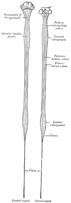
The lumbar enlargement is a widened area of the spinal cord that gives attachment to the nerves which supply the lower limbs.
The ventral ramus is the anterior division of a spinal nerve. The ventral rami supply the antero-lateral parts of the trunk and the limbs. They are mainly larger than the dorsal rami.

The fascial compartments of arm refers to the specific anatomical term of the compartments within the upper segment of the upper limb of the body. The upper limb is divided into two segments, the arm and the forearm. Each of these segments is further divided into two compartments which are formed by deep fascia – tough connective tissue septa (walls). Each compartment encloses specific muscles and nerves.
The spinal cord is a long, thin, tubular structure made up of nervous tissue that extends from the medulla oblongata in the brainstem to the lumbar region of the vertebral column (backbone) of vertebrate animals. The center of the spinal cord is hollow and contains a structure called the central canal, which contains cerebrospinal fluid. The spinal cord is also covered by meninges and enclosed by the neural arches. Together, the brain and spinal cord make up the central nervous system.

The vertebral column, also known as the spinal column, spine or backbone, is the core part of the axial skeleton in vertebrate animals. The vertebral column is the defining and eponymous characteristic of the vertebrate endoskeleton, where the notochord found in all chordates has been replaced by a segmented series of mineralized irregular bones called vertebrae, separated by fibrocartilaginous intervertebral discs. The dorsal portion of the vertebral column houses the spinal canal, an elongated cavity formed by alignment of the vertebral neural arches that encloses and protects the spinal cord, with spinal nerves exiting via the intervertebral foramina to innervate each body segments.

Each vertebra is an irregular bone with a complex structure composed of bone and some hyaline cartilage, that make up the vertebral column or spine, of vertebrates. The proportions of the vertebrae differ according to their spinal segment and the particular species.