Lateral grey column

Last updated
Lateral grey column (lateral horn)
Grey and white matter in the spinal cord.gif
Cross-Section of the Spinal Cord
Details
Identifiers
Latin cornu laterale medullae spinalis
MeSH D066152
TA98 A14.1.02.132
A14.1.02.022
TA2 6077
FMA 256536
Anatomical terminology
The four main divisions of the spinal column, from top to bottom: cervical, thoracic, lumbar, and sacral Gray 111 - Vertebral column-coloured.png
The four main divisions of the spinal column, from top to bottom: cervical, thoracic, lumbar, and sacral

The lateral grey column (lateral column, lateral cornu, lateral horn of spinal cord, intermediolateral column) is one of the three grey columns of the spinal cord (which give the shape of a butterfly); the others being the anterior and posterior grey columns. The lateral grey column is primarily involved with activity in the sympathetic division of the autonomic motor system. It projects to the side as a triangular field in the thoracic and upper lumbar [1] regions (specifically T1-L2) of the postero-lateral part of the anterior grey column.

Contents

Background information

Nervous system

The nervous system is the system of neurons, or nerve cells that relay electrical signals through the brain and body. A nerve cell receives signals from other nerve cells through tree-branch-like extensions called dendrites and passes signals through a long extension called an axon (or nerve fiber). Synapses are places where one cell's axon passes information to another cell's dendrite by sending chemicals called neurotransmitters across a small gap called a synaptic cleft. Synapses occur in various locations, including ganglia (singular: ganglion), which are masses of nerve cell bodies. Preganglionic nerve cells in the sympathetic nervous system (all of which come from the lateral grey column), use the neurotransmitter acetylcholine, while postganglionic sympathetic nerve cells use norepinephrine. [1] Grey matter in the brain and spinal cord is any accumulation of cell bodies and neuropil (neuropil is tissue rich in nerve cell bodies and dendrites). White matter consists of nerve tracts (groups of axons) and commissures (tracts that cross the brain's midline). [2]

Sympathetic nervous system

The nervous system is divided into the central nervous system (brain and spinal cord) and the peripheral nervous system (everything else). The peripheral nervous system is divided into the somatic nervous system (voluntary processes) and the autonomic nervous system (involuntary processes). The autonomic nervous system is divided into the parasympathetic nervous system (normal functioning) and the sympathetic nervous system (emergency functioning). [3] The lateral grey column mediates the functions of the sympathetic nervous system.

Spinal cord

The spinal cord is divided into 31 segments, located between the vertebrae. Each segment is defined by a posterior root entering it and an anterior root exiting it. Each of these roots is the end of a spinal nerve connecting the spinal cord to the body. The vertebral column is divided into four groups of vertebrae: (from top to bottom) cervical, thoracic, lumbar, and sacral. [4]

Structure

The lateral grey column is present at 17 levels of the spinal cord, specifically through levels T1-L2 (sympathetic outflow) as well as through levels S2-S4 (parasympathetic outflow). [5] Both these segments are located within the first thoracic vertebra to the first or second lumbar vertebra as the spinal cord ends here and the nerves form the cauda equina.

Pathway from the lateral grey column to target tissue Sympathetic Circuit from the Lateral Horn.gif
Pathway from the lateral grey column to target tissue

The lateral grey column is composed of sympathetic preganglionic visceral motor neurons which are part of the autonomic nervous system. [1] An in-depth study of the lateral grey columns in the thoracic region of rats revealed two types of cells. One cell type was characterized by many mitochondria, an indented nucleus, and a long endoplasmic reticulum, while the other cell type had an increased cytoplasmic density and a short endoplasmic reticulum. Lateral column axon terminals were found to contain noradrenaline storage granules and could be divided into four types. Type one terminals contained a few large and many small granular vesicles, and type two terminals had many large vesicles and barely noticeable granules. Type three terminals contained small, spherical vesicles and no granules, and type four were the rarest terminals that had flattened vesicles with small and large granules. [6]

The cells of the intermediolateral cell column are fusiform or star-shaped, and of a medium size. [7] The intermediolateral cell column exists at vertebral levels T1 - L2 and mediates the entire sympathetic innervation of the body. [5] In the upper part of the cervical region and lower part of the medulla oblongata, as well as in the third and fourth sacral vertebrae, this column is again differentiated.

Functions

The lateral grey column's connections mediate the functions of the sympathetic nervous system (SNS), which changes cardiac, pulmonary, hepatic (liver), and gastrointestinal activities to prepare the body for emergency situations [1] (although the sympathetic system is always active to some extent, including in the absence of a stressful environment, to maintain the appropriate level of sympathetic function). [2] When the brain responds to potential threats by sending signals to lateral grey column cells, the lateral column passes on the signals to initiate a variety of physiological changes that prepare the body for a “fight-or-flight” response. Hairs stand up to conserve heat. The gut relaxes and digestion slows so that more energy can be directed toward dealing with the threatening situation. The adrenal medulla is activated and releases epinephrine (adrenaline) into the bloodstream, where it mediates many changes, such as the preparation of muscles for emergency activity. A variety of smooth muscles relax; [1] for example, the muscles wrapped around the bronchioles of the lungs relax, allowing more oxygen to enter the bloodstream. Heart rate increases to ensure all cells are supplied quickly with the substances they need. The liver produces glucose (sugar) to fuel the muscles. [8] Blood vessels contract (vasoconstriction), which reduces bleeding and conserves body heat (the exception is blood vessels fueling large muscles that would be used in running or fighting). The pupils dilate, improving vision. [9] Perspiration increases in certain areas of the body (the purpose of this is not yet fully understood, but there is some evidence that the odor produced by this sweat serves as a signal to other individuals). [10]

Sympathetic

Sympathetic nervous system 1501 Connections of the Sympathetic Nervous System.jpg
Sympathetic nervous system

The lateral grey column plays an important role in the sympathetic division of the visceral (autonomic) motor system. Neuron cell bodies in the lateral column send their axons to synapse on sympathetic ganglia that innervate autonomic and pelvic organs; in fact, all preganglionic neurons in the sympathetic nervous system originate in the lateral grey column. [5] Neurons in the upper and middle thoracic segments control sympathetic activity in organs in the head and thorax, while neurons in the lower thoracic and upper lumbar segments control abdominal and pelvic organs and targets in the lower extremities. [2]

Slightly myelinated preganglionic fibers (aka visceral efferent fibers) leave the lateral grey column through the ventral roots and promptly form fourteen aggregates called white rami communicantes, also known as white communicating rami, which eventually enter the sympathetic trunk, a structure next to the vertebral column that consists of paired chains of sympathetic ganglia connected by nerve fibers. [5]

Most often, the fibers from the lateral grey column will travel along the sympathetic trunk until they synapse on one of the trunk's ganglia, which then passes on the information via postganglionic fibers in a gray ramus communicans. [5] 31 pairs of gray rami communicantes exit the sympathetic trunk to join the 31 pairs of spinal nerves and travel to targets such as sweat glands, hair follicles, and blood vessels. [1] Some fibers, such as those in the cardiac nerves that innervate the heart, reach the target organ directly, without first joining a gray ramus communicans.

Other fibers from lateral grey column neurons pass through the sympathetic trunk without synapsing there. The greater splanchnic nerve leaves the vertebral levels T5-T9 and synapses in the abdomen in the celiac ganglia, which innervates the celiac artery [5] (splanchnic nerves are the nerves that innervate thoracic and abdominal viscera). [2] The lesser splanchnic nerve travels from the vertebral levels T10-T11 to the abdomen's superior mesenteric ganglia, which innervates the superior mesenteric artery, and to the aorticorenal ganglion. [5] The least, or lowest, splanchnic nerve connects the T12 level to the renal plexus. [11] The lumbar splanchnic nerves from the upper two lumbar segments synapse in the abdomen on the inferior mesenteric ganglia, associated with the inferior mesenteric artery. [5] In addition, some thoracic fibers in the splanchnic nerves innervate the adrenal medulla, a ganglion in the abdomen that mediates the sympathetic response to stress. [2]

Axons from the lateral grey column release acetylcholine at their synapses. This can excite or inhibit the postsynaptic cell, depending on the type of acetylcholine receptor in its membrane. Postganglionic cells (that is, nerve cells innervated in ganglia by lateral column neurons) typically release norepinephrine (noradrenaline) on their targets; these synapses can also be either excitatory or inhibitory. [1]

The lateral grey column receives input signals from preganglionic, myelinated fibers from viscera (internal organs), which course through prevertebral ganglia (between the visceral organ and the sympathetic chain) and paravertebral ganglia (in the sympathetic chain), white rami communicantes, and dorsal roots to synapse on cells of the intermediolateral cell column in the lateral horn. [4]

Lateral grey column nerve cells also receive signals from the brainstem and from neurons in the hypothalamus, a brain area involved in mediating many physiological functions and emotional states. [12]

Clinical significance

Horner's syndrome is characterized by small pupils, sunken eyes, partially drooping eyelid, and dryness of the skin on the face. It is caused by problems in autonomic pathways such as damage to the lateral grey column. [1]

Progressive autonomic failure is a disease associated with autonomic disturbances due to selective neuronal degeneration. [13] A study estimated the number of nerve cells in the lateral grey columns of 21 people with progressive autonomic failure in comparison with a control group. This same study found that an average of 75 percent of the lateral column cells were lost in people with progressive autonomic failure. [14]

Multiple system atrophy (MSA) is an adult onset disorder that is a sporadic and progressive. MSA is characterized by a combination of ataxia, parkinsonism, and autonomic dysfunction. [15] A study did a comparison with the lateral grey column nerve cell count between 15 cases of patients with MSA and a control group. All cases of MSA had lost over 50 percent of their lateral horn cells which shows the involvement of the intermediolateral columns in MSA. [16]

Lichtheim's disease (also known as subacute combined degeneration) is the result of a vitamin B12 deficiency and is associated with pernicious anemia. [17] The disease is characterized by the degeneration of both the lateral and posterior columns, which results in symptoms such as a spastic ataxic gait and paranoia. [18] Patients may also feel tingling or weakness in their limbs and torso. [17]

Related Research Articles

<span class="mw-page-title-main">Autonomic nervous system</span> Division of the nervous system supplying internal organs, smooth muscle and glands

The autonomic nervous system (ANS), formerly referred to as the vegetative nervous system, is a division of the nervous system that operates internal organs, smooth muscle and glands. The autonomic nervous system is a control system that acts largely unconsciously and regulates bodily functions, such as the heart rate, its force of contraction, digestion, respiratory rate, pupillary response, urination, and sexual arousal. This system is the primary mechanism in control of the fight-or-flight response.

<span class="mw-page-title-main">Parasympathetic nervous system</span> Division of the autonomic nervous system

The parasympathetic nervous system (PSNS) is one of the three divisions of the autonomic nervous system, the others being the sympathetic nervous system and the enteric nervous system. The enteric nervous system is sometimes considered part of the autonomic nervous system, and sometimes considered an independent system.

<span class="mw-page-title-main">Sympathetic nervous system</span> Part of the autonomic nervous system which stimulates fight-or-flight responses

The sympathetic nervous system (SNS) is one of the three divisions of the autonomic nervous system, the others being the parasympathetic nervous system and the enteric nervous system. The enteric nervous system is sometimes considered part of the autonomic nervous system, and sometimes considered an independent system.

<span class="mw-page-title-main">Spinal nerve</span> Nerve that carries signals between the spinal cord and the body

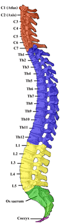
A spinal nerve is a mixed nerve, which carries motor, sensory, and autonomic signals between the spinal cord and the body. In the human body there are 31 pairs of spinal nerves, one on each side of the vertebral column. These are grouped into the corresponding cervical, thoracic, lumbar, sacral and coccygeal regions of the spine. There are eight pairs of cervical nerves, twelve pairs of thoracic nerves, five pairs of lumbar nerves, five pairs of sacral nerves, and one pair of coccygeal nerves. The spinal nerves are part of the peripheral nervous system.

<span class="mw-page-title-main">Neuromere</span>

Neuromeres are distinct groups of neural crest cells, forming segments in the neural tube of the early embryonic development of the brain. There are three classes of neuromeres in the central nervous system – prosomeres, mesomeres and rhombomeres that will develop the forebrain, midbrain, and hindbrain respectively.

<span class="mw-page-title-main">Ciliary ganglion</span> Bundle of nerves, parasympathetic ganglion

The ciliary ganglion is a bundle of nerves, parasympathetic ganglion located just behind the eye in the posterior orbit. It is 1–2 mm in diameter and in humans contains approximately 2,500 neurons. The ganglion contains postganglionic parasympathetic neurons. These neurons supply the pupillary sphincter muscle, which constricts the pupil, and the ciliary muscle which contracts to make the lens more convex. Both of these muscles are involuntary since they are controlled by the parasympathetic division of the autonomic nervous system.

<span class="mw-page-title-main">Nerve plexus</span> Network of nerve fibres

A nerve plexus is a plexus of intersecting nerves. A nerve plexus is composed of afferent and efferent fibers that arise from the merging of the anterior rami of spinal nerves and blood vessels. There are five spinal nerve plexuses, except in the thoracic region, as well as other forms of autonomic plexuses, many of which are a part of the enteric nervous system. The nerves that arise from the plexuses have both sensory and motor functions. These functions include muscle contraction, the maintenance of body coordination and control, and the reaction to sensations such as heat, cold, pain, and pressure. There are several plexuses in the body, including:

<span class="mw-page-title-main">Superior cervical ganglion</span> Largest of the cervical ganglia

The superior cervical ganglion (SCG) is the upper-most and largest of the cervical sympathetic ganglia of the sympathetic trunk. It probably formed by the union of four sympathetic ganglia of the cervical spinal nerves C1–C4. It is the only ganglion of the sympathetic nervous system that innervates the head and neck. The SCG innervates numerous structures of the head and neck.

<span class="mw-page-title-main">Sympathetic trunk</span> Bundle of nerve fibers

The sympathetic trunks are a paired bundle of nerve fibers that run from the base of the skull to the coccyx. They are a major component of the sympathetic nervous system.

<span class="mw-page-title-main">White ramus communicans</span>

The white ramus communicans from Latin ramus (branch) and communicans (communicating) is the preganglionic sympathetic outflow nerve tract from the spinal cord.

Each spinal nerve receives a branch called a gray ramus communicans from the adjacent paravertebral ganglion of the sympathetic trunk. The gray rami communicantes contain postganglionic nerve fibers of the sympathetic nervous system and are composed of largely unmyelinated neurons. This is in contrast to the white rami communicantes, in which heavily myelinated neurons give the rami their white appearance.

<span class="mw-page-title-main">Ramus communicans</span> Latin term used for a nerve which connects two other nerves

Ramus communicans is the Latin term used for a nerve which connects two other nerves, and can be translated as "communicating branch".

<span class="mw-page-title-main">Sympathetic ganglia</span> Ganglia of the sympathetic nervous system

The sympathetic ganglia, or paravertebral ganglia, are autonomic ganglia of the sympathetic nervous system. Ganglia are 20,000 to 30,000 afferent and efferent nerve cell bodies that run along on either side of the spinal cord. Afferent nerve cell bodies bring information from the body to the brain and spinal cord, while efferent nerve cell bodies bring information from the brain and spinal cord to the rest of the body. The cell bodies create long sympathetic chains that are on either side of the spinal cord. They also form para- or pre-vertebral ganglia of gross anatomy.

<span class="mw-page-title-main">Sacral splanchnic nerves</span>

Sacral splanchnic nerves are splanchnic nerves that connect the inferior hypogastric plexus to the sympathetic trunk in the pelvis.

<span class="mw-page-title-main">Esophageal plexus</span>

The esophageal plexus is formed by nerve fibers from two sources, branches of the vagus nerve, and visceral branches of the sympathetic trunk. The esophageal plexus and the cardiac plexus contain the same types of fibers and are both considered thoracic autonomic plexus.

<span class="mw-page-title-main">General visceral afferent fiber</span> Part of the visceral nervous system

The general visceral afferent (GVA) fibers conduct sensory impulses from the internal organs, glands, and blood vessels to the central nervous system. They are considered to be part of the visceral nervous system, which is closely related to the autonomic nervous system, but 'visceral nervous system' and 'autonomic nervous system' are not direct synonyms and care should be taken when using these terms. Unlike the efferent fibers of the autonomic nervous system, the afferent fibers are not classified as either sympathetic or parasympathetic.

<span class="mw-page-title-main">Lumbar ganglia</span>

The lumbar ganglia are paravertebral ganglia located in the inferior portion of the sympathetic trunk. The lumbar portion of the sympathetic trunk typically has 4 lumbar ganglia. The lumbar splanchnic nerves arise from the ganglia here, and contribute sympathetic efferent fibers to the nearby plexuses. The first two lumbar ganglia have both white and gray rami communicates.

<span class="mw-page-title-main">Spinal cord</span> Long, tubular central nervous system structure in the vertebral column

The spinal cord is a long, thin, tubular structure made up of nervous tissue that extends from the medulla oblongata in the brainstem to the lumbar region of the vertebral column (backbone) of vertebrate animals. The center of the spinal cord is hollow and contains a structure called central canal, which contains cerebrospinal fluid. The spinal cord is also covered by meninges and enclosed by the neural arches. Together, the brain and spinal cord make up the central nervous system.

<span class="mw-page-title-main">Outline of the human nervous system</span> Overview of and topical guide to the human nervous system

The following diagram is provided as an overview of and topical guide to the human nervous system:

<span class="mw-page-title-main">Roots of the ciliary ganglion</span>

The ciliary ganglion is a parasympathetic ganglion located just behind the eye in the posterior orbit. Three types of axons enter the ciliary ganglion but only the preganglionic parasympathetic axons synapse there. The entering axons are arranged into three roots of the ciliary ganglion, which join enter the posterior surface of the ganglion.

References

PD-icon.svgThis article incorporates text in the public domain from page 753 of the 20th edition of Gray's Anatomy (1918)

  1. 1 2 3 4 5 6 7 8 Pritchard, Thomas C.; Alloway, Kevin D. (1999). Medical neuroscience (1st ed.). Madison, Conn.: Fence Creek Pub. ISBN   978-1889325293.
  2. 1 2 3 4 5 Purves, Dale, ed. (2011). Neuroscience (5th ed.). Sunderland, Mass.: Sinauer. ISBN   978-0-87893-695-3.
  3. "Divisions of the Nervous System." Neuroscience for Kids. University of Washington. http://faculty.washington.edu/chudler/nsdivide.html
  4. 1 2 Dafny, N. (1997). Anatomy of the spinal cord. http://neuroscience.uth.tmc.edu/s2/chapter03.html Archived 2011-10-08 at the Wayback Machine
  5. 1 2 3 4 5 6 7 8 Dlugos, C. (1999). Autonomic nervous system. http://www.smbs.buffalo.edu/ana/newpage41.htm
  6. Katoh, Y; Isomura, G; Shimizu, N (Dec 1980). "Fine structural organization of the lateral horn of the rat spinal cord as studied by glyoxylic acid-permanganate fixation". Archivum Histologicum Japonicum. 43 (5): 445–58. doi: 10.1679/aohc1950.43.445 . PMID   7235870.
  7. Weaver, Lynne C.; Polosa, Canio, eds. (2006). Autonomic Dysfunction After Spinal Cord Injury. Progress in Brain Research. Vol. 152. doi:10.1016/s0079-6123(05)x5200-1. ISBN   9780444519252.
  8. "How Cells Communicate During the Fight or Flight Response." Genetic Science Learning Center, University of Utah. "How Cells Communicate During the Fight or Flight Response". Archived from the original on 2013-08-08. Retrieved 2013-10-23.
  9. "The Science of Stress." Weber State University. http://faculty.weber.edu/molpin/healthclasses/1110/bookchapters/stressphysiologychapter.htm Archived 2017-11-20 at the Wayback Machine
  10. Reddy, Sumathi. "Why We Sweat When We're Stressed". WSJ. Retrieved 2023-03-14.
  11. Tank, P. W. (2009). Nerves of the Thoracic Region.http://anatomy.uams.edu/anatomyhtml/nerves_thorax.html
  12. Harting, John. "Hypothalamus," Neuroscience Coursebook. University of Wisconsin. January 21, 2005. Web. http://www.neuroanatomy.wisc.edu/coursebook/neuro2(2).pdf
  13. Hasegawa, Y; Takahashi, A (Apr 1992). "[Progressive autonomic failure with Parkinson's disease]". Nihon Rinsho. 50 (4): 790–8. PMID   1619762.
  14. Oppenheimer, DR (Jun 1980). "Lateral horn cells in progressive autonomic failure". Journal of the Neurological Sciences. 46 (3): 393–404. doi:10.1016/0022-510X(80)90064-7. PMID   6247458. S2CID   46267684.
  15. "Consensus statement on the definition of orthostatic hypotension, pure autonomic failure, and multiple system atrophy". Neurology. 46 (5): 1470. 1 May 1996. doi:10.1212/WNL.46.5.1470. PMID   8628505. S2CID   219212717.
  16. Gray, F; Vincent, D; Hauw, JJ (1988). "Quantitative study of lateral horn cells in 15 cases of multiple system atrophy". Acta Neuropathologica. 75 (5): 513–8. doi:10.1007/BF00687140. PMID   3376754. S2CID   11957905.
  17. 1 2 Ilniczky, S; Jelencsik, I; Kenéz, J; Szirmai, I (Jan 2002). "MR findings in subacute combined degeneration of the spinal cord caused by nitrous oxide anaesthesia--two cases". European Journal of Neurology. 9 (1): 101–4. doi:10.1046/j.1468-1331.2002.00336.x. PMID   11784385. S2CID   29561772.
  18. Benzel, E., Waxman, C., Stephen, G., Byrne, T. N. "Diseases of the Spine and Spinal Cord." Oxford University Press, USA, 2000.