Related Research Articles

Transverse myelitis (TM) is a rare neurological condition wherein the spinal cord is inflamed. The adjective transverse implies that the spinal inflammation (myelitis) extends horizontally throughout the cross section of the spinal cord; the terms partial transverse myelitis and partial myelitis are sometimes used to specify inflammation that affects only part of the width of the spinal cord. TM is characterized by weakness and numbness of the limbs, deficits in sensation and motor skills, dysfunctional urethral and anal sphincter activities, and dysfunction of the autonomic nervous system that can lead to episodes of high blood pressure. Signs and symptoms vary according to the affected level of the spinal cord. The underlying cause of TM is unknown. The spinal cord inflammation seen in TM has been associated with various infections, immune system disorders, or damage to nerve fibers, by loss of myelin. As opposed to leukomyelitis which affects only the white matter, it affects the entire cross-section of the spinal cord. Decreased electrical conductivity in the nervous system can result.

Viral meningitis, also known as aseptic meningitis, is a type of meningitis due to a viral infection. It results in inflammation of the meninges. Symptoms commonly include headache, fever, sensitivity to light and neck stiffness.
Myelitis is inflammation of the spinal cord which can disrupt the normal responses from the brain to the rest of the body, and from the rest of the body to the brain. Inflammation in the spinal cord can cause the myelin and axon to be damaged resulting in symptoms such as paralysis and sensory loss. Myelitis is classified to several categories depending on the area or the cause of the lesion; however, any inflammatory attack on the spinal cord is often referred to as transverse myelitis.

Japanese encephalitis (JE) is an infection of the brain caused by the Japanese encephalitis virus (JEV). While most infections result in little or no symptoms, occasional inflammation of the brain occurs. In these cases, symptoms may include headache, vomiting, fever, confusion and seizures. This occurs about 5 to 15 days after infection.

Tick-borne encephalitis virus (TBEV) is a positive-strand RNA virus associated with tick-borne encephalitis in the genus Flavivirus.

Aseptic meningitis is the inflammation of the meninges, a membrane covering the brain and spinal cord, in patients whose cerebral spinal fluid test result is negative with routine bacterial cultures. Aseptic meningitis is caused by viruses, mycobacteria, spirochetes, fungi, medications, and cancer malignancies. The testing for both meningitis and aseptic meningitis is mostly the same. A cerebrospinal fluid sample is taken by lumbar puncture and is tested for leukocyte levels to determine if there is an infection and goes on to further testing to see what the actual cause is. The symptoms are the same for both meningitis and aseptic meningitis but the severity of the symptoms and the treatment can depend on the certain cause.

Oropouche fever is a tropical viral infection which can infect humans. It is transmitted by biting midges and mosquitoes, from a natural reservoir which includes sloths, non-human primates, and birds. The disease is named after the region where it was first discovered and isolated in 1955, by the Oropouche River in Trinidad and Tobago. Oropouche fever is caused by the Oropouche virus (OROV), of the Bunyavirales order of viruses.

Viral encephalitis is inflammation of the brain parenchyma, called encephalitis, by a virus. The different forms of viral encephalitis are called viral encephalitides. It is the most common type of encephalitis and often occurs with viral meningitis. Encephalitic viruses first cause infection and replicate outside of the central nervous system (CNS), most reaching the CNS through the circulatory system and a minority from nerve endings toward the CNS. Once in the brain, the virus and the host's inflammatory response disrupt neural function, leading to illness and complications, many of which frequently are neurological in nature, such as impaired motor skills and altered behavior.
HIV-associated neurocognitive disorders (HAND) are neurological disorders associated with HIV infection and AIDS. It is a syndrome of progressive deterioration of memory, cognition, behavior, and motor function in HIV-infected individuals during the late stages of the disease, when immunodeficiency is severe. HAND may include neurological disorders of various severity. HIV-associated neurocognitive disorders are associated with a metabolic encephalopathy induced by HIV infection and fueled by immune activation of macrophages and microglia. These cells are actively infected with HIV and secrete neurotoxins of both host and viral origin. The essential features of HIV-associated dementia (HAD) are disabling cognitive impairment accompanied by motor dysfunction, speech problems and behavioral change. Cognitive impairment is characterised by mental slowness, trouble with memory and poor concentration. Motor symptoms include a loss of fine motor control leading to clumsiness, poor balance and tremors. Behavioral changes may include apathy, lethargy and diminished emotional responses and spontaneity. Histopathologically, it is identified by the infiltration of monocytes and macrophages into the central nervous system (CNS), gliosis, pallor of myelin sheaths, abnormalities of dendritic processes and neuronal loss.
Granulomatous meningoencephalitis (GME) is an inflammatory disease of the central nervous system (CNS) of dogs and, rarely, cats. It is a form of meningoencephalitis. GME is likely second only to encephalitis caused by canine distemper virus as the most common cause of inflammatory disease of the canine CNS. The disease is more common in female dogs of young and middle age. It has a rapid onset. The lesions of GME exist mainly in the white matter of the cerebrum, brainstem, cerebellum, and spinal cord. The cause is only known to be noninfectious and is considered at this time to be idiopathic. Because lesions resemble those seen in allergic meningoencephalitis, GME is thought to have an immune-mediated cause, but it is also thought that the disease may be based on an abnormal response to an infectious agent. One study searched for viral DNA from canine herpesvirus, canine adenovirus, and canine parvovirus in brain tissue from dogs with GME, necrotizing meningoencephalitis, and necrotizing leukoencephalitis, but failed to find any.
A neurotropic virus is a virus that is capable of infecting nerve tissue.
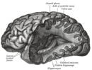
The central nervous system (CNS) controls most of the functions of the body and mind. It comprises the brain, spinal cord and the nerve fibers that branch off to all parts of the body. The CNS viral diseases are caused by viruses that attack the CNS. Existing and emerging viral CNS infections are major sources of human morbidity and mortality.
Herpes meningitis is inflammation of the meninges, the protective tissues surrounding the spinal cord and brain, due to infection from viruses of the Herpesviridae family - the most common amongst adults is HSV-2. Symptoms are self-limiting over 2 weeks with severe headache, nausea, vomiting, neck-stiffness, and photophobia. Herpes meningitis can cause Mollaret's meningitis, a form of recurrent meningitis. Lumbar puncture with cerebrospinal fluid results demonstrating aseptic meningitis pattern is necessary for diagnosis and polymerase chain reaction is used to detect viral presence. Although symptoms are self-limiting, treatment with antiviral medication may be recommended to prevent progression to Herpes Meningoencephalitis.

Retrograde tracing is a research method used in neuroscience to trace neural connections from their point of termination to their source. Retrograde tracing techniques allow for detailed assessment of neuronal connections between a target population of neurons and their inputs throughout the nervous system. These techniques allow the "mapping" of connections between neurons in a particular structure and the target neurons in the brain. The opposite technique is anterograde tracing, which is used to trace neural connections from their source to their point of termination. Both the anterograde and retrograde tracing techniques are based on the visualization of axonal transport.
The International Society for NeuroVirology (ISNV) was founded to promote research into disease-causing viruses that infect the human brain and nervous system. The ISNV membership includes scientists and clinicians from around the world who work in the fields of basic, translational, and clinical neurovirology.
Polioencephalitis is a viral infection of the brain, causing inflammation within the grey matter of the brain stem. The virus has an affinity for neuronal cell bodies and has been found to affect mostly the midbrain, pons, medulla and cerebellum of most infected patients. The infection can reach up through the thalamus and hypothalamus and possibly reach the cerebral hemispheres. The infection is caused by the poliomyelitis virus which is a single-stranded, positive sense RNA virus surrounded by a non-enveloped capsid. Humans are the only known natural hosts of this virus. The disease has been eliminated from the U.S. since the mid-twentieth century, but is still found in certain areas of the world such as Africa.
West Caucasian bat lyssavirus (WCBL) is a member of genus Lyssavirus, family Rhabdoviridae and order Mononegavirales. This virus was first isolated from Miniopterus schreibersii, in the western Caucasus Mountains of southeastern Europe in 2002. WCBL is the most divergent form of Lyssavirus, and is found in Miniopterus bats (insectivorous), Rousettus aegyptiacus, and Eidolon helvum. The latter two are both fruit bats. The virus is fragile as it can be inactivated by UV light and chemicals, such as ether, chloroform, and bleach. WCBL has not been known to infect humans thus far.

Robyn S. Klein is an American neuroimmunologist as well as the Vice Provost and Associate Dean for Graduate Education at Washington University in St. Louis. Klein is also a professor in the Departments of Medicine, Anatomy & Neurobiology, and Pathology & Immunology. Her research explores the pathogenesis of neuroinflammation in the central nervous system by probing how immune signalling molecules regulate blood brain barrier permeability. Klein is also a fervent advocate for gender equity in STEM, publishing mechanisms to improve gender equity in speakers at conferences, participating nationally on gender equity discussion panels, and through service as the president of the Academic Women’s Network at the Washington University School of Medicine.
Georgette D. Kanmogne is a Cameroonian American geneticist and molecular virologist and a full professor and vice chair for resource allocation and faculty development within the Department of Pharmacology and Experimental Neurosciences at the University of Nebraska Medical Center in Omaha, Nebraska. Kanmogne's research program focuses on exploring the pathogenesis of neuroAIDS by deciphering the mechanisms underlying blood brain barrier dysfunction and viral entry into the central nervous system. Her research also addresses the lack of HIV therapies that cross the blood brain barrier (BBB) and has played a critical role in the development of nanoparticles encapsulating HIV-drugs that can cross the BBB to prevent viral-mediated neuron death in the brain. Kanmogne collaborates with clinical and basic researchers across America, Cameroon, and West Africa, spanning disciplines from hematology to psychiatry, to explore how viral genetic diversity is correlated with the neurological impact of HIV.
References
- 1 2 3 Johnson, R (1995). "Neurovirology: evolution of a new discipline", Journal of Neurovirology, 1(2).
- 1 2 3 4 McKendall, R and Stroop, W (1994). "Handbook of Neurovirology".
- 1 2 3 4 5 Gosztonyi, G (2001). The Mechanisms of Neuronal Damage in Virus Infections of the Nervous System.
- ↑ G. M. Baer, T. R. Shanthaveerappa, G. H. Bourne (1965). "Studies on the Pathogenesis of Fixed Rabies Virus in Rats", Bull. Org. mond. Sante, 33(783).
- ↑ "International Society for NeuroVirology".
- 1 2 3 4 5 6 7 8 Nath A, Berger J (2003). Clinical Neurovirology.
- ↑ Mueller N, Gilden D, Cohrs R, (2008). "Varicella Zoster Virus Infection: Clinical Features, Molecular Pathogenesis of Disease, and Latency". Neurologic Clinics. 26(675).
- 1 2 Lincoln J, Hankiewicz K (2008). "Could Epstein-Barr Virus or Canine Distemper Virus Cause Multiple Sclerosis?". Neurologic Clinics 26(699).
- ↑ Jackson A. (2008) "Rabies". Neurologic Clinics. 26(717).
- ↑ Wright E, Brew B, Wesselingh S (2008). "Pathogenesis and Diagnosis of Viral Infections of the Nervous System". Neurologic Clinics. 26 (617).
- ↑ Wright, E et al (2008). Pathogenesis and Diagnosis of Viral Infections of the Nervous System. 26(617).
- ↑ Irani, D (2008). "Aseptic Meningitis and Viral Myelitis", Neurologic Clinics, 26(635).
- ↑ Davies NW, Brown LJ, Irish D, et al. (2005). "Factors influencing PCR detection of viruses in cerebrospinal fluid of patients with suspected CNS infections". Journal of Neurology, Neurosurgery, and Psychiatry. 76 (82).
- ↑ Ferris M, Mactutus C, Booze R (2008). "Neurotoxic profiles of HIV, psychostimulant drugs of abuse, and their concerted effect on the brain: Current status of dopamine system vulnerability in NeuroAIDS". Neuroscience and Biobehavioral Reviews 32(883).