Molecular neuroscience

Last updated

Molecular neuroscience is a branch of neuroscience that observes concepts in molecular biology applied to the nervous systems of animals. The scope of this subject covers topics such as molecular neuroanatomy, mechanisms of molecular signaling in the nervous system, the effects of genetics and epigenetics on neuronal development, and the molecular basis for neuroplasticity and neurodegenerative diseases. [1] As with molecular biology, molecular neuroscience is a relatively new field that is considerably dynamic.

Contents

Locating neurotransmitters

In molecular biology, communication between neurons typically occurs by chemical transmission across gaps between the cells called synapses. The transmitted chemicals, known as neurotransmitters, regulate a significant fraction of vital body functions. [2] It is possible to anatomically locate neurotransmitters by labeling techniques. It is possible to chemically identify certain neurotransmitters such as catecholamines by fixing neural tissue sections with formaldehyde. This can give rise to formaldehyde-induced fluorescence when exposed to ultraviolet light. Dopamine, a catecholamine, was identified in the nematode C. elegans by using this technique. [3] Immunocytochemistry, which involves raising antibodies against targeted chemical or biological entities, includes a few other techniques of interest. A targeted neurotransmitter could be specifically tagged by primary and secondary antibodies with radioactive labeling in order to identify the neurotransmitter by autoradiography. The presence of neurotransmitters (though not necessarily the location) can be observed in enzyme-linked immunocytochemistry or enzyme-linked immunosorbent assays (ELISA) in which substrate-binding in the enzymatic assays can induce precipitates, fluorophores, or chemiluminescence. In the event that neurotransmitters cannot be histochemically identified, an alternative method is to locate them by their neural uptake mechanisms. [1]

Voltage-gated ion channels

Structure of eukaryotic voltage-gated potassium ion channels 2r9r opm.png
Structure of eukaryotic voltage-gated potassium ion channels

Excitable cells in living organisms have voltage-gated ion channels. These can be observed throughout the nervous system in neurons. The first ion channels to be characterized were the sodium and potassium ion channels by A.L. Hodgkin and A.F. Huxley in the 1950s upon studying the giant axon of the squid genus Loligo . Their research demonstrated the selective permeability of cellular membranes, dependent on physiological conditions, and the electrical effects that result from these permeabilities to produce action potentials. [4]

Sodium ion channels

Sodium channels were the first voltage-gated ion channels to be isolated in 1984 from the eel Electrophorus electricus by Shosaku Numa. The pufferfish toxin tetrodotoxin (TTX), a sodium channel blocker, was used to isolate the sodium channel protein by binding it using the column chromatography technique for chemical separation. The amino acid sequence of the protein was analyzed by Edman degradation and then used to construct a cDNA library which could be used to clone the channel protein. Cloning the channel itself allowed for applications such as identifying the same channels in other animals. [1] Sodium channels are known for working in concert with potassium channels during the development of graded potentials and action potentials. Sodium channels allow an influx of Na+ ions into a neuron, resulting in a depolarization from the resting membrane potential of a neuron to lead to a graded potential or action potential, depending on the degree of depolarization. [5]

Potassium ion channels

Potassium channels come in a variety of forms, are present in most eukaryotic cells, and typically tend to stabilize the cell membrane at the potassium equilibrium potential. As with sodium ions, graded potentials and action potentials are also dependent on potassium channels. While influx of Na+ ions into a neuron induce cellular depolarization, efflux of K+ ions out of a neuron causes a cell to repolarize to resting membrane potential. The activation of potassium ion channels themselves are dependent on the depolarization resulting from Na+ influx during an action potential. [1] As with sodium channels, the potassium channels have their own toxins that block channel protein action. An example of such a toxin is the large cation, tetraethylammonium (TEA), but it is notable that the toxin does not have the same mechanism of action on all potassium channels, given the variety of channel types across species. The presence of potassium channels was first identified in Drosophila melanogaster mutant flies that shook uncontrollably upon anesthesia due to problems in cellular repolarization that led to abnormal neuron and muscle electrophysiology. Potassium channels were first identified by manipulating molecular genetics (of the flies) instead of performing channel protein purification because there were no known high-affinity ligands for potassium channels (such as TEA) at the time of discovery. [1] [6]

Calcium ion channels

Calcium channels are important for certain cell-signaling cascades as well as neurotransmitter release at axon terminals. A variety of different types of calcium ion channels are found in excitable cells. As with sodium ion channels, calcium ion channels have been isolated and cloned by chromatographic purification techniques. It is notable, as with the case of neurotransmitter release, that calcium channels can interact with intracellular proteins and plays a strong role in signaling, especially in locations such as the sarcoplasmic reticulum of muscle cells. [1]

Receptors

Various types of receptors can be used for cell signaling and communication and can include ionotropic receptors and metabotropic receptors. These cell surface receptor types are differentiated by the mechanism and duration of action with ionotropic receptors being associated with fast signal transmission and metabotropic receptors being associated with slow signal transmission. Metabotropic receptors happen to cover a wide variety of cell-surface receptors with notably different signaling cascades. [1] [5]

Ionotropic receptors

Prototypical depiction of ionotropic receptor in the case of Ca ion flow LGIC.png
Prototypical depiction of ionotropic receptor in the case of Ca ion flow

Ionotropic receptors, otherwise known as ligand-gated ion channels, are fast acting receptors that mediate neural and physiological function by ion channel flow with ligand-binding. Nicotinic, GABA, and Glutamate receptors are among some of the cell surface receptors regulated by ligand-gated ion channel flow. GABA is the brain's main inhibitory neurotransmitter and glutamate is the brain's main excitatory neurotransmitter. [1]

GABA receptors

GABAA and GABAC receptors are known to be ionotropic, while the GABAB receptor is metabotropic. GABAA receptors mediate fast inhibitory responses in the central nervous system (CNS) and are found on neurons, glial cells, and adrenal medulla cells. It is responsible for inducing Cl ion influx into cells, thereby reducing the probability that membrane depolarization will occur upon the arrival of a graded potential or an action potential. GABA receptors can also interact with non-endogenous ligands to influence activity. For example, the compound diazepam (marketed as Valium) is an allosteric agonist which increases the affinity of the receptor for GABA. The increased physiological inhibitory effects resulting from increased GABA binding make diazepam a useful tranquilizer or anticonvulsant (antiepileptic drugs). On the other hand, GABA receptors can also be targeted by decreasing Cl cellular influx with the effect of convulsants like picrotoxin. The antagonistic mechanism of action for this compound is not directly on the GABA receptor, but there are other compounds that are capable of allosteric inactivation, including T-butylbicyclophorothionate (TBPS) and pentylenetetrazole (PZT). Compared with GABAA, GABAC receptors have a higher affinity for GABA, they are likely to be longer-lasting in activity, and their responses are likely to be generated by lower GABA concentrations. [1]

Glutamate receptors

Ionotropic glutamate receptors can include NMDA, AMPA, and kainate receptors. These receptors are named after agonists that facilitate glutamate activity. NMDA receptors are notable for their excitatory mechanisms to affect neuronal plasticity in learning and memory, as well as neuropathologies such as stroke and epilepsy. NDMA receptors have multiple binding sites just like ionotropic GABA receptors and can be influenced by co-agonists such the glycine neurotransmitter or phencyclidine (PCP). The NMDA receptors carry a current by Ca2+ ions and can be blocked by extracellular Mg2+ ions depending on voltage and membrane potential. This Ca2+ influx is increased by excitatory postsynaptic potentials (EPSPs) produced by NMDA receptors, activating Ca2+-based signaling cascades (such as neurotransmitter release). AMPA generate shorter and larger excitatory postsynaptic currents than other ionotropic glutamate receptors. [5]

Nicotinic ACh receptors

Nicotinic receptors bind the acetylcholine (ACh) neurotransmitter to produce non-selective cation channel flow that generates excitatory postsynaptic responses. Receptor activity, which can be influenced by nicotine consumption, produces feelings of euphoria, relaxation, and inevitably addiction in high levels. [5]

Metabotropic receptors

G-protein-linked receptor signaling cascade GPCR cycle.jpg
G-protein-linked receptor signaling cascade

Metabotropic receptors, are slow response receptors in postsynaptic cells. Typically these slow responses are characterized by more elaborate intracellular changes in biochemistry. Responses of neurotransmitter uptake by metabotropic receptors can result in the activation of intracellualar enzymes and cascades involving second messengers, as is the case with G protein-linked receptors. Various metabotropic receptors can include certain glutamate receptors, muscarinic ACh receptors, GABAB receptors, and receptor tyrosine kinases.

G protein-linked receptors

The G protein-linked signaling cascade can significantly amplify the signal of a particular neurotransmitter to produce hundreds to thousands of second messengers in a cell. The mechanism of action by which G protein-linked receptors cause a signaling cascade is as follows:

  1. Neurotransmitter binds to the receptor
  2. The receptor undergoes a conformational change to allow G-protein complex binding
  3. GDP is exchanged with GTP upon G protein complex binding to the receptor
  4. The α-subunit of the G protein complex is bound to GTP and separates to bind with a target protein such as adenylate cyclase
  5. The binding to the target protein either increases or decreases the rate of second messenger (such as cyclic AMP) production
  6. GTPase hydrolyzes the α-subunit so that is bound to GDP and the α-subunit returns to the G protein complex inactive

Neurotransmitter release

Structure of a synapse where neurotransmitter release and uptake occurs SynapseSchematic en.svg
Structure of a synapse where neurotransmitter release and uptake occurs

Neurotransmitters are released in discrete packets known as quanta from the axon terminal of one neuron to the dendrites of another across a synapse. These quanta have been identified by electron microscopy as synaptic vesicles. Two types of vesicles are small synaptic vessicles (SSVs), which are about 40-60nm in diameter, and large dense-core vesicles (LDCVs), electron-dense vesicles approximately 120-200nm in diameter. [1] The former is derived from endosomes and houses neurotransmitters such as acetylcholine, glutamate, GABA, and glycine. The latter is derived from the Golgi apparatus and houses larger neurotransmitters such as catecholamines and other peptide neurotransmitters. [7] Neurotransmitters are released from an axon terminal and bind to postsynaptic dendrites in the following procession: [5]

  1. Mobilization/recruitment of synaptic vesicle from cytoskeleton
  2. Docking of vesicle (binding) to presynaptic membrane
  3. Priming of vesicle by ATP (relatively slow step)
  4. Fusion of primed vesicle with presynaptic membrane and exocytosis of the housed neurotransmitter
  5. Uptake of neurotransmitters in receptors of a postsynaptic cell
  6. Initiation or inhibition of action potential in postsynaptic cell depending on whether the neurotransmitters are excitatory or inhibitory (excitatory will result in depolarization of the postsynaptic membrane)

Neurotransmitter release is calcium-dependent

Neurotransmitter release is dependent on an external supply of Ca2+ ions which enter axon terminals via voltage-gated calcium channels. Vesicular fusion with the terminal membrane and release of the neurotransmitter is caused by the generation of Ca2+ gradients induced by incoming action potentials. The Ca2+ ions cause the mobilization of newly synthesized vesicles from a reserve pool to undergo this membrane fusion. This mechanism of action was discovered in squid giant axons. [8] Lowering intracellular Ca2+ ions provides a direct inhibitory effect on neurotransmitter release. [1] After release of the neurotransmitter occurs, vesicular membranes are recycled to their origins of production. Calcium ion channels can vary depending on the location of incidence. For example, the channels at an axon terminal differ from the typical calcium channels of a cell body (whether neural or not). Even at axon terminals, calcium ion channel types can vary, as is the case with P-type calcium channels located at the neuromuscular junction. [1]

Neuronal gene expression

Sex differences

Differences in sex determination are controlled by sex chromosomes. Sex hormonal releases have a significant effect on sexual dimorphisms (phenotypic differentiation of sexual characteristics) of the brain. Recent studies seem to suggest that regulating these dimorphisms has implications for understanding normal and abnormal brain function. Sexual dimorphisms may be significantly influenced by sex-based brain gene expression which varies from species to species.

Animal models such as rodents, Drosophila melanogaster , and Caenorhabditis elegans , have been used to observe the origins and/or extent of sex bias in the brain versus the hormone-producing gonads of an animal. With the rodents, studies on genetic manipulation of sex chromosomes resulted in an effect on one sex that was completely opposite of the effect in the other sex. For example, a knockout of a particular gene only resulted in anxiety-like effects in males. With studies on D. menlanogaster it was found that a large brain sex bias of expression occurred even after the gonads were removed, suggesting that sex bias could be independent of hormonal control in certain aspects. [9]

Observing sex-biased genes has the potential for clinical significance in observing brain physiology and the potential for related (whether directly or indirectly) neurological disorders. Examples of diseases with sex biases in development include Huntington's disease, cerebral ischemia, and Alzheimer's disease. [9]

Epigenetics of the brain

Many brain functions can be influenced at the cellular and molecular level by variations and changes in gene expression, without altering the sequence of DNA in an organism. This is otherwise known as epigenetic regulation. Examples of epigenetic mechanisms include histone modifications and DNA methylation. Such changes have been found to be strongly influential in the incidence of brain disease, mental illness, and addiction. [10] Epigenetic control has been shown to be involved in high levels of plasticity in early development, thereby defining its importance in the critical period of an organism. [11] Examples of how epigenetic changes can affect the human brain are as follows:

Molecular mechanisms of neurodegenerative diseases

Excitotoxicity and glutamate receptors

Excitotoxicity is phenomenon in which glutamate receptors are inappropriately activated. It can be caused by prolonged excitatory synaptic transmission in which high levels of glutamate neurotransmitter cause excessive activation in a postsynaptic neuron that can result in the death of the postsynaptic neuron. Following brain injury (such as from ischemia), it has been found that excitotoxicity is a significant cause of neuronal damage. This can be understandable in the case where sudden perfusion of blood after reduced blood flow to the brain can result in excessive synaptic activity caused by the presence of increased glutamate and aspartate during the period of ischemia. [5] [13]

Alzheimer's disease

Alzheimer's disease is the most common neurodegenerative disease and is the most common form of dementia in the elderly. The disorder is characterized by progressive loss of memory and various cognitive functions. It is hypothesized that the deposition of amyloid-β peptide (40-42 amino acid residues) in the brain is integral in the incidence of Alzheimer's disease. Accumulation is purported to block hippocampal long-term potentiation. It is also possible that a receptor for amyloid-β oligomers could be a prion protein. [14]

Parkinson's disease

Parkinson's disease is the second most common neurodegenerative disease after Alzheimer's disease. It is a hypokinetic movement basal ganglia disease caused by the loss of dopaminergic neurons in the substantia nigra of the human brain. The inhibitory outflow of the basal ganglia is thus not decreased, and so upper motor neurons, mediated by the thalamus, are not activated in a timely manner. Specific symptoms include rigidity, postural problems, slow movements, and tremors. Blocking GABA receptor input from medium spiny neurons to reticulata cells, causes inhibition of upper motor neurons similar to the inhibition that occurs in Parkinson's disease. [5]

Huntington's disease

Huntington's disease is a hyperkinetic movement basal ganglia disease caused by lack of normal inhibitory inputs from medium spiny neurons of the basal ganglia. This poses the opposite effects of those associated with Parkinson's disease, including inappropriate activation of upper motor neurons. As with the GABAergic mechanisms observed in relation to Parkinson's disease, a GABA agonist injected into the substantia nigra pars reticulata decreases inhibition of upper motor neurons, resulting in ballistic involuntary motor movements, similar to symptoms of Huntington's disease. [5]

Related Research Articles

<span class="mw-page-title-main">Neuron</span> Electrically excitable cell found in the nervous system of animals

Within a nervous system, a neuron, neurone, or nerve cell is an electrically excitable cell that fires electric signals called action potentials across a neural network. Neurons communicate with other cells via synapses, which are specialized connections that commonly use minute amounts of chemical neurotransmitters to pass the electric signal from the presynaptic neuron to the target cell through the synaptic gap.

<span class="mw-page-title-main">Chemical synapse</span> Biological junctions through which neurons signals can be sent

Chemical synapses are biological junctions through which neurons' signals can be sent to each other and to non-neuronal cells such as those in muscles or glands. Chemical synapses allow neurons to form circuits within the central nervous system. They are crucial to the biological computations that underlie perception and thought. They allow the nervous system to connect to and control other systems of the body.

<span class="mw-page-title-main">Neurotransmitter receptor</span> Type of protein

A neurotransmitter receptor is a membrane receptor protein that is activated by a neurotransmitter. Chemicals on the outside of the cell, such as a neurotransmitter, can bump into the cell's membrane, in which there are receptors. If a neurotransmitter bumps into its corresponding receptor, they will bind and can trigger other events to occur inside the cell. Therefore, a membrane receptor is part of the molecular machinery that allows cells to communicate with one another. A neurotransmitter receptor is a class of receptors that specifically binds with neurotransmitters as opposed to other molecules.

An inhibitory postsynaptic potential (IPSP) is a kind of synaptic potential that makes a postsynaptic neuron less likely to generate an action potential. The opposite of an inhibitory postsynaptic potential is an excitatory postsynaptic potential (EPSP), which is a synaptic potential that makes a postsynaptic neuron more likely to generate an action potential. IPSPs can take place at all chemical synapses, which use the secretion of neurotransmitters to create cell-to-cell signalling. EPSPs and IPSPs compete with each other at numerous synapses of a neuron. This determines whether an action potential occurring at the presynaptic terminal produces an action potential at the postsynaptic membrane. Some common neurotransmitters involved in IPSPs are GABA and glycine.

<span class="mw-page-title-main">Excitatory postsynaptic potential</span> Process causing temporary increase in postsynaptic potential

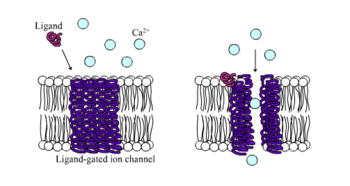
In neuroscience, an excitatory postsynaptic potential (EPSP) is a postsynaptic potential that makes the postsynaptic neuron more likely to fire an action potential. This temporary depolarization of postsynaptic membrane potential, caused by the flow of positively charged ions into the postsynaptic cell, is a result of opening ligand-gated ion channels. These are the opposite of inhibitory postsynaptic potentials (IPSPs), which usually result from the flow of negative ions into the cell or positive ions out of the cell. EPSPs can also result from a decrease in outgoing positive charges, while IPSPs are sometimes caused by an increase in positive charge outflow. The flow of ions that causes an EPSP is an excitatory postsynaptic current (EPSC).

<span class="mw-page-title-main">Excitatory synapse</span> Sort of synapse

An excitatory synapse is a synapse in which an action potential in a presynaptic neuron increases the probability of an action potential occurring in a postsynaptic cell. Neurons form networks through which nerve impulses travels, each neuron often making numerous connections with other cells of neurons. These electrical signals may be excitatory or inhibitory, and, if the total of excitatory influences exceeds that of the inhibitory influences, the neuron will generate a new action potential at its axon hillock, thus transmitting the information to yet another cell.

<span class="mw-page-title-main">End-plate potential</span>

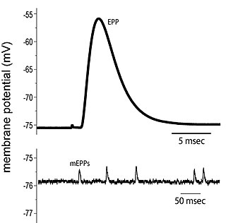
End plate potentials (EPPs) are the voltages which cause depolarization of skeletal muscle fibers caused by neurotransmitters binding to the postsynaptic membrane in the neuromuscular junction. They are called "end plates" because the postsynaptic terminals of muscle fibers have a large, saucer-like appearance. When an action potential reaches the axon terminal of a motor neuron, vesicles carrying neurotransmitters are exocytosed and the contents are released into the neuromuscular junction. These neurotransmitters bind to receptors on the postsynaptic membrane and lead to its depolarization. In the absence of an action potential, acetylcholine vesicles spontaneously leak into the neuromuscular junction and cause very small depolarizations in the postsynaptic membrane. This small response (~0.4mV) is called a miniature end plate potential (MEPP) and is generated by one acetylcholine-containing vesicle. It represents the smallest possible depolarization which can be induced in a muscle.

Neuropharmacology is the study of how drugs affect function in the nervous system, and the neural mechanisms through which they influence behavior. There are two main branches of neuropharmacology: behavioral and molecular. Behavioral neuropharmacology focuses on the study of how drugs affect human behavior (neuropsychopharmacology), including the study of how drug dependence and addiction affect the human brain. Molecular neuropharmacology involves the study of neurons and their neurochemical interactions, with the overall goal of developing drugs that have beneficial effects on neurological function. Both of these fields are closely connected, since both are concerned with the interactions of neurotransmitters, neuropeptides, neurohormones, neuromodulators, enzymes, second messengers, co-transporters, ion channels, and receptor proteins in the central and peripheral nervous systems. Studying these interactions, researchers are developing drugs to treat many different neurological disorders, including pain, neurodegenerative diseases such as Parkinson's disease and Alzheimer's disease, psychological disorders, addiction, and many others.

<span class="mw-page-title-main">Kainate receptor</span> Class of ionotropic glutamate receptors

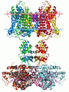
Kainate receptors, or kainic acid receptors (KARs), are ionotropic receptors that respond to the neurotransmitter glutamate. They were first identified as a distinct receptor type through their selective activation by the agonist kainate, a drug first isolated from the algae Digenea simplex. They have been traditionally classified as a non-NMDA-type receptor, along with the AMPA receptor. KARs are less understood than AMPA and NMDA receptors, the other ionotropic glutamate receptors. Postsynaptic kainate receptors are involved in excitatory neurotransmission. Presynaptic kainate receptors have been implicated in inhibitory neurotransmission by modulating release of the inhibitory neurotransmitter GABA through a presynaptic mechanism.

<span class="mw-page-title-main">Glutamate receptor</span> Cell-surface proteins that bind glutamate and trigger changes which influence the behavior of cells

Glutamate receptors are synaptic and non synaptic receptors located primarily on the membranes of neuronal and glial cells. Glutamate is abundant in the human body, but particularly in the nervous system and especially prominent in the human brain where it is the body's most prominent neurotransmitter, the brain's main excitatory neurotransmitter, and also the precursor for GABA, the brain's main inhibitory neurotransmitter. Glutamate receptors are responsible for the glutamate-mediated postsynaptic excitation of neural cells, and are important for neural communication, memory formation, learning, and regulation.

<span class="mw-page-title-main">Neurotransmission</span> Impulse transmission between neurons

Neurotransmission is the process by which signaling molecules called neurotransmitters are released by the axon terminal of a neuron, and bind to and react with the receptors on the dendrites of another neuron a short distance away. A similar process occurs in retrograde neurotransmission, where the dendrites of the postsynaptic neuron release retrograde neurotransmitters that signal through receptors that are located on the axon terminal of the presynaptic neuron, mainly at GABAergic and glutamatergic synapses.

<span class="mw-page-title-main">Synapse</span> Structure connecting neurons in the nervous system

In the nervous system, a synapse is a structure that permits a neuron to pass an electrical or chemical signal to another neuron or to the target effector cell.

<span class="mw-page-title-main">Synaptic potential</span> Potential difference across the postsynaptic membrane

Synaptic potential refers to the potential difference across the postsynaptic membrane that results from the action of neurotransmitters at a neuronal synapse. In other words, it is the “incoming” signal that a neuron receives. There are two forms of synaptic potential: excitatory and inhibitory. The type of potential produced depends on both the postsynaptic receptor, more specifically the changes in conductance of ion channels in the post synaptic membrane, and the nature of the released neurotransmitter. Excitatory post-synaptic potentials (EPSPs) depolarize the membrane and move the potential closer to the threshold for an action potential to be generated. Inhibitory postsynaptic potentials (IPSPs) hyperpolarize the membrane and move the potential farther away from the threshold, decreasing the likelihood of an action potential occurring. The Excitatory Post Synaptic potential is most likely going to be carried out by the neurotransmitters glutamate and acetylcholine, while the Inhibitory post synaptic potential will most likely be carried out by the neurotransmitters gamma-aminobutyric acid (GABA) and glycine. In order to depolarize a neuron enough to cause an action potential, there must be enough EPSPs to both depolarize the postsynaptic membrane from its resting membrane potential to its threshold and counterbalance the concurrent IPSPs that hyperpolarize the membrane. As an example, consider a neuron with a resting membrane potential of -70 mV (millivolts) and a threshold of -50 mV. It will need to be raised 20 mV in order to pass the threshold and fire an action potential. The neuron will account for all the many incoming excitatory and inhibitory signals via summative neural integration, and if the result is an increase of 20 mV or more, an action potential will occur.

<span class="mw-page-title-main">Calyx of Held</span>

The Calyx of Held is a particularly large synapse in the mammalian auditory central nervous system, so named after Hans Held who first described it in his 1893 article Die centrale Gehörleitung because of its resemblance to the calyx of a flower. Globular bushy cells in the anteroventral cochlear nucleus (AVCN) send axons to the contralateral medial nucleus of the trapezoid body (MNTB), where they synapse via these calyces on MNTB principal cells. These principal cells then project to the ipsilateral lateral superior olive (LSO), where they inhibit postsynaptic neurons and provide a basis for interaural level detection (ILD), required for high frequency sound localization. This synapse has been described as the largest in the brain.

<span class="mw-page-title-main">Summation (neurophysiology)</span>

Summation, which includes both spatial summation and temporal summation, is the process that determines whether or not an action potential will be generated by the combined effects of excitatory and inhibitory signals, both from multiple simultaneous inputs, and from repeated inputs. Depending on the sum total of many individual inputs, summation may or may not reach the threshold voltage to trigger an action potential.

<span class="mw-page-title-main">Axon terminal</span> Nerve fiber part

Axon terminals are distal terminations of the branches of an axon. An axon, also called a nerve fiber, is a long, slender projection of a nerve cell that conducts electrical impulses called action potentials away from the neuron's cell body in order to transmit those impulses to other neurons, muscle cells or glands. In the central nervous system, most presynaptic terminals are actually formed along the axons, not at their ends.

Cellular neuroscience is a branch of neuroscience concerned with the study of neurons at a cellular level. This includes morphology and physiological properties of single neurons. Several techniques such as intracellular recording, patch-clamp, and voltage-clamp technique, pharmacology, confocal imaging, molecular biology, two photon laser scanning microscopy and Ca2+ imaging have been used to study activity at the cellular level. Cellular neuroscience examines the various types of neurons, the functions of different neurons, the influence of neurons upon each other, and how neurons work together.

Anoxic depolarization is a progressive and uncontrollable depolarization of neurons during stroke or brain ischemia in which there is an inadequate supply of blood to the brain. Anoxic depolarization is induced by the loss of neuronal selective membrane permeability and the ion gradients across the membrane that are needed to support neuronal activity. Normally, the Na+/K+-ATPase pump maintains the transmembrane gradients of K+ and Na+ ions, but with anoxic brain injury, the supply of energy to drive this pump is lost. The hallmarks of anoxic depolarization are increased concentrations of extracellular K+ ions, intracellular Na+ and Ca2+ ions, and extracellular glutamate and aspartate. Glutamate and aspartate are normally present as the brain's primary excitatory neurotransmitters, but high concentrations activate a number of downstream apoptotic and necrotic pathways. This results in neuronal dysfunction and brain death.

Communication between neurons happens primarily through chemical neurotransmission at the synapse. Neurotransmitters are packaged into synaptic vesicles for release from the presynaptic cell into the synapse, from where they diffuse and can bind to postsynaptic receptors. While most presynaptic cells are historically thought to release one vesicle at a time per active site, more recent research has pointed towards the possibility of multiple vesicles being released from the same active site in response to an action potential.

Ionotropic GABA receptors (iGABARs) are ligand-gated ion channel of the GABA receptors class which are activated by gamma-aminobutyric acid (GABA), and include:

References

  1. 1 2 3 4 5 6 7 8 9 10 11 12 Longstaff, Alan; Revest, Patricia (1998). Molecular Neuroscience. Garland Science. ISBN   978-1859962503.
  2. "What are Neurotransmitters?". Archived from the original on 25 September 2019. Retrieved 1 November 2013.
  3. Riddle, Donald (1998). C. Elegans II. New York: Cold Spring Harbor Laboratory Press. ISBN   978-0879695323.
  4. Hodgkin, Allan L.; Andrew F. Huxley (1952). "The dual effect of membrane potential on sodium conductance in the giant axon of Loligo". The Journal of Physiology. 116 (4): 497–506. doi:10.1113/jphysiol.1952.sp004719. PMC   1392212 . PMID   14946715.
  5. 1 2 3 4 5 6 7 8 Purves, Dale (2012). Neuroscience (5th ed.). Massachusetts, USA: Sinauer Associates, Inc. p. 80. ISBN   978-0-87893-695-3.
  6. Kamb, Alexander; Linda E. Iverson; Mark A. Tanouye (31 July 1987). "Molecular characterization of Shaker, a Drosophila gene that encodes a potassium channel". Cell. 50 (3): 405–413. doi:10.1016/0092-8674(87)90494-6. PMID   2440582. S2CID   31247228.
  7. Davies, R. Wayne; Brian J. Morris (1997). Molecular Biology of the Neuron. Oxford, UK: BIOS Scientific Publishers Ltd. ISBN   978-1859962404.
  8. Dipolo, R.; C. Caputo; F. Bezanilla (March 1983). "Voltage-dependent calcium channel in the squid axon". Proc Natl Acad Sci U S A. 80 (6): 1743–1745. Bibcode:1983PNAS...80.1743D. doi: 10.1073/pnas.80.6.1743 . PMC   393680 . PMID   6300873.
  9. 1 2 Jazin, E.; Cahill, L. (January 2010). "Sex differences in molecular neuroscience: from fruit flies to humans". Nature Reviews Neuroscience. 11 (1): 9–17. doi:10.1038/nrn2754. PMID   20019686. S2CID   2545405.
  10. "Epigenetics and the Human Brain". Genetics Science and Learning Center at The University of Utah. Retrieved 10 November 2013.
  11. 1 2 Fagiolini, Michela; Catherin L. Jensen; Frances A. Champagne (2009). "Epigenetic Influences on brain development and plasticity" (PDF). Current Opinion in Neurobiology. 19 (2): 1–6. doi:10.1016/j.conb.2009.05.009. PMC   2745597 . PMID   19545993. Archived from the original (PDF) on 22 June 2010. Retrieved 14 November 2013.
  12. 1 2 3 Tsankova, Nadia; Renthal, William; Kumar, Arvind; Nestler, Eric J. (2007). "Epigenetic Regulation in Psychiatric Disorders". Nature Reviews Neuroscience. 8 (5): 355–367. doi:10.1038/nrn2132. PMID   17453016. S2CID   34125955.
  13. Lau, A.; M. Tymianski (2010). "Glutamate receptors, neurotoxicity and neurodegeneration". Pflügers Arch. 460 (2): 525–542. doi:10.1007/s00424-010-0809-1. PMID   20229265. S2CID   12421120.
  14. Laren, Juha; David A. Gimbel; Haakon B. Nygaard; John W. Gilbert (February 2009). "Cellular prion protein mediates impairment of synaptic plasticity by amyloid-β oligomers". Nature. 457 (7233): 1128–1132. Bibcode:2009Natur.457.1128L. doi:10.1038/nature07761. PMC   2748841 . PMID   19242475.