"},"Pfam":{"wt":"PF03494"},"Pfam_clan":{"wt":""},"InterPro":{"wt":"IPR013803"},"SMART":{"wt":""},"PROSITE":{"wt":""},"MEROPS":{"wt":""},"SCOP":{"wt":"2lfm"},"TCDB":{"wt":"1.C.50"},"OPM family":{"wt":"304"},"OPM protein":{"wt":"2y3k"},"CAZy":{"wt":""},"CDD":{"wt":""},"Membranome superfamily":{"wt":"45"}},"i":0}}]}" id="mwBg">Protein family
Amyloid beta peptide (beta-APP) | |||||||||
---|---|---|---|---|---|---|---|---|---|
![]() A partially folded structure of amyloid beta(1 40) in an aqueous environment (pdb 2lfm) [1] | |||||||||
Identifiers | |||||||||
Symbol | APP | ||||||||
Pfam | PF03494 | ||||||||
InterPro | IPR013803 | ||||||||
SCOP2 | 2lfm / SCOPe / SUPFAM | ||||||||
TCDB | 1.C.50 | ||||||||
OPM superfamily | 304 | ||||||||
OPM protein | 2y3k | ||||||||
Membranome | 45 | ||||||||
|
amyloid beta (A4) precursor protein (peptidase nexin-II, Alzheimer disease) | |||||||
---|---|---|---|---|---|---|---|
![]() Processing of the amyloid precursor protein | |||||||
Identifiers | |||||||
Symbol | APP | ||||||
Alt. symbols | AD1 | ||||||
NCBI gene | 351 | ||||||
HGNC | 620 | ||||||
OMIM | 104760 | ||||||
RefSeq | NM_000484 | ||||||
UniProt | P05067 | ||||||
Other data | |||||||
Locus | Chr. 21 q21.2 | ||||||
|
Amyloid beta (Aβ, Abeta or beta-amyloid) denotes peptides of 36–43 amino acids that are the main component of the amyloid plaques found in the brains of people with Alzheimer's disease. [2] The peptides derive from the amyloid-beta precursor protein (APP), which is cleaved by beta secretase and gamma secretase to yield Aβ in a cholesterol-dependent process and substrate presentation. [3] Both neurons and oligodendrocytes produce and release Aβ in the brain, contributing to formation of amyloid plaques. [4] Aβ molecules can aggregate to form flexible soluble oligomers which may exist in several forms. It is now believed that certain misfolded oligomers (known as "seeds") can induce other Aβ molecules to also take the misfolded oligomeric form, leading to a chain reaction akin to a prion infection. The oligomers are toxic to nerve cells. [5] The other protein implicated in Alzheimer's disease, tau protein, also forms such prion-like misfolded oligomers, and there is some evidence that misfolded Aβ can induce tau to misfold. [6] [7]
A study has suggested that APP and its amyloid potential is of ancient origins, dating as far back as early deuterostomes. [8]
The normal function of Aβ is not yet known. [9] Though some animal studies have shown that the absence of Aβ does not lead to any obvious loss of physiological function, [10] [11] several potential activities have been discovered for Aβ, including activation of kinase enzymes, [12] [13] protection against oxidative stress, [14] [15] regulation of cholesterol transport, [16] [17] functioning as a transcription factor, [18] [19] and anti-microbial activity (potentially associated with Aβ's pro-inflammatory activity). [20] [21] [22]
The glymphatic system clears metabolic waste from the mammalian brain, and in particular amyloid beta. [23] A number of proteases have been implicated by both genetic and biochemical studies as being responsible for the recognition and degradation of amyloid beta; these include insulin degrading enzyme [24] and presequence protease. [25] The rate of removal is significantly increased during sleep. [26] However, the significance of the glymphatic system in Aβ clearance in Alzheimer's disease is unknown. [27]
Aβ is the main component of amyloid plaques, extracellular deposits found in the brains of people with Alzheimer's disease. [28] Aβ can also form the deposits that line cerebral blood vessels in cerebral amyloid angiopathy. The plaques are composed of a tangle of Aβ oligomers [29] and regularly ordered aggregates called amyloid fibrils, [30] a protein fold shared by other peptides such as the prions associated with protein misfolding disease, also known as proteinopathy.[ citation needed ]
Research suggests that soluble oligomeric forms of the amyloid beta may be causative agents in the development of Alzheimer's disease. [31] It is generally believed that Aβ oligomers are the most toxic. [32] Several genetic, cell biology, biochemical and animal studies using experimental models support the concept that Aβ plays a central role in the development of Alzheimer's disease pathology. [33] [34]
Brain Aβ is elevated in people with sporadic Alzheimer's disease. Aβ is the main constituent of brain parenchymal and vascular amyloid; it contributes to cerebrovascular lesions and is neurotoxic. [33] [34] [35] It is unresolved how Aβ accumulates in the central nervous system and subsequently initiates the disease of cells. Significant efforts have been focused on the mechanisms responsible for Aβ production, including the proteolytic enzymes gamma- and β-secretases which generate Aβ from its precursor protein, APP (amyloid precursor protein). [36] [37] Aβ circulates in plasma, cerebrospinal fluid (CSF) and brain interstitial fluid (ISF) mainly as soluble Aβ40. [33] [38] Amyloid plaques contain both Aβ40 and Aβ42, [39] while vascular amyloid is predominantly the shorter Aβ40. Several sequences of Aβ were found in both lesions. [40] [41]
Increases in either total Aβ levels or the relative concentration of both Aβ40 and Aβ42 (where the former is more concentrated in cerebrovascular plaques and the latter in neuritic plaques) [42] have been implicated in the pathogenesis of both familial and sporadic Alzheimer's disease. Due to its more hydrophobic nature, the Aβ42 is the most amyloidogenic form of the peptide. However the central sequence KLVFFAE is known to form amyloid on its own, and probably forms the core of the fibril. [43] One study further correlated Aβ42 levels in the brain not only with onset of Alzheimer's disease, but also reduced cerebrospinal fluid pressure, suggesting that a build-up or inability to clear Aβ42 fragments may play a role into the pathology. [44]
The "amyloid hypothesis" — that the plaques are responsible for the pathology of Alzheimer's disease — is accepted by the majority of researchers, but is not conclusively established. An alternative hypothesis is that amyloid oligomers rather than plaques are responsible for the disease. [32] [45] This more recent variation of the amyloid hypothesis identifies the cytotoxic species as an intermediate misfolded form of amyloid beta, neither a soluble monomer nor a mature aggregated polymer but an oligomeric species. This ion channel hypothesis postulates that oligomers of soluble, non-fibrillar Aβ form membrane ion channels allowing unregulated calcium influx into neurons. [46]
This cytotoxic-fibril hypothesis presents a clear target for drug development: inhibit the fibrillization process. Much early development work on lead compounds has focused on this inhibition; [47] [48] [49] most are also reported to reduce neurotoxicity, but the toxic-oligomer theory suggests that prevention of oligomeric assembly is more important [50] [51] For example, apomorphine was seen to significantly improve memory function through the increased successful completion of the Morris Water Maze. [50]
While Aβ has been implicated in cancer development, prompting studies on a variety of cancers to elucidate the nature of its possible effects, results are largely inconclusive. Aβ levels have been assessed in relation to a number of cancers, including esophageal, colorectal, lung, and hepatic, in response to observed reductions in risk for developing Alzheimer's disease in survivors of these cancers.[ citation needed ] All cancers were shown to be associated positively with increased Aβ levels, particularly hepatic cancers. [52] This direction of association however has not yet been established. Studies focusing on human breast cancer cell lines have further demonstrated that these cancerous cells display an increased level of expression of amyloid precursor protein. [53]
Adults with Down syndrome had accumulation of amyloid in association with evidence of Alzheimer's disease, including declines in cognitive functioning, memory, fine motor movements, executive functioning, and visuospatial skills. [54]
Aβ is formed after sequential cleavage of the amyloid precursor protein (APP), a transmembrane glycoprotein of undetermined function. APP can be cleaved by the proteolytic enzymes α-, β- and γ-secretase; Aβ protein is generated by successive action of the β and γ secretases. The γ secretase, which produces the C-terminal end of the Aβ peptide, cleaves within the transmembrane region of APP and can generate a number of isoforms of 30–51 amino acid residues in length. [55] The most common isoforms are Aβ40 and Aβ42; the longer form is typically produced by cleavage that occurs in the endoplasmic reticulum, while the shorter form is produced by cleavage in the trans-Golgi network. [56]
Autosomal-dominant mutations in APP cause hereditary early-onset Alzheimer's disease (familial AD, fAD). This form of AD accounts for no more than 10% of all cases, and the vast majority of AD is not accompanied by such mutations. [57] However, familial Alzheimer's disease is likely to result from altered proteolytic processing. This is evidenced by the fact that many mutations that lead to fAD occur near γ-secretase cleavage sites on APP. [58] One of the most common mutations causing fAD, London Mutation, occurs at codon 717 of the APP gene, [59] [60] and results in a valine to isoleucine amino acid substitution. Histochemical analysis of the APP V717I mutation has revealed extensive Aβ pathology throughout neuroaxis as well as widespread cerebral amyloid angiopathy (CAA). [61]
The gene for the amyloid precursor protein is located on chromosome 21, and accordingly people with Down syndrome have a very high incidence of Alzheimer's disease. [62]
Amyloid beta is commonly thought to be intrinsically unstructured, meaning that in solution it does not acquire a unique tertiary fold but rather populates a set of structures. As such, it cannot be crystallized and most structural knowledge on amyloid beta comes from NMR and molecular dynamics. Early NMR-derived models of a 26-aminoacid polypeptide from amyloid beta (Aβ 10–35) show a collapsed coil structure devoid of significant secondary structure content. [63] However, the most recent (2012) NMR structure of (Aβ 1-40) has significant secondary and tertiary structure. [1] Replica exchange molecular dynamics studies suggested that amyloid beta can indeed populate multiple discrete structural states; [64] more recent studies identified a multiplicity of discrete conformational clusters by statistical analysis. [65] By NMR-guided simulations, amyloid beta 1-40 and amyloid beta 1-42 also seem to feature highly different conformational states, [66] with the C-terminus of amyloid beta 1-42 being more structured than that of the 1-40 fragment.
Low-temperature and low-salt conditions allowed to isolate pentameric disc-shaped oligomers devoid of beta structure. [67] In contrast, soluble oligomers prepared in the presence of detergents seem to feature substantial beta sheet content with mixed parallel and antiparallel character, different from fibrils; [68] computational studies suggest an antiparallel beta-turn-beta motif instead for membrane-embedded oligomers. [69]
Immunotherapy may stimulate the host immune system to recognize and attack Aβ, or provide antibodies that either prevent plaque deposition or enhance clearance of plaques or Aβ oligomers. Oligomerization is a chemical process that converts individual molecules into a chain consisting of a finite number of molecules. Prevention of oligomerization of Aβ has been exemplified by active or passive Aβ immunization. In this process antibodies to Aβ are used to decrease cerebral plaque levels. This is accomplished by promoting microglial clearance and/or redistributing the peptide from the brain to systemic circulation. Antibodies that target Aβ and were tested in clinical trials included aducanumab, bapineuzumab, crenezumab, gantenerumab, lecanemab, and solanezumab. [70] [71]
Imaging compounds, notably Pittsburgh compound B, (6-OH-BTA-1, a thioflavin), can selectively bind to amyloid beta in vitro and in vivo. This technique, combined with PET imaging, is used to image areas of plaque deposits in those with Alzheimer's. [72]
Amyloid beta can be measured semiquantitatively with immunostaining, which also allows one to determine location. Amyloid beta may be primarily vascular, as in cerebral amyloid angiopathy, or in amyloid plaques in white matter. [73]
One sensitive method is ELISA which is an immunosorbent assay which utilizes a pair of antibodies that recognize amyloid beta. [74] [75]
Atomic force microscopy, which can visualize nanoscale molecular surfaces, can be used to determine the aggregation state of amyloid beta in vitro. [76]
Vibrational microspectroscopy is a label-free method that measures the vibration of molecules in tissue samples. [77] Amyloid proteins like Aβ can be detected with this technique because of their high content of β-sheet structures. [78] Recently, the formation of Aβ fibrils was resolved in different plaque-types in Alzheimer's disease, indicating that plaques transit different stages in their development. [29]
Dual polarisation interferometry is an optical technique which can measure early stages of aggregation by measuring the molecular size and densities as the fibrils elongate. [79] [80] These aggregate processes can also be studied on lipid bilayer constructs. [81]
Amyloids are aggregates of proteins characterised by a fibrillar morphology of typically 7–13 nm in diameter, a β-sheet secondary structure and ability to be stained by particular dyes, such as Congo red. In the human body, amyloids have been linked to the development of various diseases. Pathogenic amyloids form when previously healthy proteins lose their normal structure and physiological functions (misfolding) and form fibrous deposits within and around cells. These protein misfolding and deposition processes disrupt the healthy function of tissues and organs.
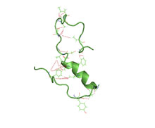
Amyloid-beta precursor protein (APP) is an integral membrane protein expressed in many tissues and concentrated in the synapses of neurons. It functions as a cell surface receptor and has been implicated as a regulator of synapse formation, neural plasticity, antimicrobial activity, and iron export. It is coded for by the gene APP and regulated by substrate presentation. APP is best known as the precursor molecule whose proteolysis generates amyloid beta (Aβ), a polypeptide containing 37 to 49 amino acid residues, whose amyloid fibrillar form is the primary component of amyloid plaques found in the brains of Alzheimer's disease patients.
Amyloid plaques are extracellular deposits of amyloid beta (Aβ) protein that present mainly in the grey matter of the brain. Degenerative neuronal elements and an abundance of microglia and astrocytes can be associated with amyloid plaques. Some plaques occur in the brain as a result of aging, but large numbers of plaques and neurofibrillary tangles are characteristic features of Alzheimer's disease. The plaques are highly variable in shape and size; in tissue sections immunostained for Aβ, they comprise a log-normal size distribution curve, with an average plaque area of 400-450 square micrometers (μm2). The smallest plaques, which often consist of diffuse deposits of Aβ, are particularly numerous. Plaques form when Aβ misfolds and aggregates into oligomers and longer polymers, the latter of which are characteristic of amyloid.
Pittsburgh compound B (PiB) is a radioactive analog of thioflavin T, which can be used in positron emission tomography scans to image beta-amyloid plaques in neuronal tissue. Due to this property, Pittsburgh compound B may be used in investigational studies of Alzheimer's disease.
Thioflavins are fluorescent dyes that are available as at least two compounds, namely Thioflavin T and Thioflavin S. Both are used for histology staining and biophysical studies of protein aggregation. In particular, these dyes have been used since 1989 to investigate amyloid formation. They are also used in biophysical studies of the electrophysiology of bacteria. Thioflavins are corrosive, irritant, and acutely toxic, causing serious eye damage. Thioflavin T has been used in research into Alzheimer's disease and other neurodegenerative diseases.
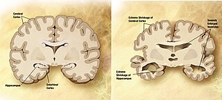
A neurodegenerative disease is caused by the progressive loss of neurons, in the process known as neurodegeneration. Neuronal damage may also ultimately result in their death. Neurodegenerative diseases include amyotrophic lateral sclerosis, multiple sclerosis, Parkinson's disease, Alzheimer's disease, Huntington's disease, multiple system atrophy, tauopathies, and prion diseases. Neurodegeneration can be found in the brain at many different levels of neuronal circuitry, ranging from molecular to systemic. Because there is no known way to reverse the progressive degeneration of neurons, these diseases are considered to be incurable; however research has shown that the two major contributing factors to neurodegeneration are oxidative stress and inflammation. Biomedical research has revealed many similarities between these diseases at the subcellular level, including atypical protein assemblies and induced cell death. These similarities suggest that therapeutic advances against one neurodegenerative disease might ameliorate other diseases as well.
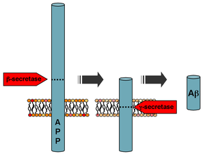
Beta-secretase 1, also known as beta-site amyloid precursor protein cleaving enzyme 1, beta-site APP cleaving enzyme 1 (BACE1), membrane-associated aspartic protease 2, memapsin-2, aspartyl protease 2, and ASP2, is an enzyme that in humans is encoded by the BACE1 gene. Expression of BACE1 is observed mainly in neurons and oligodendrocytes.
The biochemistry of Alzheimer's disease, the most common cause of dementia, is not yet very well understood. Alzheimer's disease (AD) has been identified as a proteopathy: a protein misfolding disease due to the accumulation of abnormally folded amyloid beta (Aβ) protein in the brain. Amyloid beta is a short peptide that is an abnormal proteolytic byproduct of the transmembrane protein amyloid-beta precursor protein (APP), whose function is unclear but thought to be involved in neuronal development. The presenilins are components of proteolytic complex involved in APP processing and degradation.
Presenilins are a family of related multi-pass transmembrane proteins which constitute the catalytic subunits of the gamma-secretase intramembrane protease protein complex. They were first identified in screens for mutations causing early onset forms of familial Alzheimer's disease by Peter St George-Hyslop. Vertebrates have two presenilin genes, called PSEN1 that codes for presenilin 1 (PS-1) and PSEN2 that codes for presenilin 2 (PS-2). Both genes show conservation between species, with little difference between rat and human presenilins. The nematode worm C. elegans has two genes that resemble the presenilins and appear to be functionally similar, sel-12 and hop-1.
APH-1 is a protein originally identified in the round worm Caenorhabditis elegans as a regulator of the cell-surface localization of nicastrin in the Notch signaling pathway.
In medicine, proteinopathy, or proteopathy, protein conformational disorder, or protein misfolding disease, is a class of diseases in which certain proteins become structurally abnormal, and thereby disrupt the function of cells, tissues and organs of the body.
Insulin-degrading enzyme, also known as IDE, is an enzyme.
Collagen alpha-1(XXV) chain is a protein that in humans is encoded by the COL25A1 gene.
Early-onset Alzheimer's disease (EOAD), also called younger-onset Alzheimer's disease (YOAD), is Alzheimer's disease diagnosed before the age of 65. It is an uncommon form of Alzheimer's, accounting for only 5–10% of all Alzheimer's cases. About 60% have a positive family history of Alzheimer's and 13% of them are inherited in an autosomal dominant manner. Most cases of early-onset Alzheimer's share the same traits as the "late-onset" form and are not caused by known genetic mutations. Little is understood about how it starts.
Solanezumab is a monoclonal antibody being investigated by Eli Lilly as a neuroprotector for patients with Alzheimer's disease. The drug originally attracted extensive media coverage proclaiming it a breakthrough, but it has failed to show promise in Phase III trials.
The biomarkers of Alzheimer's disease are neurochemical indicators used to assess the risk or presence of the disease. The biomarkers can be used to diagnose Alzheimer's disease (AD) in a very early stage, but they also provide objective and reliable measures of disease progress. It is imperative to diagnose AD disease as soon as possible, because neuropathologic changes of AD precede the symptoms by years. It is well known that amyloid beta (Aβ) is a good indicator of AD disease, which has facilitated doctors to accurately pre-diagnose cases of AD. When Aβ peptide is released by proteolytic cleavage of amyloid-beta precursor protein, some Aβ peptides that are solubilized are detected in CSF and blood plasma which makes AB peptides a promising candidate for biological markers. It has been shown that the amyloid beta biomarker shows 80% or above sensitivity and specificity, in distinguishing AD from dementia. It is believed that amyloid beta as a biomarker will provide a future for diagnosis of AD and eventually treatment of AD.
p3 peptide also known as amyloid β- peptide (Aβ)17–40/42 is the peptide resulting from the α- and γ-secretase cleavage from the amyloid precursor protein (APP). It is known to be the major constituent of diffuse plaques observed in Alzheimer's disease (AD) brains and pre-amyloid plaques in people affected by Down syndrome. However, p3 peptide's role in these diseases is not truly known yet.
The ion channel hypothesis of Alzheimer's disease (AD), also known as the channel hypothesis or the amyloid beta ion channel hypothesis, is a more recent variant of the amyloid hypothesis of AD, which identifies amyloid beta (Aβ) as the underlying cause of neurotoxicity seen in AD. While the traditional formulation of the amyloid hypothesis pinpoints insoluble, fibrillar aggregates of Aβ as the basis of disruption of calcium ion homeostasis and subsequent apoptosis in AD, the ion channel hypothesis in 1993 introduced the possibility of an ion-channel-forming oligomer of soluble, non-fibrillar Aβ as the cytotoxic species allowing unregulated calcium influx into neurons in AD.
Dennis J. Selkoe is an American physician (neurologist) known for his research into the molecular basis of Alzheimer's disease. In 1985 he became Co-Director of the Center for Neurological Diseases and from 1990, Vincent and Stella Coates Professor of Neurological Diseases at Harvard Medical School. He is also a Fellow of the AAAS and a member of the National Academy of Medicine.
Buntanetap is an orally-administered small molecule inhibitor of several neurotoxic proteins that is under investigation in the treatment of Alzheimer's disease, frontotemporal dementia, chronic traumatic encephalopathy and Parkinson's disease. It is the (+) enantiomer of phenserine, as the (-) enantiomer also has unwanted anticholinergic effects. It is currently in phase III trials for the treatment of Parkinson's.
{{cite journal}}
: CS1 maint: overridden setting (link)