Atrial natriuretic peptide (ANP) or atrial natriuretic factor (ANF) is a natriuretic peptide hormone secreted from the cardiac atria that in humans is encoded by the NPPA gene. [5] Natriuretic peptides (ANP, BNP, and CNP) are a family of hormone/paracrine factors that are structurally related. [6] The main function of ANP is causing a reduction in expanded extracellular fluid (ECF) volume by increasing renal sodium excretion. ANP is synthesized and secreted by cardiac muscle cells in the walls of the atria in the heart. These cells contain volume receptors which respond to increased stretching of the atrial wall due to increased atrial blood volume.
Reduction of blood volume by ANP can result in secondary effects such as reduction of extracellular fluid (ECF) volume, improved cardiac ejection fraction with resultant improved organ perfusion, decreased blood pressure, and increased serum potassium. These effects may be blunted or negated by various counter-regulatory mechanisms operating concurrently on each of these secondary effects.
Brain natriuretic peptide (BNP) – a misnomer; it is secreted by cardiac muscle cells in the heart ventricles – is similar to ANP in its effect. It acts via the same receptors as ANP does, but with 10-fold lower affinity than ANP. The biological half-life of BNP, however, is twice as long as that of ANP, and that of NT-proBNP is even longer, making these peptides better choices than ANP for diagnostic blood testing.
A member of the natriuretic peptide gene family, NPPA encodes an important cardiac signaling molecule known as atrial natriuretic peptide/factor (ANP). [7] ANP carries out endocrine functions of the heart. It acts as a diuretic by inhibiting sodium reabsorption in the kidneys. ANP also acts in the heart to prevent cardiac hypertrophy and to regulate vascular remodeling and energy metabolism. [8] NPPA expression is varied throughout mammalian development into adulthood. Fetal expression of NPPA is associated with the formation of chamber myocardium, muscle cells of the atria and ventricles in the early developing heart. [9] Early expression of this gene has been associated with ventricular hypertrophy in both in vitro and in vivo models. [10] NPPA variants affect plasma ANP concentrations, blood pressure levels, and cardiovascular diseases such as atrial fibrillation (AF). [11] ANP-deficient mice were found to have a large increase in heart and left ventricular weight in response to volume overload, which is normally prevented by proper regulation of blood pressure. [12] Using a knock-in (KI) rat model, researchers found an AF-associated human variant in NPPA caused inflammation, fibroblast activation, atrial fibrosis, and AF in KI rats. [13] These findings suggest NPPA is a critical gene in cardiac development and dysfunction of this gene can lead to heart problems via altered ANP levels.
The discovery of a natriuretic factor (one that promotes kidney excretion of salt and water) was first reported by Adolfo José de Bold in 1981 when rat atrial extracts were found to contain a substance that increased salt and urine output in the kidney. [14] Later, the substance was purified from heart tissue by several groups and named atrial natriuretic factor (ANF) or ANP. [15]
ANP is a 28-amino acid peptide with a 17-amino acid ring in the middle of the molecule. The ring is formed by a disulfide bond between two cysteine residues at positions 7 and 23. ANP is closely related to BNP (brain natriuretic peptide) and CNP (C-type natriuretic peptide), which all share a similar amino acid ring structure. ANP is one of a family of nine structurally similar natriuretic hormones: seven are atrial in origin. [16]
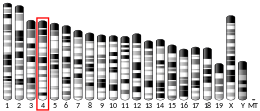
ANP is synthesized as an inactive preprohormone, encoded by the human NPPA gene located on the short arm of chromosome 1. [6] The NPPA gene is expressed primarily in atrial myocytes and consists of 2 introns and three exons, with translation of this gene yielding a high molecular mass 151 amino acid polypeptide known as preproANP. [17] The preprohormone is activated via post-translational modification that involves cleavage of the 25 amino acid signal sequence to produce proANP, a 126 amino acid peptide that is the major form of ANP stored in intracellular granules of the atria. [17] Following stimulation of atrial cells, proANP is released and rapidly converted to the 28-amino-acid C-terminal mature ANP on the cell surface by the cardiac transmembrane serine protease corin. [18] [19] Recently, it was discovered that ANP also can be O-glycosylated. [20]
ANP is secreted in response to:
Three types of atrial natriuretic peptide receptors have been identified on which natriuretic peptides act. They are all cell surface receptors and designated:
NPR-A and NPR-B have a single membrane-spanning segment with an extracellular domain that binds the ligand.[ citation needed ] The intracellular domain maintains two consensus catalytic domains for guanylyl cyclase activity. Binding of a natriuretic peptide induces a conformational change in the receptor that causes receptor dimerization and activation.[ citation needed ]
The binding of ANP to its receptor causes the conversion of GTP to cGMP and raises intracellular cGMP. As a consequence, cGMP activates a cGMP-dependent kinase (PKG or cGK) that phosphorylates proteins at specific serine and threonine residues. In the medullary collecting duct, the cGMP generated in response to ANP may act not only through PKG but also via direct modulation of ion channels. [22]
NPR-C functions mainly as a clearance receptor by binding and sequestering ANP from the circulation. All natriuretic peptides are bound by the NPR-C.[ citation needed ]
Maintenance of the ECF volume (space), and its subcompartment the vascular space, is crucial for survival.[ citation needed ] These compartments are maintained within a narrow range, despite wide variations in dietary sodium intake. There are three volume regulating systems: two salt saving systems, the renin angiotensin aldosterone system (RAAS) and the renal sympathetic system (RSS); and the salt excreting natriuretic peptide (NP) hormone system. When the vascular space contracts, the RAAS and RSS are "turned on"; when the atria expand, NP's are "turned on". Each system also suppresses its counteracting system(s). NP's are made in cardiac, intestinal, renal, and adrenal tissue: ANP in one of a family of cardiac NP's: others at BNP, CNP, and DNP. [16]
ANP binds to a specific set of receptors – ANP receptors. Receptor-agonist binding causes the increase in renal sodium excretion, which results in a decreased ECF and blood volume. Secondary effects may be an improvement in cardiac ejection fraction and reduction of systemic blood pressure.[ citation needed ]
ANP acts on the kidney to increase sodium and water excretion (natriuresis) in the following ways: [23] [24]
ANP has the opposite effect of angiotensin II on the kidney: angiotensin II increases renal sodium retention and ANP increases renal sodium loss.
Relaxes vascular smooth muscle in arterioles and venules by:
Promotes uterine spiral artery remodeling, which is important for preventing pregnancy-induced hypertension. [27]
ANP is produced locally by several immune cells. ANP is shown to regulate several functions of innate and adaptive immune system as well as shown to have cytoprotective effects. [29]
Modulation of the effects of ANP is achieved through gradual degradation of the peptide by the enzyme neutral endopeptidase (NEP). Recently, NEP inhibitors have been developed, such as Sacubitril and Sacubitril/valsartan. They may be clinically useful in treating patients in heart failure with reduced ejection fraction .
Fragments derived from the ANP precursor, including the signal peptide, N-terminal pro-ANP and ANP, have been detected in human blood. [30] ANP and related peptides are used as biomarkers for cardiovascular diseases such as stroke, coronary artery disease, myocardial infarction and heart failure. [31] [32] [33] [34] A specific ANP precursor called mid-regional pro-atrial natriuretic peptide (MRproANP) is a highly sensitive biomarker in heart failure. [35] MRproANP levels below 120 pmol/L can be used to effectively rule out acute heart failure. [35]
Large amounts of ANP secretion has been noted to cause electrolyte disturbances (hyponatremia) and polyuria. These indications can be a marker of a large atrial myxoma. [36]
Opinions regarding the use of ANP for the treatment of acute heart failure and kidney disease are varied. [37] While this molecule has been shown to successfully restore some hemodynamic parameters following heart failure, and yield clinical improvement for kidney injury, whether it ultimately reduces mortality and its long-term effects are unknown. [38] Therefore, more studies need to be conducted to better understand the therapeutic effects of ANP. [38] Newly synthesized homologues of ANP molecule are being assessed for the treatment of acute heart failure. [39] Preliminary research on one of such molecules, ularitide, has shown that this drug is safe, well tolerated, and effective in the treatment of acute heart failure. [39]
Brain natriuretic peptide (BNP) – a misnomer; it is secreted by ventricular myocytes – is similar to ANP in its effect. It acts via atrial natriuretic peptide receptors but with 10-fold lower affinity than ANP. The biological half-life of BNP, however, is twice as long as that of ANP, and that of NT-proBNP is even longer, making these peptides better choices than ANP for diagnostic blood testing.
In addition to the mammalian natriuretic peptides (ANP, BNP, CNP), other natriuretic peptides with similar structure and properties have been isolated elsewhere in the animal kingdom. A salmon natriuretic peptide known as salmon cardiac peptide has been described, [40] and dendroaspis natriuretic peptide (DNP) has been found in the venom of the green mamba, as well as an NP in a species of African snake. [41]
Beside these four, five additional natriuretic peptides have been identified: long-acting natriuretic peptide (LANP), vessel dilator, kaliuretic peptide, urodilatin, and adrenomedullin. [16]
Neutral endopeptidase (NEP) also known as neprilysin is the enzyme that metabolizes natriuretic peptides. Several inhibitors of NEP are currently being developed to treat disorders ranging from hypertension to heart failure. Most of them are dual inhibitors (NEP and ACE). In 2014, PARADIGM-HF study was published in NEJM. This study considered as a landmark study in treatment of heart failure. The study was double blinded; compared LCZ696 versus enalapril in patients with heart failure. The study showed lower all cause mortality, cardiovascular mortality and hospitalization in LCZ696 arm. [42] Omapatrilat (dual inhibitor of NEP and angiotensin-converting enzyme) developed by BMS did not receive FDA approval due to angioedema safety concerns. Other dual inhibitors of NEP with ACE/angiotensin receptor are (in 2003) being developed by pharmaceutical companies. [43]
ANP is also called atrial natriuretic factor (ANF), atrial natriuretic hormone (ANH), cardionatrine, cardiodilatin (CDD), and atriopeptin.
The 2015 version of this article was updated by an external expert under a dual publication model. The corresponding academic peer reviewed article was published in Gene and can be cited as: Wei Song, Hao Wang, Qingyu Wu (12 June 2015). "Atrial natriuretic peptide in cardiovascular biology and disease (NPPA)". Gene . Gene Wiki Review Series. 569 (1): 1–6. doi:10.1016/J.GENE.2015.06.029. ISSN 0378-1119. PMC 4496260 . PMID 26074089. Wikidata Q28082833. |
Angiotensin-converting-enzyme inhibitors are a class of medication used primarily for the treatment of high blood pressure and heart failure. This class of medicine works by causing relaxation of blood vessels as well as a decrease in blood volume, which leads to lower blood pressure and decreased oxygen demand from the heart.
Human vasopressin, also called antidiuretic hormone (ADH), arginine vasopressin (AVP) or argipressin, is a hormone synthesized from the AVP gene as a peptide prohormone in neurons in the hypothalamus, and is converted to AVP. It then travels down the axon terminating in the posterior pituitary, and is released from vesicles into the circulation in response to extracellular fluid hypertonicity (hyperosmolality). AVP has two primary functions. First, it increases the amount of solute-free water reabsorbed back into the circulation from the filtrate in the kidney tubules of the nephrons. Second, AVP constricts arterioles, which increases peripheral vascular resistance and raises arterial blood pressure.
Renin, also known as an angiotensinogenase, is an aspartic protease protein and enzyme secreted by the kidneys that participates in the body's renin-angiotensin-aldosterone system (RAAS)—also known as the renin-angiotensin-aldosterone axis—that increases the volume of extracellular fluid and causes arterial vasoconstriction. Thus, it increases the body's mean arterial blood pressure.
The renin-angiotensin system (RAS), or renin-angiotensin-aldosterone system (RAAS), is a hormone system that regulates blood pressure, fluid, and electrolyte balance, and systemic vascular resistance.
Angiotensin is a peptide hormone that causes vasoconstriction and an increase in blood pressure. It is part of the renin–angiotensin system, which regulates blood pressure. Angiotensin also stimulates the release of aldosterone from the adrenal cortex to promote sodium retention by the kidneys.
The baroreflex or baroreceptor reflex is one of the body's homeostatic mechanisms that helps to maintain blood pressure at nearly constant levels. The baroreflex provides a rapid negative feedback loop in which an elevated blood pressure causes the heart rate to decrease. Decreased blood pressure decreases baroreflex activation and causes heart rate to increase and to restore blood pressure levels. Their function is to sense pressure changes by responding to change in the tension of the arterial wall. The baroreflex can begin to act in less than the duration of a cardiac cycle and thus baroreflex adjustments are key factors in dealing with postural hypotension, the tendency for blood pressure to decrease on standing due to gravity.
Natriuresis is the process of sodium excretion in the urine through the action of the kidneys. It is promoted by ventricular and atrial natriuretic peptides as well as calcitonin, and inhibited by chemicals such as aldosterone. Natriuresis lowers the concentration of sodium in the blood and also tends to lower blood volume because osmotic forces drag water out of the body's blood circulation and into the urine along with the sodium. Many diuretic drugs take advantage of this mechanism to treat medical conditions like hypernatremia and hypertension, which involve excess blood volume.
The adenosine A1 receptor (A1AR) is one member of the adenosine receptor group of G protein-coupled receptors with adenosine as endogenous ligand.
Atrial volume receptors are low pressure baroreceptors that are found in the atria of the heart. They are myelinated vagal fibres in the endocardium found at the junction between atria and the vena cava/pulmonary vein.
An atrial natriuretic peptide receptor is a receptor for atrial natriuretic peptide.
A natriuretic peptide is a hormone molecule that plays a crucial role in the regulation of the cardiovascular system. These hormones were first discovered in the 1980s and were found to have very strong diuretic, natriuretic, and vasodilatory effects. There are three main types of natriuretic peptides: atrial natriuretic peptide (ANP), brain natriuretic peptide (BNP), and C-type natriuretic peptide (CNP). Two minor hormones include urodilatin (URO) which is processed in the kidney and encoded by the same gene as ANP, and dendroaspis NP (DNP) that was discovered through isolation of the venom from the green mamba snake. Since they are activated during heart failure, they are important for the protection of the heart and its tissues.
The median preoptic nucleus is located dorsal to the other three nuclei of the preoptic area of the anterior hypothalamus. The hypothalamus is located just beneath the thalamus, the main sensory relay station of the nervous system, and is considered part of the limbic system, which also includes structures such as the hippocampus and the amygdala. The hypothalamus is highly involved in maintaining homeostasis of the body, and the median preoptic nucleus is no exception, contributing to regulation of blood composition, body temperature, and non-REM sleep.
Urodilatin (URO) is a hormone that causes natriuresis by increasing renal blood flow. It is secreted in response to increased mean arterial pressure and increased blood volume from the cells of the distal tubule and collecting duct. It is important in oliguric patients as it lowers serum creatinine and increases urine output.
Natriuretic peptide precursor C, also known as NPPC, is a protein that in humans is encoded by the NPPC gene. The precursor NPPC protein is cleaved to the 22 amino acid peptide C-type natriuretic peptide (CNP).
Natriuretic peptide receptor A/guanylate cyclase A , also known as NPR1, is an atrial natriuretic peptide receptor. In humans it is encoded by the NPR1 gene.
Natriuretic peptide receptor C/guanylate cyclase C , also known as NPR3, is an atrial natriuretic peptide receptor. In humans it is encoded by the NPR3 gene.
Corin, also called atrial natriuretic peptide-converting enzyme, is a protein that in humans is encoded by the CORIN gene.
Management of heart failure requires a multimodal approach. It involves a combination of lifestyle modifications, medications, and possibly the use of devices or surgery.
Cenderitide is a natriuretic peptide developed by the Mayo Clinic as a potential treatment for heart failure. Cenderitide is created by the fusion of the 15 amino acid C-terminus of the snake venom dendroaspis natriuretic peptide (DNP) with the full C-type natriuretic peptide (CNP) structure. This peptide chimera is a dual activator of the natriuretic peptide receptors NPR-A and NPR-B and therefore exhibits the natriuretic and diuretic properties of DNP, as well as the antiproliferative and antifibrotic properties of CNP.
Brain natriuretic peptide (BNP), also known as B-type natriuretic peptide, is a hormone secreted by cardiomyocytes in the heart ventricles in response to stretching caused by increased ventricular blood volume. BNP is one of the three natriuretic peptides, in addition to atrial natriuretic peptide (ANP) and C-type natriuretic peptide ( CNP). BNP was first discovered in porcine brain tissue in 1988, which led to its initial naming as "brain natriuretic peptide", although subsequent research revealed that BNP is primarily produced and secreted by the ventricular myocardium in response to increased ventricular blood volume and stretching. To reflect its true source, BNP is now often referred to as "B-type natriuretic peptide" while retaining the same acronym.
question number 14