Baroreflex

Last updated
Flowchart showing baroreceptor reflex 2116 Baroreceptor Reflex Flow Art.jpg
Flowchart showing baroreceptor reflex

The baroreflex or baroreceptor reflex is one of the body's homeostatic mechanisms that helps to maintain blood pressure at nearly constant levels. The baroreflex provides a rapid negative feedback loop in which an elevated blood pressure causes the heart rate to decrease. Decreased blood pressure decreases baroreflex activation and causes heart rate to increase and to restore blood pressure levels. Their function is to sense pressure changes by responding to change in the tension of the arterial wall [1] The baroreflex can begin to act in less than the duration of a cardiac cycle (fractions of a second) and thus baroreflex adjustments are key factors in dealing with postural hypotension, the tendency for blood pressure to decrease on standing due to gravity.

Contents

The system relies on specialized neurons, known as baroreceptors, chiefly in the aortic arch and carotid sinuses, to monitor changes in blood pressure and relay them to the medulla oblongata. Baroreceptors are stretch receptors and respond to the pressure induced stretching of the blood vessel in which they are found. Baroreflex-induced changes in blood pressure are mediated by both branches of the autonomic nervous system: the parasympathetic and sympathetic nerves. Baroreceptors are active even at normal blood pressures so their activity informs the brain about both increases and decreases in blood pressure.

The body contains two other, slower-acting systems to regulate blood pressure: the heart releases atrial natriuretic peptide when blood pressure is too high, and the kidneys sense and correct low blood pressure with the renin–angiotensin system. [2]

Anatomy

Baroreceptors are present in the atria of the heart and vena cavae, but the most sensitive baroreceptors are in the carotid sinuses and aortic arch. While the carotid sinus baroreceptor axons travel within the glossopharyngeal nerve (CN IX), the aortic arch baroreceptor axons travel within the vagus nerve (CN X). Baroreceptor activity travels along these nerves directly into the central nervous system to excite glutamatergic neurons within the solitary nucleus (SN) in the brainstem. [3] Baroreceptor information flows from these NSS neurons to both parasympathetic and sympathetic neurons within the brainstem.[ citation needed ]

The SN neurons send excitatory fibers (glutamatergic) to the caudal ventrolateral medulla (CVLM), activating the CVLM. The activated CVLM then sends inhibitory fibers (GABAergic) to the rostral ventrolateral medulla (RVLM), thus inhibiting the RVLM. The RVLM is the primary regulator of the sympathetic nervous system, sending excitatory fibers (glutamatergic) to the sympathetic preganglionic neurons located in the intermediolateral nucleus of the spinal cord. Hence, when the baroreceptors are activated (by an increased blood pressure), the NTS activates the CVLM, which in turn inhibits the RVLM, thus decreasing the activity of the sympathetic branch of the autonomic nervous system, leading to a relative decrease in blood pressure. Likewise, low blood pressure activates baroreceptors less and causes an increase in sympathetic tone via "disinhibition" (less inhibition, hence activation) of the RVLM. Cardiovascular targets of the sympathetic nervous system includes both blood vessels and the heart.[ citation needed ]

Even at resting levels of blood pressure, arterial baroreceptor discharge activates SN neurons. Some of these SN neurons are tonically activated by this resting blood pressure and thus activate excitatory fibers to the nucleus ambiguus and dorsal nucleus of vagus nerve to regulate the parasympathetic nervous system. These parasympathetic neurons send axons to the heart and parasympathetic activity slows cardiac pacemaking and thus heart rate. This parasympathetic activity is further increased during conditions of elevated blood pressure. The parasympathetic nervous system is primarily directed toward the heart.[ citation needed ]

Activation

The baroreceptors are stretch-sensitive mechanoreceptors. At low pressures, baroreceptors become inactive. When blood pressure rises, the carotid and aortic sinuses are distended further, resulting in increased stretch and, therefore, a greater degree of activation of the baroreceptors. At normal resting blood pressures, many baroreceptors are actively reporting blood pressure information and the baroreflex is actively modulating autonomic activity. Active baroreceptors fire action potentials ("spikes") more frequently. The greater the stretch the more rapidly baroreceptors fire action potentials. Many individual baroreceptors are inactive at normal resting pressures and only become activated when their stretch or pressure threshold is exceeded.[ citation needed ]

Baroreceptor mechanosensitivity is hypothesised to be linked to the expression of PIEZO1 and PIEZO2 on neurons in the petrosal and nodose ganglia.

Baroreceptor action potentials are relayed to the solitary nucleus, which uses frequency as a measure of blood pressure. Increased activation of the solitary nucleus inhibits the vasomotor center and stimulates the vagal nuclei. The end-result of baroreceptor activation is inhibition of the sympathetic nervous system and activation of the parasympathetic nervous system.[ citation needed ]

The sympathetic and parasympathetic branches of the autonomic nervous system have opposing effects on blood pressure. Sympathetic activation leads to an elevation of total peripheral resistance and cardiac output via increased contractility of the heart, heart rate, and arterial vasoconstriction, which tends to increase blood pressure. Conversely, parasympathetic activation leads to decreased cardiac output via decrease in heart rate, resulting in a tendency to lower blood pressure.[ citation needed ]

By coupling sympathetic inhibition and parasympathetic activation, the baroreflex maximizes blood pressure reduction. Sympathetic inhibition leads to a drop in peripheral resistance, while parasympathetic activation leads to a depressed heart rate (reflex bradycardia) and contractility. The combined effects will dramatically decrease blood pressure. In a similar manner, sympathetic activation with parasympathetic inhibition allows the baroreflex to elevate blood pressure.[ citation needed ]

Set point and tonic activation

Baroreceptor firing has an inhibitory effect on sympathetic outflow. The sympathetic neurons fire at different rates which determines the release of norepinephrine onto cardiovascular targets. Norepinephrine constricts blood vessels to increase blood pressure. When baroreceptors are stretched (due to an increased blood pressure) their firing rate increases which in turn decreases the sympathetic outflow resulting in reduced norepinephrine and thus blood pressure. When the blood pressure is low, baroreceptor firing is reduced and this in turn results in augmented sympathetic outflow and increased norepinephrine release on the heart and blood vessels, increasing blood pressure.[ citation needed ]

Effect on heart rate variability

The baroreflex may be responsible for a part of the low-frequency component of heart rate variability, the so-called Mayer waves, at 0.1 Hz. [4]

Baroreflex activation therapy

Baroreflex activation is distinct from vagal stimulation. It works through an afferent limb which has the double effect of stimulating vagal outflow and attenuating global sympathetic outflow. Baroreflex activation.svg
Baroreflex activation is distinct from vagal stimulation. It works through an afferent limb which has the double effect of stimulating vagal outflow and attenuating global sympathetic outflow.

High blood pressure

The baroreflex can be used to treat resistant hypertension. [5] This stimulation is provided by a pacemaker-like device. While the devices appears to lower blood pressure, evidence remains very limited as of 2018. [5]

Heart failure

The ability of baroreflex activation therapy to reduce sympathetic nerve activity suggests a potential in the treatment of chronic heart failure, because in this condition there is often intense sympathetic activation and patients with such sympathetic activation show a markedly increased risk of fatal arrhythmias and death.[ citation needed ]

One trial [6] has already shown that baroreflex activation therapy improves functional status, quality of life, exercise capacity and N-terminal pro-brain natriuretic peptide.[ citation needed ]

See also

Related Research Articles

<span class="mw-page-title-main">Autonomic nervous system</span> Division of the nervous system supplying internal organs, smooth muscle and glands

The autonomic nervous system (ANS), formerly referred to as the vegetative nervous system, is a division of the nervous system that operates internal organs, smooth muscle and glands. The autonomic nervous system is a control system that acts largely unconsciously and regulates bodily functions, such as the heart rate, its force of contraction, digestion, respiratory rate, pupillary response, urination, and sexual arousal. This system is the primary mechanism in control of the fight-or-flight response.

<span class="mw-page-title-main">Parasympathetic nervous system</span> Division of the autonomic nervous system

The parasympathetic nervous system (PSNS) is one of the three divisions of the autonomic nervous system, the others being the sympathetic nervous system and the enteric nervous system. The enteric nervous system is sometimes considered part of the autonomic nervous system, and sometimes considered an independent system.

<span class="mw-page-title-main">Sympathetic nervous system</span> Part of the autonomic nervous system which stimulates fight-or-flight responses

The sympathetic nervous system (SNS) is one of the three divisions of the autonomic nervous system, the others being the parasympathetic nervous system and the enteric nervous system. The enteric nervous system is sometimes considered part of the autonomic nervous system, and sometimes considered an independent system.

Baroreceptors are sensors located in the carotid sinus and in the aortic arch. They sense the blood pressure and relay the information to the brain, so that a proper blood pressure can be maintained.

<span class="mw-page-title-main">Heart rate</span> Speed of the heartbeat, measured in beats per minute

Heart rate is the frequency of the heartbeat measured by the number of contractions of the heart per minute. The heart rate can vary according to the body's physical needs, including the need to absorb oxygen and excrete carbon dioxide, but is also modulated by numerous factors, including genetics, physical fitness, stress or psychological status, diet, drugs, hormonal status, environment, and disease/illness as well as the interaction between and among these factors. It is usually equal or close to the pulse measured at any peripheral point.

<span class="mw-page-title-main">Nucleus ambiguus</span>

The nucleus ambiguus is a group of large motor neurons, situated deep in the medullary reticular formation named by Jacob Clarke. The nucleus ambiguus contains the cell bodies of neurons that innervate the muscles of the soft palate, pharynx, and larynx which are associated with speech and swallowing. As well as motor neurons, the nucleus ambiguus contains preganglionic parasympathetic neurons which innervate postganglionic parasympathetic neurons in the heart.

The cardiovascular centre is a part of the human brain which regulates heart rate through the nervous and endocrine systems. It is considered one of the vital centres of the medulla oblongata.

Cushing reflex is a physiological nervous system response to increased intracranial pressure (ICP) that results in Cushing's triad of increased blood pressure, irregular breathing, and bradycardia. It is usually seen in the terminal stages of acute head injury and may indicate imminent brain herniation. It can also be seen after the intravenous administration of epinephrine and similar drugs. It was first described in detail by American neurosurgeon Harvey Cushing in 1901.

<span class="mw-page-title-main">Carotid sinus</span> Dilated area near internal carotid artery above bifurcation

In human anatomy, the carotid sinus is a dilated area at the base of the internal carotid artery just superior to the bifurcation of the internal carotid and external carotid at the level of the superior border of thyroid cartilage. The carotid sinus extends from the bifurcation to the "true" internal carotid artery. The carotid sinus is sensitive to pressure changes in the arterial blood at this level. It is the major baroreception site in humans and most mammals.

The Bainbridge reflex or Bainbridge effect, also called the atrial reflex, is an increase in heart rate due to an increase in central venous pressure. Increased blood volume is detected by stretch receptors located in both sides of atria at the venoatrial junctions.

Vagal tone is activity of the vagus nerve, the 10th cranial nerve and a fundamental component of the parasympathetic branch of the autonomic nervous system. This branch of the nervous system is not under conscious control and is largely responsible for the regulation of several body compartments at rest. Vagal activity results in various effects, including: heart rate reduction, vasodilation/constriction of vessels, glandular activity in the heart, lungs, and digestive tract, liver, immune system regulation as well as control of gastrointestinal sensitivity, motility and inflammation.

<span class="mw-page-title-main">Median preoptic nucleus</span> Nucleus in the anterior hypothalamus

The median preoptic nucleus is located dorsal to the other three nuclei of the preoptic area of the anterior hypothalamus. The hypothalamus is located just beneath the thalamus, the main sensory relay station of the nervous system, and is considered part of the limbic system, which also includes structures such as the hippocampus and the amygdala. The hypothalamus is highly involved in maintaining homeostasis of the body, and the median preoptic nucleus is no exception, contributing to regulation of blood composition, body temperature, and non-REM sleep.

Reflex bradycardia is a bradycardia in response to the baroreceptor reflex, one of the body's homeostatic mechanisms for preventing abnormal increases in blood pressure. In the presence of high mean arterial pressure, the baroreceptor reflex produces a reflex bradycardia as a method of decreasing blood pressure by decreasing cardiac output.

The Bezold–Jarisch reflex involves a variety of cardiovascular and neurological processes which cause hypopnea, hypotension and bradycardia in response to noxious stimuli detected in the cardiac ventricles. The reflex is named after Albert von Bezold and Adolf Jarisch Junior. The significance of the discovery is that it was the first recognition of a chemical (non-mechanical) reflex.

The vasomotor center (VMC) is a portion of the medulla oblongata. Together with the cardiovascular center and respiratory center, it regulates blood pressure. It also has a more minor role in other homeostatic processes. Upon increase in carbon dioxide level at central chemoreceptors, it stimulates the sympathetic system to constrict vessels. This is opposite to carbon dioxide in tissues causing vasodilatation, especially in the brain. Cranial nerves IX and X both feed into the vasomotor centre and are themselves involved in the regulation of blood pressure.

<span class="mw-page-title-main">Pathophysiology of hypertension</span>

Pathophysiology is a study which explains the function of the body as it relates to diseases and conditions. The pathophysiology of hypertension is an area which attempts to explain mechanistically the causes of hypertension, which is a chronic disease characterized by elevation of blood pressure. Hypertension can be classified by cause as either essential or secondary. About 90–95% of hypertension is essential hypertension. Some authorities define essential hypertension as that which has no known explanation, while others define its cause as being due to overconsumption of sodium and underconsumption of potassium. Secondary hypertension indicates that the hypertension is a result of a specific underlying condition with a well-known mechanism, such as chronic kidney disease, narrowing of the aorta or kidney arteries, or endocrine disorders such as excess aldosterone, cortisol, or catecholamines. Persistent hypertension is a major risk factor for hypertensive heart disease, coronary artery disease, stroke, aortic aneurysm, peripheral artery disease, and chronic kidney disease.

Low pressure baroreceptors are baroreceptors that relay information derived from blood pressure within the autonomic nervous system. They are stimulated by stretching of the vessel wall. They are located in large systemic veins and in the walls of the atria of the heart, and pulmonary vasculature. Low pressure baroreceptors are also referred to as volume receptors and cardiopulmonary baroreceptors.

Cardiac physiology or heart function is the study of healthy, unimpaired function of the heart: involving blood flow; myocardium structure; the electrical conduction system of the heart; the cardiac cycle and cardiac output and how these interact and depend on one another.

Neurocardiology is the study of the neurophysiological, neurological and neuroanatomical aspects of cardiology, including especially the neurological origins of cardiac disorders. The effects of stress on the heart are studied in terms of the heart's interactions with both the peripheral nervous system and the central nervous system.

<span class="mw-page-title-main">Baroreflex activation therapy</span>

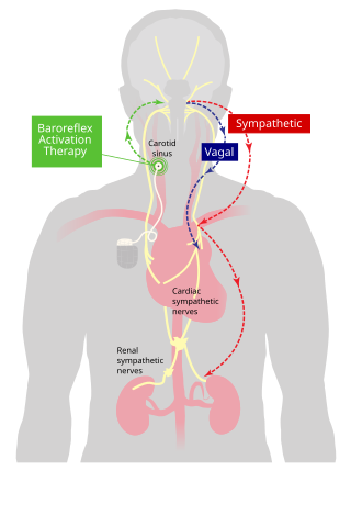
Baroreflex activation therapy is an approach to treating high blood pressure and the symptoms of heart failure. It uses an implanted device to electrically stimulate baroreceptors in the carotid sinus region. This elicits a reflex response through the sympathetic and vagal nervous systems that reduces blood pressure.

References

  1. Bär, Karl-Jürgen (2015-06-24). "Cardiac Autonomic Dysfunction in Patients with Schizophrenia and Their Healthy Relatives – A Small Review". Frontiers in Neurology. Frontiers Media SA. 6: 139. doi: 10.3389/fneur.2015.00139 . ISSN   1664-2295. PMC   4478389 . PMID   26157417.
  2. Fu, Shihui; Ping, Ping; Wang, Fengqi; Luo, Leiming (2018-01-12). "Synthesis, secretion, function, metabolism and application of natriuretic peptides in heart failure". Journal of Biological Engineering. Springer Nature. 12 (1): 2. doi: 10.1186/s13036-017-0093-0 . ISSN   1754-1611. PMC   5766980 . PMID   29344085. They are mainly produced by cardiovascular, brain and renal tissues in response to wall stretch and other causes. NPs provide natriuresis, diuresis, vasodilation, antiproliferation, antihypertrophy, antifibrosis and other cardiometabolic protection. NPs represent body's own antihypertensive system, and provide compensatory protection to counterbalance vasoconstrictor-mitogenic-sodium retaining hormones, released by renin-angiotensin-aldosterone system (RAAS) and sympathetic nervous system (SNS).
  3. Yuan, Jason; Brooks, Heddwen L.; Barman, Susan M.; Barrett, Kim E. (2019). Ganong's Review of Medical Physiology. McGraw-Hill Education. ISBN   978-1-26-012240-4.
  4. Sleight, Peter; La Rovere, Maria Teresa; Mortara, Andrea; Pinna, Gianni; Maestri, Roberto; Leuzzi, Stefano; Bianchini, Beatrice; Tavazzi, Luigi; Bernardi, Luciano (1 January 1995). "Physiology and Pathophysiology of Heart Rate and Blood Pressure Variability in Humans: Is Power Spectral Analysis Largely An Index of Baroreflex Gain?". Clinical Science. 88 (1): 103–109. doi:10.1042/cs0880103. PMID   7677832.
  5. 1 2 Wallbach, M; Koziolek, MJ (9 November 2017). "Baroreceptors in the carotid and hypertension-systematic review and meta-analysis of the effects of baroreflex activation therapy on blood pressure". Nephrology, Dialysis, Transplantation. 33 (9): 1485–1493. doi: 10.1093/ndt/gfx279 . PMID   29136223.
  6. Abraham, WT; Zile, MR; Weaver, FA; Butter, C; Ducharme, A; Halbach, M; Klug, D; Lovett, EG; Müller-Ehmsen, J; Schafer, JE; Senni, M; Swarup, V; Wachter, R; Little, WC (June 2015). "Baroreflex Activation Therapy for the Treatment of Heart Failure With a Reduced Ejection Fraction". JACC: Heart Failure. 3 (6): 487–496. doi: 10.1016/j.jchf.2015.02.006 . PMID   25982108.