A Wiggers diagram, named after its developer, Carl Wiggers, is a unique diagram that has been used in teaching cardiac physiology for more than a century. [1] [2] In the Wiggers diagram, the X-axis is used to plot time subdivided into the cardiac phases, while the Y-axis typically contains the following on a single grid:
Contents
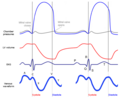
- Blood pressure
- Aortic pressure
- Ventricular pressure
- Atrial pressure
- Ventricular volume
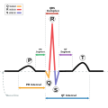
- Electrocardiogram
- Arterial flow (optional)
- Heart sounds (optional)
The Wiggers diagram clearly illustrates the coordinated variation of these values as the heart beats, assisting one in understanding the entire cardiac cycle. [1]