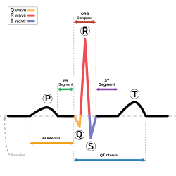
P wave (electrocardiography)

Last updated
Normal P wave, shown in darker red Normal P wave (ECG).svg
Normal P wave, shown in darker red
Diagram demonstrating features of a normal sinus rhythm electrocardiogram wave SinusRhythmLabels.svg
Diagram demonstrating features of a normal sinus rhythm electrocardiogram wave

The P wave on the ECG represents atrial depolarization, which results in atrial contraction, or atrial systole.

Contents

Physiology

The P wave is a summation wave generated by the depolarization front as it transits the atria. Normally the right atrium depolarizes slightly earlier than left atrium since the depolarization wave originates in the sinoatrial node, in the high right atrium and then travels to and through the left atrium. The depolarization front is carried through the atria along semi-specialized conduction pathways including Bachmann's bundle resulting in uniform shaped waves. Depolarization originating elsewhere in the atria (atrial ectopics) result in P waves with a different morphology from normal.

Pathology

Characteristic peaked P wave of cor pulmonale P pulmonale.svg
Characteristic peaked P wave of cor pulmonale

Peaked P waves (> 0.25 mV) suggest right atrial enlargement, cor pulmonale, (P pulmonale rhythm), [1] but have a low predictive value (~20%). [2]

A P wave with increased amplitude can indicate hypokalemia. [3] It can also indicate right atrial enlargement. [4]

A P wave with decreased amplitude can indicate hyperkalemia. [5]

P-wave changes in left and right atrial hypertrophy De-P wave morphology (CardioNetworks ECGpedia).png
P-wave changes in left and right atrial hypertrophy

Bifid P waves (known as P mitrale) indicate left-atrial abnormality - e.g. dilatation [6] or hypertrophy. [1]

If at least three different shaped P waves can be seen in a given ECG lead tracing, this implies that even if one of them arises from the SA node, at least two others are arising elsewhere. This is taken as evidence of multiple (i.e. at least two) ectopic foci, and is called multifocal (or more correctly, multiform) atrial rhythm if the rate is ≤100) or multifocal atrial tachycardia if the rate is over 100. [7] This appears particularly commonly in exacerbations of chronic obstructive lung disease. [8]

If the baseline has a totally irregular form, this suggests fibrillatory waves of atrial fibrillation or possibly artefact; a saw tooth shaped baseline suggests the flutter waves of atrial flutter. With either of these rhythms, if the ventricular rate is fast, the fibrillatory or flutter waves can easily be misinterpreted as P waves.

Absence of the P wave with a flat baseline may indicate:

If P waves are not clearly delineated in the surface ECG, a Lewis lead may be used to better visualize P waves.

Atrial repolarization

This occurs a mean of 320 ms after the end of the P wave, with a duration of 2-3 times that of the P wave and a polarity always opposite to that of the P wave. It is represented on the surface ECG by a so-called Ta wave. The clinical relevance of this is that, although a normal phenomenon, the nadir of the Ta wave can occur just after the QRS complex and cause ST depression similar to (and easily mistaken with) that occurring with disease states such as cardiac ischaemia. [9]

Related Research Articles

<span class="mw-page-title-main">Electrocardiography</span> Examination of the hearts electrical activity

Electrocardiography is the process of producing an electrocardiogram, a recording of the heart's electrical activity through repeated cardiac cycles. It is an electrogram of the heart which is a graph of voltage versus time of the electrical activity of the heart using electrodes placed on the skin. These electrodes detect the small electrical changes that are a consequence of cardiac muscle depolarization followed by repolarization during each cardiac cycle (heartbeat). Changes in the normal ECG pattern occur in numerous cardiac abnormalities, including cardiac rhythm disturbances, inadequate coronary artery blood flow, and electrolyte disturbances.

<span class="mw-page-title-main">Premature ventricular contraction</span> Skipped beat with ventricular origin

A premature ventricular contraction (PVC) is a common event where the heartbeat is initiated by Purkinje fibers in the ventricles rather than by the sinoatrial node. PVCs may cause no symptoms or may be perceived as a "skipped beat" or felt as palpitations in the chest. PVCs do not usually pose any danger.

Systole is the part of the cardiac cycle during which some chambers of the heart contract after refilling with blood.

<span class="mw-page-title-main">Wolff–Parkinson–White syndrome</span> Medical condition

Wolff–Parkinson–White syndrome (WPWS) is a disorder due to a specific type of problem with the electrical system of the heart involving an accessory pathway able to conduct electrical current between the atria and the ventricles, thus bypassing the atrioventricular node. About 60% of people with the electrical problem developed symptoms, which may include an abnormally fast heartbeat, palpitations, shortness of breath, lightheadedness, or syncope. Rarely, cardiac arrest may occur. The most common type of irregular heartbeat that occurs is known as paroxysmal supraventricular tachycardia.

<span class="mw-page-title-main">Atrial flutter</span> Medical condition

Atrial flutter (AFL) is a common abnormal heart rhythm that starts in the atrial chambers of the heart. When it first occurs, it is usually associated with a fast heart rate and is classified as a type of supraventricular tachycardia. Atrial flutter is characterized by a sudden-onset (usually) regular abnormal heart rhythm on an electrocardiogram (ECG) in which the heart rate is fast. Symptoms may include a feeling of the heart beating too fast, too hard, or skipping beats, chest discomfort, difficulty breathing, a feeling as if one's stomach has dropped, a feeling of being light-headed, or loss of consciousness.

<span class="mw-page-title-main">Cardiac conduction system</span> Aspect of heart function

The cardiac conduction system transmits the signals generated by the sinoatrial node – the heart's pacemaker, to cause the heart muscle to contract, and pump blood through the body's circulatory system. The pacemaking signal travels through the right atrium to the atrioventricular node, along the bundle of His, and through the bundle branches to Purkinje fibers in the walls of the ventricles. The Purkinje fibers transmit the signals more rapidly to stimulate contraction of the ventricles.

<span class="mw-page-title-main">Ostium primum atrial septal defect</span> Medical condition

The ostium primum atrial septal defect is a defect in the atrial septum at the level of the tricuspid and mitral valves. This is sometimes known as an endocardial cushion defect because it often involves the endocardial cushion, which is the portion of the heart where the atrial septum meets the ventricular septum and the mitral valve meets the tricuspid valve.

<span class="mw-page-title-main">Supraventricular tachycardia</span> Abnormally fast heart rhythm

Supraventricular tachycardia (SVT) is an umbrella term for fast heart rhythms arising from the upper part of the heart. This is in contrast to the other group of fast heart rhythms – ventricular tachycardia, which start within the lower chambers of the heart. There are four main types of SVT: atrial fibrillation, atrial flutter, paroxysmal supraventricular tachycardia (PSVT), and Wolff–Parkinson–White syndrome. The symptoms of SVT include palpitations, feeling of faintness, sweating, shortness of breath, and/or chest pain.

<span class="mw-page-title-main">AV nodal reentrant tachycardia</span> Medical condition

AV-nodal reentrant tachycardia (AVNRT) is a type of abnormal fast heart rhythm. It is a type of supraventricular tachycardia (SVT), meaning that it originates from a location within the heart above the bundle of His. AV nodal reentrant tachycardia is the most common regular supraventricular tachycardia. It is more common in women than men. The main symptom is palpitations. Treatment may be with specific physical maneuvers, medications, or, rarely, synchronized cardioversion. Frequent attacks may require radiofrequency ablation, in which the abnormally conducting tissue in the heart is destroyed.

Premature atrial contraction (PAC), also known as atrial premature complexes (APC) or atrial premature beats (APB), are a common cardiac dysrhythmia characterized by premature heartbeats originating in the atria. While the sinoatrial node typically regulates the heartbeat during normal sinus rhythm, PACs occur when another region of the atria depolarizes before the sinoatrial node and thus triggers a premature heartbeat, in contrast to escape beats, in which the normal sinoatrial node fails, leaving a non-nodal pacemaker to initiate a late beat.

Lown–Ganong–Levine syndrome (LGL) is a pre-excitation syndrome of the heart. Those with LGL syndrome have episodes of abnormal heart racing with a short PR interval and normal QRS complexes seen on their electrocardiogram when in a normal sinus rhythm. LGL syndrome was originally thought to be due to an abnormal electrical connection between the atria and the ventricles, but is now thought to be due to accelerated conduction through the atrioventricular node in the majority of cases. The syndrome is named after Bernard Lown, William Francis Ganong, Jr., and Samuel A. Levine.

<span class="mw-page-title-main">Multifocal atrial tachycardia</span> Fast heart rhythm associated with exacerbations of COPD

Multifocal atrial tachycardia (MAT) is an abnormal heart rhythm, specifically a type of supraventricular tachycardia, that is particularly common in older people and is associated with exacerbations of chronic obstructive pulmonary disease (COPD). Normally, the heart rate is controlled by a cluster of cells called the sinoatrial node. When a number of different clusters of cells outside the SA node take over control of the heart rate, and the rate exceeds 100 beats per minute, this is called multifocal atrial tachycardia.

<span class="mw-page-title-main">Wandering atrial pacemaker</span> Medical condition

Wandering atrial pacemaker (WAP) is an atrial rhythm where the pacemaking activity of the heart originates from different locations within the atria. This is different from normal pacemaking activity, where the sinoatrial node is responsible for each heartbeat and keeps a steady rate and rhythm. Causes of wandering atrial pacemaker are unclear, but there may be factors leading to its development. It is often seen in the young, the old, and in athletes, and rarely causes symptoms or requires treatment. Diagnosis of wandering atrial pacemaker is made by an ECG.

<span class="mw-page-title-main">Accelerated idioventricular rhythm</span> Medical condition

Accelerated idioventricular rhythm is a ventricular rhythm with a rate of between 40 and 120 beats per minute. Idioventricular means “relating to or affecting the cardiac ventricle alone” and refers to any ectopic ventricular arrhythmia. Accelerated idioventricular arrhythmias are distinguished from ventricular rhythms with rates less than 40 and those faster than 120. Though some other references limit to between 60 and 100 beats per minute. It is also referred to as AIVR and "slow ventricular tachycardia."

<span class="mw-page-title-main">Junctional rhythm</span> Medical condition

Junctional rhythm describes an abnormal heart rhythm resulting from impulses coming from a locus of tissue in the area of the atrioventricular node(AV node), the "junction" between atria and ventricles.

<span class="mw-page-title-main">Ectopic pacemaker</span> Cardiac condition

An ectopic pacemaker, also known as ectopic focus or ectopic foci, is an excitable group of cells that causes a premature heart beat outside the normally functioning SA node of the heart. It is thus a cardiac pacemaker that is ectopic, producing an ectopic beat. Acute occurrence is usually non-life-threatening, but chronic occurrence can progress into tachycardia, bradycardia or ventricular fibrillation. In a normal heart beat rhythm, the SA node usually suppresses the ectopic pacemaker activity due to the higher impulse rate of the SA node. However, in the instance of either a malfunctioning SA node or an ectopic focus bearing an intrinsic rate superior to SA node rate, ectopic pacemaker activity may take over the natural heart rhythm. This phenomenon is called an escape rhythm, the lower rhythm having escaped from the dominance of the upper rhythm. As a rule, premature ectopic beats indicate increased myocyte or conducting tissue excitability, whereas late ectopic beats indicate proximal pacemaker or conduction failure with an escape 'ectopic' beat.

<span class="mw-page-title-main">Junctional ectopic tachycardia</span> Medical condition

Junctional ectopic tachycardia (JET) is a rare syndrome of the heart that manifests in patients recovering from heart surgery. It is characterized by cardiac arrhythmia, or irregular beating of the heart, caused by abnormal conduction from or through the atrioventricular node. In newborns and infants up to 6 weeks old, the disease may also be referred to as His bundle tachycardia or congenital JET.

Atrial tachycardia is a type of heart rhythm problem in which the heart's electrical impulse comes from an ectopic pacemaker in the upper chambers (atria) of the heart, rather than from the sinoatrial node, the normal origin of the heart's electrical activity. As with any other form of tachycardia, the underlying mechanism can be either the rapid discharge of an abnormal focus, the presence of a ring of cardiac tissue that gives rise to a circle movement (reentry), or a triggered rapid rhythm due to other pathological circumstances.

<span class="mw-page-title-main">Right atrial enlargement</span> Medical condition

Right atrial enlargement (RAE) is a form of cardiomegaly, or heart enlargement. It can broadly be classified as either right atrial hypertrophy (RAH), overgrowth, or dilation, like an expanding balloon. Common causes include pulmonary hypertension, which can be the primary defect leading to RAE, or pulmonary hypertension secondary to tricuspid stenosis; pulmonary stenosis or Tetralogy of Fallot i.e. congenital diseases; chronic lung disease, such as cor pulmonale. Other recognised causes are: right ventricular failure, tricuspid regurgitation, and atrial septal defect. Right atrial enlargement (RAE) is clinically significant due to its prevalence in diagnosing supraventricular arrhythmias. Further, early diagnosis using risk factors like RAE may decrease mortality because patients with RAE are at 9x more risk of arrhythmias and other cardiac conditions compared to their healthy counterparts.

<span class="mw-page-title-main">Arrhythmia</span> Group of medical conditions characterized by irregular heartbeat

Arrhythmias, also known as cardiac arrhythmias, heart arrhythmias, or dysrhythmias, are irregularities in the heartbeat, including when it is too fast or too slow. A resting heart rate that is too fast – above 100 beats per minute in adults – is called tachycardia, and a resting heart rate that is too slow – below 60 beats per minute – is called bradycardia. Some types of arrhythmias have no symptoms. Symptoms, when present, may include palpitations or feeling a pause between heartbeats. In more serious cases, there may be lightheadedness, passing out, shortness of breath, chest pain, or decreased level of consciousness. While most cases of arrhythmia are not serious, some predispose a person to complications such as stroke or heart failure. Others may result in sudden death.

References

  1. 1 2 3 Longmore, Murray (2004). Oxford Handbook of Clinical Medicine 8th edition page 90. Oxford University Press. ISBN   978-0-19-852558-5.
  2. Reeves WC, Hallahan W, Schwiter EJ, Ciotola TJ, Buonocore E, Davidson W (1981). "Two-dimensional echocardiographic assessment of electrocardiographic criteria for right atrial enlargement". Circulation. 64 (2): 387–391. doi: 10.1161/01.CIR.64.2.387 . PMID   6454512.
  3. "Hypokalaemia".
  4. Yanowitz, Frank G. "VII. Atrial Enlargement". ECG Learning Center. Archived from the original on 2010-03-29. Retrieved 2009-09-05.
  5. Levis, Joel T (2013). "ECG Diagnosis: Hyperkalemia". The Permanente Journal. 17 (1): 69. doi:10.7812/TPP/12-088. ISSN   1552-5767. PMC   3627796 . PMID   23596374.
  6. Munuswamy K, Alpert MA, Martin RH, Whiting RB, Mechlin NJ (1983). "Sensitivity and specificity of commonly used electrocardiographic criteria for left atrial enlargement determined by m-mode echocardiography". Am J Cardiol. 53 (6): 829–832. doi:10.1016/0002-9149(84)90413-2. PMID   6230922.
  7. Kastor JA (1990). "Multifocal Atrial Tachycardia". N Engl J Med. 322 (24): 1713–1717. doi:10.1056/NEJM199006143222405. PMID   2188131.
  8. Kothari SA, Apiyasawat S, Asad N, Spodick DH (2006). "Evidence supporting a new rate threshold for multifocal atrial tachycardia". Clin Cardiol. 28 (12): 3561–3563. doi:10.1002/clc.4960281205. PMC   6654295 . PMID   16405199.
  9. Smith, SW. "Atrial Repolarization Wave Mimicking ST Depression" . Retrieved 22 October 2014.