In animals, including humans, the startle response is a largely unconscious defensive response to sudden or threatening stimuli, such as sudden noise or sharp movement, and is associated with negative affect. [1] Usually the onset of the startle response is a startle reflex reaction. The startle reflex is a brainstem reflectory reaction (reflex) that serves to protect vulnerable parts, such as the back of the neck (whole-body startle) and the eyes (eyeblink) and facilitates escape from sudden stimuli. It is found across many different species, throughout all stages of life. A variety of responses may occur depending on the affected individual's emotional state, [2] body posture, [3] preparation for execution of a motor task, [4] or other activities. [5] The startle response is implicated in the formation of specific phobias.[ citation needed ]
A startle reflex can occur in the body through a combination of actions. A reflex from hearing a sudden loud noise will happen in the primary acoustic startle reflex pathway consisting of three main central synapses, or signals that travel through the brain.
First, there is a synapse from the auditory nerve fibers in the ear to the cochlear root neurons (CRN). These are the first acoustic neurons of the central nervous system. Studies have shown a direct correlation to the amount of decrease of the startle to the number of CRNs that were killed. Second, there is a synapse from the CRN axons to the cells in the nucleus reticularis pontis caudalis (PnC) of the brain. These are neurons that are located in the pons of the brainstem. A study done to disrupt this portion of the pathway by the injection of PnC inhibitory chemicals has shown a dramatic decrease in the amount of startle by about 80 to 90 percent. Third, a synapse occurs from the PnC axons to the motor neurons in the facial motor nucleus or the spinal cord that will directly or indirectly control the movement of muscles. The activation of the facial motor nucleus causes a jerk of the head while an activation in the spinal cord causes the whole body to startle. [6]
During neuromotor examinations of newborns, it is noted that, for a number of techniques, the patterns of the startle reaction and the Moro reflex may significantly overlap, the notable distinction being the absence of arm abduction (spreading) during startle responses. [7]
There are many various reflexes that can occur simultaneously during a startle response. The fastest reflex recorded in humans happens within the masseter muscle or jaw muscle. The reflex was measured by electromyography which records the electrical activity during movement of the muscles. This also showed the response latency, or the delay between the stimulus and the response recorded, was found to be about 14 milliseconds. The blink of the eye which is the reflex of the orbicularis oculi muscle was found to have a latency of about 20 to 40 milliseconds. Out of larger body parts, the head is quickest in a movement latency in a range from 60 to 120 milliseconds. The neck then moves almost simultaneously with a latency of 75 to 121 milliseconds. Next, the shoulder jerks at 100 to 121 milliseconds along with the arms at 125 to 195 milliseconds. Lastly the legs respond with a latency of 145 to 395 milliseconds. This type of cascading response correlates to how the synapses travel from the brain and down the spinal cord to activate each motor neuron. [8]
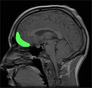
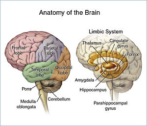
The acoustic startle reflex is thought to be caused by an auditory stimulus greater than 80 decibels. [1] The reflex is typically measured by electromyography, brain imaging or sometimes positron emission tomography. [9] [10] There are many brain structures and pathways thought to be involved in the reflex. The amygdala, hippocampus, bed nucleus of the stria terminalis (BNST) and anterior cingulate cortex are all thought to play a role in modulating the reflex. [11] [12] The anterior cingulate cortex in the brain is largely thought to be the main area associated with emotional response and awareness, which can contribute to the way an individual reacts to startle-inducing stimuli. [11] Along with the anterior cingulate cortex, the amygdala and the hippocampus are known to have implications in this reflex.
The amygdala is known to have a role in the "fight-or-flight response", and the hippocampus functions to form memories of the stimulus and the emotions associated with it. [13] The role of the BNST in the acoustic startle reflex may be attributed to specific areas within the nucleus responsible for stress and anxiety responses. [12] Activation of the BNST by certain hormones is thought to promote a startle response [12] The auditory pathway for this response was largely elucidated in rats in the 1980s. [14] The basic pathway follows the auditory pathway from the ear up to the nucleus of the lateral lemniscus (LLN) from where it activates a motor centre in the reticular formation. This centre sends descending projections to lower motor neurones of the limbs[ clarification needed ].
In slightly more detail this corresponds to ear (cochlea) → cranial nerve VIII (auditory) → cochlear nucleus (ventral/inferior) → LLN → caudal pontine reticular nucleus (PnC). The whole process has a less than 10ms[ clarification needed ] latency. There is no involvement of the superior/rostral or inferior/caudal colliculus in the reaction that "twitches" the hindlimbs, but these may be important for adjustment of pinnae and gaze towards the direction of the sound, or for the associated blink. [15]
A study undertaken in 2005 by researchers at the Department of Aviation and Logistics, University of Southern Queensland, looked at the performance of aircraft pilots following unexpected critical events. Analysing a number of recent aircraft accidents, the authors identified the negative impact of the startle response as causal or contributory in these accidents. The authors argued that fear resulting from threat, especially if life-threatening, [16] [17] prompted startle effects which had a serious negative impact on pilots' performances. The study considered training strategies to address this, including exposing pilots to unexpected critical events more often, enabling them to improve their responses. [18]
Articles related to anatomy include:
The olfactory bulb is a neural structure of the vertebrate forebrain involved in olfaction, the sense of smell. It sends olfactory information to be further processed in the amygdala, the orbitofrontal cortex (OFC) and the hippocampus where it plays a role in emotion, memory and learning.
In neurophysiology, long-term depression (LTD) is an activity-dependent reduction in the efficacy of neuronal synapses lasting hours or longer following a long patterned stimulus. LTD occurs in many areas of the CNS with varying mechanisms depending upon brain region and developmental progress.
Pavlovian fear conditioning is a behavioral paradigm in which organisms learn to predict aversive events. It is a form of learning in which an aversive stimulus is associated with a particular neutral context or neutral stimulus, resulting in the expression of fear responses to the originally neutral stimulus or context. This can be done by pairing the neutral stimulus with an aversive stimulus. Eventually, the neutral stimulus alone can elicit the state of fear. In the vocabulary of classical conditioning, the neutral stimulus or context is the "conditional stimulus" (CS), the aversive stimulus is the "unconditional stimulus" (US), and the fear is the "conditional response" (CR).
In neuroanatomy, a neural pathway is the connection formed by axons that project from neurons to make synapses onto neurons in another location, to enable neurotransmission. Neurons are connected by a single axon, or by a bundle of axons known as a nerve tract, or fasciculus. Shorter neural pathways are found within grey matter in the brain, whereas longer projections, made up of myelinated axons, constitute white matter.
The pyramidal tracts include both the corticobulbar tract and the corticospinal tract. These are aggregations of efferent nerve fibers from the upper motor neurons that travel from the cerebral cortex and terminate either in the brainstem (corticobulbar) or spinal cord (corticospinal) and are involved in the control of motor functions of the body.
Dopaminergic pathways in the human brain are involved in both physiological and behavioral processes including movement, cognition, executive functions, reward, motivation, and neuroendocrine control. Each pathway is a set of projection neurons, consisting of individual dopaminergic neurons.
The ventral tegmental area (VTA), also known as the ventral tegmental area of Tsai, or simply ventral tegmentum, is a group of neurons located close to the midline on the floor of the midbrain. The VTA is the origin of the dopaminergic cell bodies of the mesocorticolimbic dopamine system and other dopamine pathways; it is widely implicated in the drug and natural reward circuitry of the brain. The VTA plays an important role in a number of processes, including reward cognition and orgasm, among others, as well as several psychiatric disorders. Neurons in the VTA project to numerous areas of the brain, ranging from the prefrontal cortex to the caudal brainstem and several regions in between.
The stria terminalis is a structure in the brain consisting of a band of fibers running along the lateral margin of the ventricular surface of the thalamus. Serving as a major output pathway of the amygdala, the stria terminalis runs from its centromedial division to the ventromedial nucleus of the hypothalamus.
Affective neuroscience is the study of how the brain processes emotions. This field combines neuroscience with the psychological study of personality, emotion, and mood. The basis of emotions and what emotions are remains an issue of debate within the field of affective neuroscience.
The orbitofrontal cortex (OFC) is a prefrontal cortex region in the frontal lobes of the brain which is involved in the cognitive process of decision-making. In non-human primates it consists of the association cortex areas Brodmann area 11, 12 and 13; in humans it consists of Brodmann area 10, 11 and 47.
The facial motor nucleus is a collection of neurons in the brainstem that belong to the facial nerve. These lower motor neurons innervate the muscles of facial expression and the stapedius.
The amygdalofugal pathway is one of the three major efferent pathways of the amygdala, meaning that it is one of the three principal pathways by which fibers leave the amygdala. It leads from the basolateral nucleus and central nucleus of the amygdala. The amygdala is a limbic structure in the medial temporal lobe of the brain. The other main efferent pathways from the amygdala are the stria terminalis and anterior commissure.
The reward system is a group of neural structures responsible for incentive salience, associative learning, and positively-valenced emotions, particularly ones involving pleasure as a core component. Reward is the attractive and motivational property of a stimulus that induces appetitive behavior, also known as approach behavior, and consummatory behavior. A rewarding stimulus has been described as "any stimulus, object, event, activity, or situation that has the potential to make us approach and consume it is by definition a reward". In operant conditioning, rewarding stimuli function as positive reinforcers; however, the converse statement also holds true: positive reinforcers are rewarding.The reward system motivates animals to approach stimuli or engage in behaviour that increases fitness. Survival for most animal species depends upon maximizing contact with beneficial stimuli and minimizing contact with harmful stimuli. Reward cognition serves to increase the likelihood of survival and reproduction by causing associative learning, eliciting approach and consummatory behavior, and triggering positively-valenced emotions. Thus, reward is a mechanism that evolved to help increase the adaptive fitness of animals. In drug addiction, certain substances over-activate the reward circuit, leading to compulsive substance-seeking behavior resulting from synaptic plasticity in the circuit.
The medial dorsal nucleus is a large nucleus in the thalamus. It is separated from the other thalamic nuclei by the internal medullary lamina.
Synaptic gating is the ability of neural circuits to gate inputs by either suppressing or facilitating specific synaptic activity. Selective inhibition of certain synapses has been studied thoroughly, and recent studies have supported the existence of permissively gated synaptic transmission. In general, synaptic gating involves a mechanism of central control over neuronal output. It includes a sort of gatekeeper neuron, which has the ability to influence transmission of information to selected targets independently of the parts of the synapse upon which it exerts its action.
The basolateral amygdala, or basolateral complex, consists of the lateral, basal and accessory-basal nuclei of the amygdala. The lateral nuclei receives the majority of sensory information, which arrives directly from the temporal lobe structures, including the hippocampus and primary auditory cortex. The basolateral amygdala also receives dense neuromodulatory inputs from ventral tegmental area (VTA), locus coeruleus (LC), and basal forebrain, whose integrity are important for associative learning. The information is then processed by the basolateral complex and is sent as output to the central nucleus of the amygdala. This is how most emotional arousal is formed in mammals.
The trisynaptic circuit or trisynaptic loop is a relay of synaptic transmission in the hippocampus. The trisynaptic circuit is a neural circuit in the hippocampus, which is made up of three major cell groups: granule cells in the dentate gyrus, pyramidal neurons in CA3, and pyramidal neurons in CA1. The hippocampal relay involves 3 main regions within the hippocampus which are classified according to their cell type and projection fibers. The first projection of the hippocampus occurs between the entorhinal cortex (EC) and the dentate gyrus (DG). The entorhinal cortex transmits its signals from the parahippocampal gyrus to the dentate gyrus via granule cell fibers known collectively as the perforant path. The dentate gyrus then synapses on pyramidal cells in CA3 via mossy cell fibers. CA3 then fires to CA1 via Schaffer collaterals which synapse in the subiculum and are carried out through the fornix. Collectively the dentate gyrus, CA1 and CA3 of the hippocampus compose the trisynaptic loop.
The Mauthner cells are a pair of big and easily identifiable neurons located in the rhombomere 4 of the hindbrain in fish and amphibians that are responsible for a very fast escape reflex. The cells are also notable for their unusual use of both chemical and electrical synapses.
Many experiments have been done to find out how the brain interprets stimuli and how animals develop fear responses. The emotion, fear, has been hard-wired into almost every individual, due to its vital role in the survival of the individual. Researchers have found that fear is established unconsciously and that the amygdala is involved with fear conditioning.