This article needs additional citations for verification .(May 2015) |
| Masseter | |
|---|---|
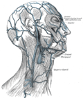
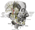
 The left masseter muscle (red highlight), partially covered by superficial muscles such as the platysma muscle (below) and both the zygomaticus major and minor muscles | |
| Details | |
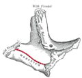
| Origin | Zygomatic arch and maxillary process of zygomatic bone |
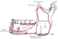
| Insertion | Angle surface of ramus of mandible, coronoid process |
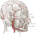
| Artery | Masseteric artery |
| Nerve | Mandibular nerve (V3) |
| Actions | Elevation (as in closing of the mouth) and protrusion of mandible |
| Identifiers | |
| Latin | musculus masseter |
| MeSH | D008406 |
| TA98 | A04.1.04.002 |
| TA2 | 2105 |
| FMA | 48996 |
| Anatomical terms of muscle | |
In anatomy, the masseter [help 1] is one of the muscles of mastication. Found only in mammals, it is particularly powerful in herbivores to facilitate chewing of plant matter. [5] The most obvious muscle of mastication is the masseter muscle, since it is the most superficial and one of the strongest.