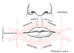
In human anatomy, the orbicularis oris muscle is a complex of muscles in the lips that encircles the mouth. It is not a true sphincter, as was once thought, as it is actually composed of four independent quadrants that interlace and give only an appearance of circularity.

The lips are a horizontal pair of soft appendages attached to the jaws and are the most visible part of the mouth of many animals, including humans. Vertebrate lips are soft, movable and serve to facilitate the ingestion of food and the articulation of sound and speech. Human lips are also a somatosensory organ, and can be an erogenous zone when used in kissing and other acts of intimacy.

The depressor labii inferioris is a facial muscle. It helps to lower the bottom lip.

The zygomaticus major muscle is a muscle of the face. It arises from either zygomatic arch (cheekbone); it inserts at the corner of the mouth. It is innervated by branches of the facial nerve.

The levator labii superioris is a muscle of the human body used in facial expression. It is a broad sheet, the origin of which extends from the side of the nose to the zygomatic bone.

The depressor anguli oris muscle is a facial muscle. It originates from the mandible and inserts into the angle of the mouth. It is associated with frowning, as it depresses the corner of the mouth.

The facial artery, formerly called the external maxillary artery, is a branch of the external carotid artery that supplies blood to superficial structures of the medial regions of the face.
In facial anatomy, the modiolus is a dense, compact, mobile, fibromuscular tissue mass of facial muscles formed by the interlacing of a number of muscles just lateral to the angle of the mouth opposite the second upper premolar tooth.
Asymmetric crying facies (ACF), also called partial unilateral facial paresis and hypoplasia of depressor angula oris muscle, is a minor congenital anomaly caused by agenesis or hypoplasia of the depressor anguli oris muscle, one of the muscles that control the movements of the lower lip. This unilateral facial weakness is first noticed when the infant cries or smiles, affecting only one corner of the mouth and occurs on the left side in nearly 80% of cases.
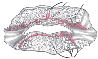
The inferior labial artery arises near the angle of the mouth as a branch of the facial artery; it passes upward and forward beneath the triangularis and, penetrating the orbicularis oris, runs in a tortuous course along the edge of the lower lip between this muscle and the mucous membrane.

The infraorbital artery is a small artery in the head that arises from the maxillary artery and passes through the inferior orbital fissure to enter the orbit, then passes forward along the floor of the orbit, finally exiting the orbit through the infraorbital foramen to reach the face.

The marginal mandibular branch of the facial nerve arises from the facial nerve in the parotid gland at the parotid plexus. It passes anterior-ward deep to the platysma and depressor anguli oris muscles. It provides motor innervation to muscles of the lower lip and chin: the depressor labii inferioris muscle, depressor anguli oris muscle, and mentalis muscle. It communicates with the mental branch of the inferior alveolar nerve.
The buccal branches of the facial nerve, are of larger size than the rest of the branches, pass horizontally forward to be distributed below the orbit and around the mouth.

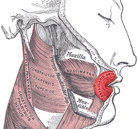
The buccal space is a fascial space of the head and neck. It is a potential space in the cheek, and is paired on each side. The buccal space is superficial to the buccinator muscle and deep to the platysma muscle and the skin. The buccal space is part of the subcutaneous space, which is continuous from head to toe.

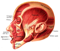
The facial muscles are a group of striated skeletal muscles supplied by the facial nerve that, among other things, control facial expression. These muscles are also called mimetic muscles. They are only found in mammals, although they derive from neural crest cells found in all vertebrates. They are the only muscles that attach to the dermis.
The transversus menti, or transverse muscle of the chin, is a facial muscle that is often considered to be the superficial fibers of the depressor anguli oris muscle which cross to the other side of the face.

The following outline is provided as an overview of and topical guide to human anatomy:
Levator muscle can refer to:

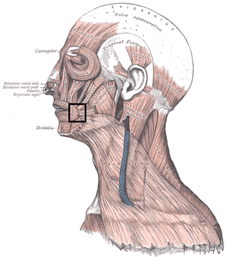
The canine space is a fascial space of the head and neck. It is a thin potential space on the face, and is paired on either side. It is located between the levator anguli oris muscle inferiorly and the levator labii superioris muscle superiorly. The term is derived from the fact that the space is in the region of the canine fossa, and that infections originating from the maxillary canine tooth may spread to involve the space. Infra-orbital is derived from infra- meaning below and orbit which refers to the eye socket.