The maxilla in vertebrates is the upper fixed bone of the jaw formed from the fusion of two maxillary bones. In humans, the upper jaw includes the hard palate in the front of the mouth. The two maxillary bones are fused at the intermaxillary suture, forming the anterior nasal spine. This is similar to the mandible, which is also a fusion of two mandibular bones at the mandibular symphysis. The mandible is the movable part of the jaw.

The inferior alveolar nerve (IAN) (also the inferior dental nerve) is a sensory branch of the mandibular nerve (CN V3) (which is itself the third branch of the trigeminal nerve (CN V)). The nerve provides sensory innervation to the lower/mandibular teeth and their corresponding gingiva as well as a small area of the face (via its mental nerve).

The levator anguli oris (caninus) is a facial muscle of the mouth arising from the canine fossa, immediately below the infraorbital foramen. It elevates angle of mouth medially. Its fibers are inserted into the angle of the mouth, intermingling with those of the zygomaticus, triangularis, and orbicularis oris. Specifically, the levator anguli oris is innervated by the buccal branches of the facial nerve.
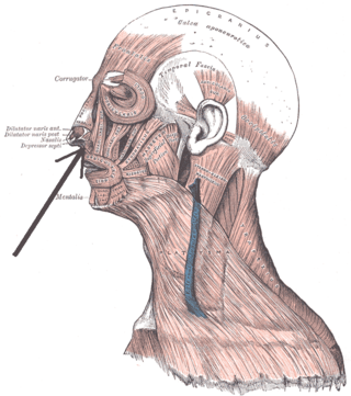
The depressor septi nasi muscle is a muscle of the face. It connects the incisive fossa of the maxilla and the orbicularis oris muscle to the nasal septum of the nose. It draws the ala of the nose downwards, reducing the size of the nostrils.
The jugular fossa is a deep depression in the inferior part of the temporal bone at the base of the skull. It lodges the bulb of the internal jugular vein.

The inferior alveolar artery is an artery of the head. It is a branch of the maxillary artery. It descends through the infratemporal fossa as part of a neurovascular bundle with the inferior alveolar nerve and vein to the mandibular foramen where it enters and passes anteriorly inside the mandible, suplying the body of mandible and the dental pulp of the lower molar and premolar teeth. Its terminal incisor branch supplies the rest of the lower teeth. Its mental branch exits the mandibula anteriorly through the mental foramen to supply adjacent lip and skin.
The nasopalatine nerve (also long sphenopalatine nerve) is a nerve of the head. It is a sensory branch of the maxillary nerve (CN V2) that passes through the pterygopalatine ganglion (without synapsing) and then through the sphenopalatine foramen to enter the nasal cavity, and finally out of the nasal cavity through the incisive canal and then the incisive fossa to enter the hard palate. It provides sensory innervation to the posteroinferior part of the nasal septum, and gingiva just posterior to the upper incisor teeth.

The carotid canal is a passage in the petrous part of the temporal bone of the skull through which the internal carotid artery and its internal carotid (nervous) plexus pass from the neck into the cranial cavity.
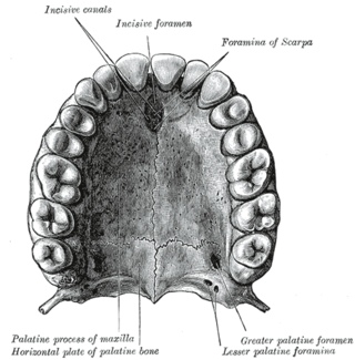
The incisive canals are two bony canals of the anterior hard palate connecting the nasal cavity and the oral cavity. An incisive canal courses through each maxilla. Below, the two incisive canals typically converge medially.
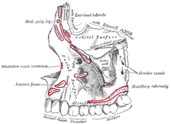
In the human mouth, the incisive foramen is the opening of the incisive canals on the hard palate immediately behind the incisor teeth. It gives passage to blood vessels and nerves. The incisive foramen is situated within the incisive fossa of the maxilla.
The greater palatine artery is a branch of the descending palatine artery and contributes to the blood supply of the hard palate and nasal septum.
The infraorbital artery is a small artery in the head that arises from the maxillary artery and passes through the inferior orbital fissure to enter the orbit, then passes forward along the floor of the orbit, finally exiting the orbit through the infraorbital foramen to reach the face.

In human anatomy of the mouth, the palatine process of maxilla, is a thick, horizontal process of the maxilla. It forms the anterior three quarters of the hard palate, the horizontal plate of the palatine bone making up the rest.

The olecranon fossa is a deep triangular depression on the posterior side of the humerus, superior to the trochlea. It provides space for the olecranon of the ulna during extension of the forearm.
The incisive papilla is an oval midline mucosal prominence of the anterior hard palate overlying the incisive fossa. It is situated posteriorly to the central incisors, and represents the anterior extremity of the palatine raphe.
Euchambersia is an extinct genus of therocephalian therapsids that lived during the Late Permian in what is now South Africa and China. The genus contains two species. The type species E. mirabilis was named by paleontologist Robert Broom in 1931 from a skull missing the lower jaw. A second skull, belonging to a probably immature individual, was later described. In 2022, a second species, E. liuyudongi, was named by Jun Liu and Fernando Abdala from a well-preserved skull. It is a member of the family Akidnognathidae, which historically has also been referred by as the synonymous Euchambersiidae.

The following outline is provided as an overview of and topical guide to human anatomy:
The inferior dental plexus is a nerve plexus formed by sensory branches of the inferior alveolar nerve. The plexus issues dental branches and gingival branches; the small dental branches provide sensory innervation to the lower/mandibular teeth.

The mandibular incisive canal is a bilaterally paired bony canal within the anterior portion of the mandible that extends from the mental foramen (usually) to near the ipsilateral lateral incisor teeth.
Boreogomphodon is an extinct genus of traversodontid cynodonts from the Late Triassic of the eastern United States. Fossils have been found from the Turkey Branch Formation in Virginia and the Pekin Formation of North Carolina.