
Paranasal sinuses are a group of four paired air-filled spaces that surround the nasal cavity. The maxillary sinuses are located under the eyes; the frontal sinuses are above the eyes; the ethmoidal sinuses are between the eyes and the sphenoidal sinuses are behind the eyes. The sinuses are named for the facial bones in which they are located.

In mammalian oral anatomy, the canine teeth, also called cuspids, dog teeth, fangs, or eye teeth, are relatively long, pointed teeth. However, they can appear more flattened, causing them to resemble incisors and leading them to be called incisiform. They developed and are used primarily for firmly holding food in order to tear it apart, and occasionally as weapons. They are often the largest teeth in a mammal's mouth. Individuals of most species that develop them normally have four, two in the upper jaw and two in the lower, separated within each jaw by incisors; humans and dogs are examples. In most species, canines are the anterior-most teeth in the maxillary bone.

Incisors are the front teeth present in most mammals. They are located in the premaxilla above and on the mandible below. Humans have a total of eight. Opossums have 18, whereas armadillos have none.

The nasal septum separates the left and right airways in the nose, dividing the two nostrils.

The pterygopalatine ganglion is a parasympathetic ganglion found in the pterygopalatine fossa. It is largely innervated by the greater petrosal nerve ; and its axons project to the lacrimal glands and nasal mucosa. The flow of blood to the nasal mucosa, in particular the venous plexus of the conchae, is regulated by the pterygopalatine ganglion and heats or cools the air in the nose. It is one of four parasympathetic ganglia of the head and neck, the others being the submandibular ganglion, otic ganglion, and ciliary ganglion.
The maxillary nerve (CN V2) is one of the three branches or divisions of the trigeminal nerve, the fifth (V) cranial nerve. It comprises the principal functions of sensation from the maxillary, nasal cavity, sinuses, the palate and subsequently that of the mid-face, and is intermediate, both in position and size, between the ophthalmic nerve and the mandibular nerve.

Permanent teeth or adult teeth are the second set of teeth formed in diphyodont mammals. In humans and old world simians, there are thirty-two permanent teeth, consisting of six maxillary and six mandibular molars, four maxillary and four mandibular premolars, two maxillary and two mandibular canines, four maxillary and four mandibular incisors.

In human anatomy, the pterygopalatine fossa is a fossa in the skull. A human skull contains two pterygopalatine fossae—one on the left side, and another on the right side. Each fossa is a cone-shaped paired depression deep to the infratemporal fossa and posterior to the maxilla on each side of the skull, located between the pterygoid process and the maxillary tuberosity close to the apex of the orbit. It is the indented area medial to the pterygomaxillary fissure leading into the sphenopalatine foramen. It communicates with the nasal and oral cavities, infratemporal fossa, orbit, pharynx, and middle cranial fossa through eight foramina.

The maxillary veins consist of a short trunk which accompanies the first part of the internal maxillary artery.
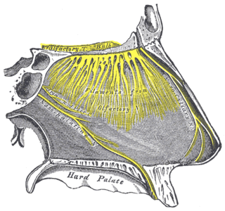
One branch of the pterygopalatine ganglion, longer and larger than the others, is named the nasopalatine nerve.

The pterygomaxillary fissure is a fissure of the human skull. It is vertical, and descends at right angles from the medial end of the inferior orbital fissure. It is a triangular interval, formed by the divergence of the maxilla from the pterygoid process of the sphenoid.

Below the bulla ethmoidalis, and partly hidden by the inferior end of the uncinate process of ethmoid bone, is the maxillary hiatus ; in a frontal section this opening is seen to be placed near the roof of the sinus.In the articulated skull this aperture is much reduced in size by the following bones: the uncinate process of the ethmoid above, the ethmoidal process of the inferior nasal concha below, the vertical part of the palatine behind, and a small part of the lacrimal above and in front; the sinus communicates with the middle meatus of the nose, generally by two small apertures left between the above-mentioned bones.

The maxillary artery supplies deep structures of the face. It branches from the external carotid artery just deep to the neck of the mandible.

The infraorbital artery is an artery in the head that branches off the maxillary artery, emerging through the infraorbital foramen, just under the orbit of the eye.

The posterior superior alveolar artery is given off from the maxillary, frequently in conjunction with the infraorbital artery just as the trunk of the vessel is passing into the pterygopalatine fossa.

The posterior superior alveolar branches arise from the trunk of the maxillary nerve just before it enters the infraorbital groove; they are generally two in number, but sometimes arise by a single trunk.
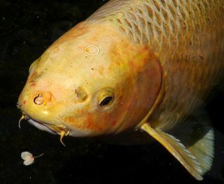
In fish anatomy and turtle anatomy, a barbel is a slender, whiskerlike sensory organ near the mouth. Fish that have barbels include the catfish, the carp, the goatfish, the hagfish, the sturgeon, the zebrafish, the black dragonfish and some species of shark such as the sawshark. Barbels house the taste buds of such fish and are used to search for food in murky water.
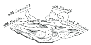
Behind the lacrimal process of the inferior nasal conchae lies a broad, thin plate, the ethmoidal process, which ascends to join the uncinate process of the ethmoid; from its lower border a thin lamina, the maxillary process, curves downward and lateralward; it articulates with the maxilla and forms a part of the medial wall of the maxillary sinus.


