
The tongue is a muscular organ in the mouth of a typical tetrapod. It manipulates food for mastication and swallowing as part of the digestive process, and is the primary organ of taste. The tongue's upper surface (dorsum) is covered by taste buds housed in numerous lingual papillae. It is sensitive and kept moist by saliva and is richly supplied with nerves and blood vessels. The tongue also serves as a natural means of cleaning the teeth. A major function of the tongue is the enabling of speech in humans and vocalization in other animals.

The facial nerve, also known as the seventh cranial nerve, cranial nerve VII, or simply CN VII, is a cranial nerve that emerges from the pons of the brainstem, controls the muscles of facial expression, and functions in the conveyance of taste sensations from the anterior two-thirds of the tongue. The nerve typically travels from the pons through the facial canal in the temporal bone and exits the skull at the stylomastoid foramen. It arises from the brainstem from an area posterior to the cranial nerve VI and anterior to cranial nerve VIII.
Articles related to anatomy include:

The paired submandibular glands are major salivary glands located beneath the floor of the mouth. They each weigh about 15 grams and contribute some 60–67% of unstimulated saliva secretion; on stimulation their contribution decreases in proportion as the parotid secretion rises to 50%. The average length of the normal human submandibular salivary gland is approximately 27mm, while the average width is approximately 14.3mm.
The paired sublingual glands are major salivary glands in the mouth. They are the smallest, most diffuse, and the only unencapsulated major salivary glands. They provide only 3-5% of the total salivary volume. There are also two other types of salivary glands; they are submandibular and parotid glands.

The internal jugular vein is a paired jugular vein that collects blood from the brain and the superficial parts of the face and neck. This vein runs in the carotid sheath with the common carotid artery and vagus nerve.

The hyoglossus, thin and quadrilateral, arises from the side of the body and from the whole length of the greater cornu of the hyoid bone, and passes almost vertically upward to enter the side of the tongue, between the styloglossus and the inferior longitudinal muscle of the tongue. It forms a part of the floor of submandibular triangle.
Sublingual, from the Latin for "under the tongue", refers to the pharmacological route of administration by which substances diffuse into the blood through tissues under the tongue.

The lingual nerve carries sensory innervation from the anterior two-thirds of the tongue. It contains fibres from both the mandibular division of the trigeminal nerve (CN V3) and from the facial nerve (CN VII). The fibres from the trigeminal nerve are for touch, pain and temperature (general sensation), and the ones from the facial nerve are for taste (special sensation).

The lingual artery arises from the external carotid artery between the superior thyroid artery and facial artery. It can be located easily in the tongue.

The obturator artery is a branch of the internal iliac artery that passes antero-inferiorly on the lateral wall of the pelvis, to the upper part of the obturator foramen, and, escaping from the pelvic cavity through the obturator canal, it divides into both an anterior and a posterior branch.

In human anatomy, the mandibular canal is a canal within the mandible that contains the inferior alveolar nerve, inferior alveolar artery, and inferior alveolar vein. It runs obliquely downward and forward in the ramus, and then horizontally forward in the body, where it is placed under the alveoli and communicates with them by small openings.

The maxillary artery supplies deep structures of the face. It branches from the external carotid artery just deep to the neck of the mandible.
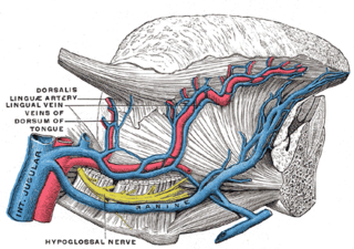
The lingual veins begin on the dorsum, sides, and under surface of the tongue, and, passing backward along the course of the lingual artery, end in the internal jugular vein.
The submandibular ganglion is part of the human autonomic nervous system. It is one of four parasympathetic ganglia of the head and neck..

The middle cranial fossa, deeper than the anterior cranial fossa, is narrow medially and widens laterally to the sides of the skull. It is separated from the posterior fossa by the clivus and the petrous crest.

The infratemporal fossa is an irregularly shaped cavity that is a part of the skull. It is situated below and medial to the zygomatic arch. It is not fully enclosed by bone in all directions. It contains superficial muscles, including the lower part of the temporalis muscle, the lateral pterygoid muscle, and the medial pterygoid muscle. It also contains important blood vessels such as the middle meningeal artery, the pterygoid plexus, and the retromandibular vein, and nerves such as the mandibular nerve (CN V3) and its branches.

This article describes the anatomy of the head and neck of the human body, including the brain, bones, muscles, blood vessels, nerves, glands, nose, mouth, teeth, tongue, and throat.

The following outline is provided as an overview of and topical guide to human anatomy:

The sublingual space is a fascial space of the head and neck. It is a potential space located below the mouth and above the mylohyoid muscle, and is part of the suprahyoid group of fascial spaces.
