Palatine process of maxilla

Last updated
Palatine process of maxilla
Sobo 1909 100 - Palatine process of maxilla.png
Inferior surface of maxilla. The bony palate and alveolar arch. (Palatine process labeled at bottom right.)
Palatine process.jpg
Inferior surface of maxilla.
Details
Identifiers
Latin processus palatinus ossis maxillae, processus palatinus maxillae
TA98 A02.1.12.029
TA2 786
FMA 52896
Anatomical terms of bone

In human anatomy of the mouth, the palatine process of maxilla (palatal process), is a thick, horizontal process of the maxilla. It forms the anterior three quarters of the hard palate, the horizontal plate of the palatine bone making up the rest. It is the most important bone in the midface. It provides structural support for the viscerocranium. [1]

Contents

Structure

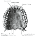
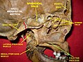
It is perforated by numerous foramina for the passage of the nutrient vessels; is channelled at the back part of its lateral border by a groove, sometimes a canal, for the transmission of the descending palatine vessels and the anterior palatine nerve from the spheno-palatine ganglion; and presents little depressions for the lodgement of the palatine glands.

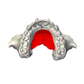
When the two maxillae are articulated, a funnel-shaped opening, the incisive foramen, is seen in the middle line, immediately behind the incisor teeth.

In this opening the orifices of two lateral canals are visible; they are named the incisive canals or foramina of Stenson; through each of them passes the terminal branch of the descending palatine artery and the nasopalatine nerve.

On the under surface of the palatine process, a delicate linear suture, well seen in young skulls, may sometimes be noticed extending laterally and forward on either side from the incisive foramen to the interval between the lateral incisor and the canine tooth.

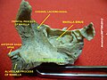
The small part in front of this suture constitutes the premaxilla (os incisivum), which in most vertebrates forms an independent bone; it includes the whole thickness of the alveolus, the corresponding part of the floor of the nose and the anterior nasal spine, and contains the sockets of the incisor teeth.

The upper surface of the palatine process is concave from side to side, smooth, and forms the greater part of the floor of the nasal cavity. It presents, close to its medial margin, the upper orifice of the incisive canal.

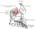
The lateral border of the process is incorporated with the rest of the bone.

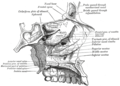
The medial border is thicker in front than behind, and is raised above into a ridge, the nasal crest, which, with the corresponding ridge of the opposite bone, forms a groove for the reception of the vomer. The front part of this ridge rises to a considerable height, and is named the incisor crest; it is prolonged forward into a sharp process, which forms, together with a similar process of the opposite bone, the anterior nasal spine.

The posterior border is serrated for articulation with the horizontal part of the palatine bone.

Variation

Occasionally two additional canals are present in the middle line; they are termed the foramina of Scarpa, and when present transmit the nasopalatine nerves, the left passing through the anterior, and the right through the posterior canal.

Clinical significance

Additional images

Related Research Articles

<span class="mw-page-title-main">Maxilla</span> Upper jaw bone

In vertebrates, the maxilla is the upper fixed bone of the jaw formed from the fusion of two maxillary bones. In humans, the upper jaw includes the hard palate in the front of the mouth. The two maxillary bones are fused at the intermaxillary suture, forming the anterior nasal spine. This is similar to the mandible, which is also a fusion of two mandibular bones at the mandibular symphysis. The mandible is the movable part of the jaw.

<span class="mw-page-title-main">Zygomatic bone</span> Facial bone

In the human skull, the zygomatic bone, also called cheekbone or malar bone, is a paired irregular bone, situated at the upper and lateral part of the face and forms part of the lateral wall and floor of the orbit, of the temporal fossa and the infratemporal fossa. It presents a malar and a temporal surface; four processes, and four borders.

<span class="mw-page-title-main">Palatine bone</span> Bone of the facial skeleton

In anatomy, the palatine bones are two irregular bones of the facial skeleton in many animal species, located above the uvula in the throat. Together with the maxilla, they comprise the hard palate.

<span class="mw-page-title-main">Inferior nasal concha</span> Facial bone

The inferior nasal concha is one of the three paired nasal conchae in the nose. It extends horizontally along the lateral wall of the nasal cavity and consists of a lamina of spongy bone, curled upon itself like a scroll,. The inferior nasal conchae are considered a pair of facial bones. As the air passes through the turbinates, the air is churned against these mucosa-lined bones in order to receive warmth, moisture and cleansing. Superior to inferior nasal concha are the middle nasal concha and superior nasal concha which both arise from the ethmoid bone, of the cranial portion of the skull. Hence, these two are considered as a part of the cranial bones.

<span class="mw-page-title-main">Vomer</span> Unpaired facial bone of the skull

The vomer is one of the unpaired facial bones of the skull. It is located in the midsagittal line, and articulates with the sphenoid, the ethmoid, the left and right palatine bones, and the left and right maxillary bones. The vomer forms the inferior part of the nasal septum in humans, with the superior part formed by the perpendicular plate of the ethmoid bone. The name is derived from the Latin word for a ploughshare and the shape of the bone.

<span class="mw-page-title-main">Orbit (anatomy)</span> Cavity or socket of the skull in which the eye and its appendages are situated

In anatomy, the orbit is the cavity or socket/hole of the skull in which the eye and its appendages are situated. "Orbit" can refer to the bony socket, or it can also be used to imply the contents. In the adult human, the volume of the orbit is 30 millilitres, of which the eye occupies 6.5 ml. The orbital contents comprise the eye, the orbital and retrobulbar fascia, extraocular muscles, cranial nerves II, III, IV, V, and VI, blood vessels, fat, the lacrimal gland with its sac and duct, the eyelids, medial and lateral palpebral ligaments, cheek ligaments, the suspensory ligament, septum, ciliary ganglion and short ciliary nerves.

<span class="mw-page-title-main">Maxillary nerve</span> Branch of the trigeminal nerve responsible for the mid-face

In neuroanatomy, the maxillary nerve (V2) is one of the three branches or divisions of the trigeminal nerve, the fifth (CN V) cranial nerve. It comprises the principal functions of sensation from the maxilla, nasal cavity, sinuses, the palate and subsequently that of the mid-face, and is intermediate, both in position and size, between the ophthalmic nerve and the mandibular nerve.

<i>Galesaurus</i> Extinct genus of cynodonts from the Triassic of South Africa

Galesaurus is an extinct genus of carnivorous cynodont therapsid that lived between the Induan and the Olenekian stages of the Early Triassic in what is now South Africa. It was incorrectly classified as a dinosaur by Sir Richard Owen in 1859.

<span class="mw-page-title-main">Pterygopalatine fossa</span> Fossa in the skull

In human anatomy, the pterygopalatine fossa is a fossa in the skull. A human skull contains two pterygopalatine fossae—one on the left side, and another on the right side. Each fossa is a cone-shaped paired depression deep to the infratemporal fossa and posterior to the maxilla on each side of the skull, located between the pterygoid process and the maxillary tuberosity close to the apex of the orbit. It is the indented area medial to the pterygomaxillary fissure leading into the sphenopalatine foramen. It communicates with the nasal and oral cavities, infratemporal fossa, orbit, pharynx, and middle cranial fossa through eight foramina.

<span class="mw-page-title-main">Nasopalatine nerve</span> Nerve of the head

The nasopalatine nerve (also long sphenopalatine nerve) is a nerve of the head. It is a sensory branch of the maxillary nerve (CN V2) that passes through the pterygopalatine ganglion (without synapsing) and then through the sphenopalatine foramen to enter the nasal cavity, and finally out of the nasal cavity through the incisive canal and then the incisive fossa to enter the hard palate. It provides sensory innervation to the posteroinferior part of the nasal septum, and gingiva just posterior to the upper incisor teeth.

<span class="mw-page-title-main">Incisive canals</span> Two lateral canals in the incisive foramen

The incisive canals are two bony canals of the anterior hard palate connecting the nasal cavity and the oral cavity. An incisive canal courses through each maxilla. Below, the two incisive canals typically converge medially.

<span class="mw-page-title-main">Incisive foramen</span> Opening of the incisive canals on the hard palate immediately behind the incisor teeth

In the human mouth, the incisive foramen is the opening of the incisive canals on the hard palate immediately behind the incisor teeth. It gives passage to blood vessels and nerves. The incisive foramen is situated within the incisive fossa of the maxilla.

<span class="mw-page-title-main">Petrous part of the temporal bone</span> Feature at the base of the human skull

The petrous part of the temporal bone is pyramid-shaped and is wedged in at the base of the skull between the sphenoid and occipital bones. Directed medially, forward, and a little upward, it presents a base, an apex, three surfaces, and three angles, and houses in its interior, the components of the inner ear. The petrous portion is among the most basal elements of the skull and forms part of the endocranium. Petrous comes from the Latin word petrosus, meaning "stone-like, hard". It is one of the densest bones in the body. In other mammals, it is a separate bone, the petrosal bone.

<span class="mw-page-title-main">Sphenoidal process of palatine bone</span> Thin, compressed plate

The sphenoidal process of palatine bone is a thin, superomedially directed plate of bone. It is smaller and more inferior compared to the orbital process of palatine bone.

<span class="mw-page-title-main">Perpendicular plate of palatine bone</span>

The perpendicular plate of palatine bone is the vertical part of the palatine bone, and is thin, of an oblong form, and presents two surfaces and four borders.

<span class="mw-page-title-main">Horizontal plate of palatine bone</span> Part of the facial skeleton

The horizontal plate of palatine bone is a quadrilateral part of the palatine bone, and has two surfaces and four borders.

<span class="mw-page-title-main">Greater palatine nerve</span> Branch of the pterygopalatine ganglion

The greater palatine nerve is a branch of the pterygopalatine ganglion. This nerve is also referred to as the anterior palatine nerve, due to its location anterior to the lesser palatine nerve. It carries both general sensory fibres from the maxillary nerve, and parasympathetic fibers from the nerve of the pterygoid canal. It may be anaesthetised for procedures of the mouth and maxillary (upper) teeth.

<span class="mw-page-title-main">Outline of human anatomy</span> Overview of and topical guide to human anatomy

The following outline is provided as an overview of and topical guide to human anatomy:

<span class="mw-page-title-main">Premaxilla</span> Cranial bones at the very tip of the upper jaw of many animals

The premaxilla is one of a pair of small cranial bones at the very tip of the upper jaw of many animals, usually, but not always, bearing teeth. In humans, they are fused with the maxilla. The "premaxilla" of therian mammals has been usually termed as the incisive bone. Other terms used for this structure include premaxillary bone or os premaxillare, intermaxillary bone or os intermaxillare, and Goethe's bone.

<i>Polonosuchus</i> Extinct genus of reptiles

Polonosuchus is a genus of rauisuchid known from the late Triassic of Poland. It was a huge predator about 5–6 metres in length and, like all rauisuchians, was equipped with a large head of long sharp teeth. The legs were placed almost underneath the body, unlike most reptiles, which would have made it quite fast and a powerful runner. The appearance was very similar to that of the more known Postosuchus, of North America, and shared with the latter the ecological niche of the apex predator.

References

  1. Dalgorf D, Higgins K. Reconstruction of the midface and maxilla. Curr Opin Otolaryngol Head Neck Surg. 2008 Aug;16(4):303-11