 Position of condyloid process (shown in red)
 Mandible. Position of condyloid process is shown in red.
 Mandible. Outer surface. Side view. (Condyle and neck labeled at upper right.)
 Inner surface of mandible. Condyloid process is at upper left.
 The Pterygoidei; the zygomatic arch and a portion of the ramus of the mandible have been removed.
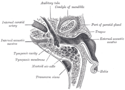 Horizontal section through left ear; upper half of section.
 Frequency of mandibular fractures by location.
| Condyloid process | |
|---|---|
 Position of condyloid process (shown in red). | |
 Mandible. Condyloid processes are shown in red. | |
| Details | |
| Identifiers | |
| Latin | processus condylaris mandibulae |
| MeSH | D008335 |
| TA98 | A02.1.15.035 |
| TA2 | 872 |
| FMA | 52836 |
| Anatomical terms of bone | |
The condyloid process or condylar process is the process on the human and other mammalian species' mandibles that ends in a condyle, the mandibular condyle. It is thicker than the coronoid process of the mandible and consists of two portions: the condyle and the constricted portion which supports it, the neck.


