Mandibular canal

Last updated
Mandibular canal
Gray1003.png
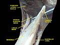
The permanent teeth, viewed from the right. The external layer of bone has been partly removed and the maxillary sinus has been opened.
Details
Identifiers
Latin canalis mandibulae
MeSH D000088263
TA98 A02.1.15.030
TA2 867
FMA 59473
Anatomical terms of bone
The mandibular incisive canal (indicated here by coral green arrows) continuing anteriorly (to the right) from the mandibular canal (purple arrows) after the mental foramen (light green circle) Mandibular Incisive Canal Highlighted.jpg
The mandibular incisive canal (indicated here by coral green arrows) continuing anteriorly (to the right) from the mandibular canal (purple arrows) after the mental foramen (light green circle)

In human anatomy, the mandibular canal is a canal within the mandible that contains the inferior alveolar nerve, inferior alveolar artery, and inferior alveolar vein. It runs obliquely downward and forward in the ramus, and then horizontally forward in the body, where it is placed under the alveoli and communicates with them by small openings.

Contents

On arriving at the incisor teeth, it turns back to communicate with the mental foramen, giving off a small canal known as the mandibular incisive canal, which run to the cavities containing the incisor teeth. [1] It carries branches of the inferior alveolar nerve and artery.

The mandibular canal is continuous with tow foramina: the mental foramen which opens in the mental region of the mandible and carried the distal fibres of the inferior alveolar nerve as the mental nerve; and the mandibular foramen on medial aspect of ramus, into which the mandibular nerve enters to become the inferior alveolar nerve. The mandibular canal often runs close to the apices of the third molar tooth, and the inferior alveolar nerve can become damaged during removal of this tooth, causing sensory disturbance in the distribution of the nerve. This is sometimes the case for the second or first molar teeth, and care must be taken during removal or root canal treatment in such cases to prevent nerve injury or extrusion of root canal filling materials. [2]

Variations

Several variations of the mandibular canal exist with varying frequency. The most common variant is the retromolar canal (~10 % of canals), whereby a branch is given off in the mandibular ramus which terminates in the retromolar region of the mandible. The retromolar canal may cause bleeding during surgery in the retromolar region such as removal of mandibular third molar teeth. Other variants include a bifid canal with a branch (~41%): [3] following the course of the main mandibular canal before re-joining it (forward or buccolingual type); terminating at the apex of a tooth, usually the molar teeth (dental type); opening as an accessory mental foramen. [4] A trifid mandibular canal variation has also been described. [5] [6]

Additional Images

See also

Related Research Articles

<span class="mw-page-title-main">Mandibular foramen</span> Opening on the inner surface of the mandible, allowing for passage of mandibular neurovasculature

The mandibular foramen is an opening on the internal surface of the ramus of the mandible. It allows for divisions of the mandibular nerve and blood vessels to pass through.

<span class="mw-page-title-main">Inferior alveolar nerve</span> Branch of the mandibular nerve

The inferior alveolar nerve (IAN) (also the inferior dental nerve) is a sensory branch of the mandibular nerve (CN V3) (which is itself the third branch of the trigeminal nerve (CN V)). The nerve provides sensory innervation to the lower/mandibular teeth and their corresponding gingiva as well as a small area of the face (via its mental nerve).

<span class="mw-page-title-main">Dental extraction</span> Operation to remove a tooth

A dental extraction is the removal of teeth from the dental alveolus (socket) in the alveolar bone. Extractions are performed for a wide variety of reasons, but most commonly to remove teeth which have become unrestorable through tooth decay, periodontal disease, or dental trauma, especially when they are associated with toothache. Sometimes impacted wisdom teeth cause recurrent infections of the gum (pericoronitis), and may be removed when other conservative treatments have failed. In orthodontics, if the teeth are crowded, healthy teeth may be extracted to create space so the rest of the teeth can be straightened.

<span class="mw-page-title-main">Buccal nerve</span> Nerve in the human face

The buccal nerve is a sensory nerve of the face arising from the mandibular nerve. It conveys sensory information from the skin of the cheek, and parts of the oral mucosa, periodontium, and gingiva.

<span class="mw-page-title-main">Mental foramen</span> Opening on the anterior aspect of the mandible through which the mental nerve and vessels exit

The mental foramen is one of two foramina (openings) located on the anterior surface of the mandible. It is part of the mandibular canal. It transmits the terminal branches of the inferior alveolar nerve and the mental vessels.

<span class="mw-page-title-main">Lingual nerve</span> Human nerve relaying sense to the tongue

The lingual nerve carries sensory innervation from the anterior two-thirds of the tongue. It contains fibres from both the mandibular division of the trigeminal nerve (CN V3) and from the facial nerve (CN VII). The fibres from the trigeminal nerve are for touch, pain and temperature (general sensation), and the ones from the facial nerve are for taste (special sensation).

<span class="mw-page-title-main">Inferior alveolar artery</span>

The inferior alveolar artery is an artery of the head. It is a branch of the maxillary artery. It descends through the infratemporal fossa as part of a neurovascular bundle with the inferior alveolar nerve and vein to the mandibular foramen where it enters and passes anteriorly inside the mandible, suplying the body of mandible and the dental pulp of the lower molar and premolar teeth. Its terminal incisor branch supplies the rest of the lower teeth. Its mental branch exits the mandibula anteriorly through the mental foramen to supply adjacent lip and skin.

<span class="mw-page-title-main">Mental nerve</span> Sensory nerve of the face

The mental nerve is a sensory nerve of the face. It is a branch of the posterior trunk of the inferior alveolar nerve, itself a branch of the mandibular nerve (CN V3), itself a branch of the trigeminal nerve (CN V). It provides sensation to the front of the chin and the lower lip, as well as the gums of the anterior mandibular (lower) teeth. It can be blocked with local anaesthesia for procedures on the chin, lower lip, and mucous membrane of the inner cheek. Problems with the nerve cause chin numbness.

<span class="mw-page-title-main">Mylohyoid nerve</span> Nerve of the head

The mylohyoid nerve is a mixed nerve of the head. It is a branch of the inferior alveolar nerve. It provides motor innervation the mylohyoid muscle, and the anterior belly of the digastric muscle. It provides sensory innervation to part of the submental area, and sometimes also the mandibular (lower) molar teeth, requiring local anaesthesia for some oral procedures.

<span class="mw-page-title-main">Maxillary artery</span> Artery supplying face structures in humans

The maxillary artery supplies deep structures of the face. It branches from the external carotid artery just deep to the neck of the mandible.

<span class="mw-page-title-main">Infratemporal fossa</span> Cavity that is part of the skull

The infratemporal fossa is an irregularly shaped cavity that is a part of the skull. It is situated below and medial to the zygomatic arch. It is not fully enclosed by bone in all directions. It contains superficial muscles, including the lower part of the temporalis muscle, the lateral pterygoid muscle, and the medial pterygoid muscle. It also contains important blood vessels such as the middle meningeal artery, the pterygoid plexus, and the retromandibular vein, and nerves such as the mandibular nerve (CN V3) and its branches.

<span class="mw-page-title-main">Outline of human anatomy</span> Overview of and topical guide to human anatomy

The following outline is provided as an overview of and topical guide to human anatomy:

<span class="mw-page-title-main">Inferior dental plexus</span> Nerve

The inferior dental plexus is a nerve plexus formed by sensory branches of the inferior alveolar nerve. The plexus issues dental branches and gingival branches; the small dental branches provide sensory innervation to the lower/mandibular teeth.

<span class="mw-page-title-main">Coronectomy</span> Removal of the crown of a tooth

When extracting lower wisdom teeth, coronectomy is a treatment option involving removing the crown of the lower wisdom tooth, whilst keeping the roots in place in healthy patients. This option is given to patients as an alternative to extraction when the wisdom teeth are in close association with the inferior alveolar nerve, and so used to prevent damage to the nerve which may occur during extraction.

<span class="mw-page-title-main">Mandibular fracture</span> Medical condition

Mandibular fracture, also known as fracture of the jaw, is a break through the mandibular bone. In about 60% of cases the break occurs in two places. It may result in a decreased ability to fully open the mouth. Often the teeth will not feel properly aligned or there may be bleeding of the gums. Mandibular fractures occur most commonly among males in their 30s.

<span class="mw-page-title-main">Mandibular incisive canal</span>

The mandibular incisive canal is a bilaterally paired bony canal within the anterior portion of the mandible that extends from the mental foramen (usually) to near the ipsilateral lateral incisor teeth.

<span class="mw-page-title-main">Mandible</span> Lower jaw bone

In jawed vertebrates, the mandible, lower jaw, or jawbone is a bone that makes up the lower – and typically more mobile – component of the mouth.

<span class="mw-page-title-main">Human mouth</span> Part of human anatomy

In human anatomy, the mouth is the first portion of the alimentary canal that receives food and produces saliva. The oral mucosa is the mucous membrane epithelium lining the inside of the mouth.

<span class="mw-page-title-main">Pterygomandibular space</span>

The pterygomandibular space is a fascial space of the head and neck. It is a potential space in the head and is paired on each side. It is located between the lateral pterygoid muscle and the medial surface of the ramus of the mandible. The pterygomandibular space is one of the four compartments of the masticator space.

<span class="mw-page-title-main">Mandibular setback surgery</span>

Mandibular setback surgery is a surgical procedure performed along the occlusal plane to prevent bite opening on the anterior or posterior teeth and retract the lower jaw for both functional and aesthetic effects in patients with mandibular prognathism. It is an orthodontic surgery that is a form of reconstructive plastic surgery. There are three main types of procedures for mandibular setback surgery: Bilateral Sagittal Split Osteotomy (BSSO), Intraoral Vertical Ramus Osteotomy (IVRO) and Extraoral Ramus Osteotomy (EVRO), depending on the magnitude of mandibular setback for each patient. Postoperative care aims to minimise postoperative complications, complications includes bite changes, relapse and nerve injury.

References

PD-icon.svgThis article incorporates text in the public domain from page 173 of the 20th edition of Gray's Anatomy (1918)

  1. Greenstein, G; Cavallaro, J; Tarnow, D: "Practical Application of Anatomy the Dental Implant Surgeon," J Perio October 2008, pg 1837
  2. https://www.webcitation.org/query?url=http://www.geocities.com/akramjfr/mandbCanal.html&date=2009-10-25+12:10:14 [ dead link ]
  3. Yang, Mengyao; Yang, Jifeng; Deng, Shuli (2023-08-14). "Prevalence and characteristics of accessory mandibular canals in an eastern Chinese population by cone beam computed tomography". Surgical and Radiologic Anatomy. doi:10.1007/s00276-023-03215-7. ISSN   1279-8517.
  4. Allison, J.R.; Carr, A. (November 2017). "A prevalence study of bifid mandibular canals using cone beam computed tomography". Oral Surgery. 10 (4): e55–e61. doi:10.1111/ors.12267.
  5. Rashsuren, Oyuntugs; Choi, Jin-Woo; Han, Won-Jeong; Kim, Eun-Kyung (2014-09-01). "Assessment of bifid and trifid mandibular canals using cone-beam computed tomography". Imaging Science in Dentistry. 44 (3): 229–236. doi:10.5624/isd.2014.44.3.229. ISSN   2233-7822. PMC   4182358 . PMID   25279344.
  6. Mizbah, K.; Gerlach, N.; Maal, T. J.; Bergé, S. J.; Meijer, Gert J. (March 2012). "The clinical relevance of bifid and trifid mandibular canals". Oral and Maxillofacial Surgery. 16 (1): 147–151. doi:10.1007/s10006-011-0278-5. ISSN   1865-1550. PMC   3294215 . PMID   21698363.