
In anatomy, the palatine bones are two irregular bones of the facial skeleton in many animal species, located above the uvula in the throat. Together with the maxillae, they comprise the hard palate.
In mammalian anatomy, the cribriform plate, horizontal lamina or lamina cribrosa is part of the ethmoid bone. It is received into the ethmoidal notch of the frontal bone and roofs in the nasal cavities. It supports the olfactory bulb, and is perforated by olfactory foramina for the passage of the olfactory nerves to the roof of the nasal cavity to convey smell to the brain. The foramina at the medial part of the groove allow the passage of the nerves to the upper part of the nasal septum while the foramina at the lateral part transmit the nerves to the superior nasal concha.
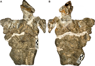
Galesaurus is an extinct genus of carnivorous cynodont therapsid that lived between the Induan and the Olenekian stages of the Early Triassic in what is now South Africa. It was incorrectly classified as a dinosaur by Sir Richard Owen in 1859.
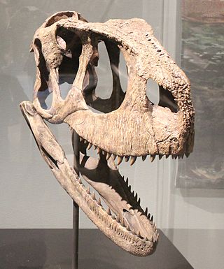
Rugops is a monospecific genus of basal abelisaurid theropod dinosaur from Niger that lived during the Late Cretaceous period in what is now the Echkar Formation. The type and only species, Rugops primus, is known only from a partial skull. It was named and described in 2004 by Paul Sereno, Jeffery Wilson and Jack Conrad. Rugops has an estimated length of 4.4–5.3 metres and weight of 410 kilograms. The top of its skull bears several pits which correlates with overlaying scale and the front of the snout would have had an armour-like dermis.

In human anatomy, the pterygopalatine fossa is a fossa in the skull. A human skull contains two pterygopalatine fossae—one on the left side, and another on the right side. Each fossa is a cone-shaped paired depression deep to the infratemporal fossa and posterior to the maxilla on each side of the skull, located between the pterygoid process and the maxillary tuberosity close to the apex of the orbit. It is the indented area medial to the pterygomaxillary fissure leading into the sphenopalatine foramen. It communicates with the nasal and oral cavities, infratemporal fossa, orbit, pharynx, and middle cranial fossa through eight foramina.

The ethmoidal labyrinth or lateral mass of the ethmoid bone consists of a number of thin-walled cellular cavities, the ethmoid air cells, arranged in three groups, anterior, middle, and posterior, and interposed between two vertical plates of bone; the lateral plate forms part of the orbit, the medial plate forms part of the nasal cavity. In the disarticulated bone many of these cells are opened into, but when the bones are articulated, they are closed in at every part, except where they open into the nasal cavity.
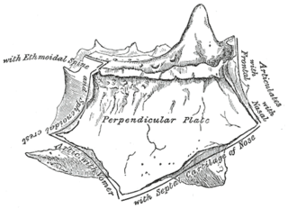
The perpendicular plate of the ethmoid bone is a thin, flattened lamina, polygonal in form, which descends from the under surface of the cribriform plate, and assists in forming the septum of the nose; it is generally deflected a little to one or other side. The anterior border articulates with the spine of the frontal bone and the crest of the nasal bones.
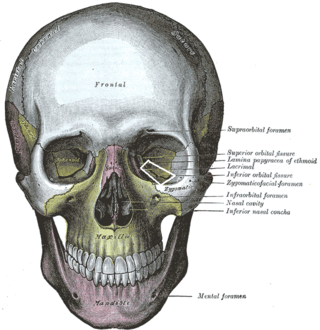
The inferior orbital fissure is a gap between the greater wing of sphenoid bone, and the maxilla. It connects the orbit (anteriorly) with the infratemporal fossa and pterygopalatine fossa (posteriorly).

The anterior ethmoidal artery is a branch of the ophthalmic artery in the orbit. It exits the orbit through the anterior ethmoidal foramen alongside the anterior ethmoidal nerve. It contributes blood supply to the ethmoid sinuses, frontal sinuses, the dura mater, lateral nasal wall, and nasal septum. It issues a meningeal branch, and nasal branches.
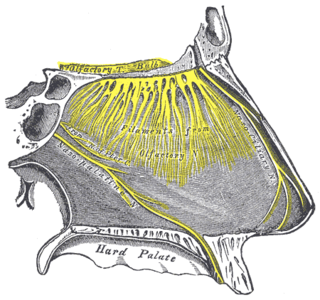
The nasopalatine nerve (also long sphenopalatine nerve) is a nerve of the head. It is a sensory branch of the maxillary nerve (CN V2) that passes through the pterygopalatine ganglion (without synapsing) and then through the sphenopalatine foramen to enter the nasal cavity, and finally out of the nasal cavity through the incisive canal and then the incisive fossa to enter the hard palate. It provides sensory innervation to the posteroinferior part of the nasal septum, and gingiva just posterior to the upper incisor teeth.
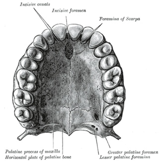
The incisive canals are two bony canals of the anterior hard palate connecting the nasal cavity and the oral cavity. An incisive canal courses through each maxilla. Below, the two incisive canals typically converge medially.

The anterior cranial fossa is a depression in the floor of the cranial base which houses the projecting frontal lobes of the brain. It is formed by the orbital plates of the frontal, the cribriform plate of the ethmoid, and the small wings and front part of the body of the sphenoid; it is limited behind by the posterior borders of the small wings of the sphenoid and by the anterior margin of the chiasmatic groove. The lesser wings of the sphenoid separate the anterior and middle fossae.

In human anatomy of the mouth, the palatine process of maxilla, is a thick, horizontal process of the maxilla. It forms the anterior three quarters of the hard palate, the horizontal plate of the palatine bone making up the rest.

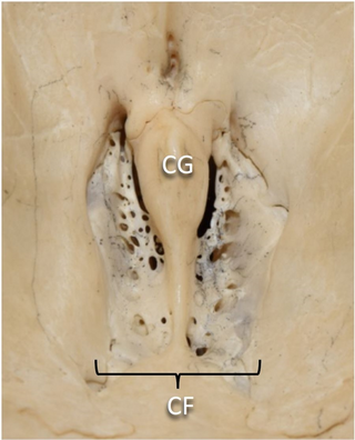
The olfactory foramina, also known as the cribriform foramina, is the grouping of holes located on the cribriform plate. The cribriform plate forms the roof of the nasal cavity, and the olfactory foramina are in the two depressions lateral to the median blade of the cribriform plate called the crista galli. There is a pair of olfactory bulbs of the brain that rest in these two depressions. These holes that make up the olfactory foramina allow passage for about 20 bundles of nerve fibers that make up the olfactory nerve, also known as Cranial Nerve I (CNI), from the nasal cavity to meet with the olfactory bulbs. Therefore, the olfactory foramina are necessary for the human sense of smell. These foramina vary in size and number with age.
The Inferior posterior nasal branches of greater palatine nerve are small nerves which largely supply the posterior aspect of the nasal cavity. They pass through small foramina in the palatine canal to supply the lateral walls of the nasal cavity - including the superior, middle, and inferior nasal concha. These nerves run alongside the lateral posterior nasal arteries, which are branches of the sphenopalatine artery.

Polonosuchus is a genus of rauisuchid known from the late Triassic of Poland. It was a huge predator about 5–6 metres in length and, like all rauisuchians, was equipped with a large head of long sharp teeth. The legs were placed almost underneath the body, unlike most reptiles, which would have made it quite fast and a powerful runner. The appearance was very similar to that of the more known Postosuchus, of North America, and shared with the latter the ecological niche of the apex predator.

Xixiasaurus is a genus of troodontid dinosaur that lived during the Late Cretaceous Period in what is now China. The only known specimen was discovered in Xixia County, Henan Province, in central China, and became the holotype of the new genus and species Xixiasaurus henanensis in 2010. The names refer to the areas of discovery, and can be translated as "Henan Xixia lizard". The specimen consists of an almost complete skull, part of the lower jaw, and teeth, as well as a partial right forelimb.

Pervushovisaurus is a genus of platypterygiine ichthyosaur from the Late Cretaceous (Cenomanian) of the Saratov region in western Russia, the La Penthiève Beds of France and the Cambridge area of the UK.

Nexuotapirus is an extinct genus of tapir from the Late Oligocene and Early Miocene of North America.

Spectrovenator is a genus of basal abelisaurid theropod dinosaur that lived during the early Cretaceous period in what is now Brazil. It is known by a single species, S. ragei, recovered from the Quiricó Formation.
