Posterior lacrimal crest

Last updated
Posterior lacrimal crest
Gray163.png
Left lacrimal bone. Orbital surface. Enlarged. (Posterior lacrimal crest visible but not labeled.)
Details
Part of lacrimal bone
System skeletal
Identifiers
Latin crista lacrimalis posterior
TA98 A02.1.09.002
TA2 745
FMA 57609
Anatomical terms of bone

The posterior lacrimal crest is a vertical bony ridge on the orbital surface of the lacrimal bone. It divides the bone into two parts. It gives origin to the lacrimal part of the orbicularis oculi muscle.

Contents

Structure

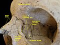
The posterior lacrimal crest is a vertical bony ridge on the orbital (lateral) surface of the lacrimal bone. It divides the lacrimal bone into two parts. It is quite thin and fragile in most people. [1]

The lacrimal groove is in front of this crest. The inner margin of it unites with the frontal process of the maxilla to complete the fossa for the lacrimal sac. [2] The portion of the lacrimal bone behind the posterior lacrimal crest is smooth, and forms part of the medial wall of the orbit. The lacrimal crest ends below in the lacrimal hamulus (a small hook-like projection), which articulates with the lacrimal tubercle of the maxilla.

Relations

The posterior lacrimal crest is just behind the lacrimal sac, and its upper part lodges the lacrimal sac. [3] [4] The lower part lodges the nasolacrimal duct. Horner's muscle, part of the orbicularis oris muscle, inserts between 2 mm and 4 mm from the posterior lacrimal crest. [5] Together with the thicker and more prominent anterior lacrimal crest, it forms the fossa for the lacrimal sac. [1]

Variation

In most people, the posterior lacrimal crest is fairly prominent. [6] However, in around 20% of people, it is fairly shallow. [6] In contrast, the anterior lacrimal crest is almost always very prominent. [6]

Function

The posterior lacrimal crest gives origin to the lacrimal part of the orbicularis oculi muscle. It also helps to protect the lacrimal sac. [3]

Clinical significance

The posterior lacrimal crest may be vulnerable to avulsion fractures. [7] It is generally quite thin and fragile. [1] 25% of such avulsion fractures are related to a Le Fort III skull fracture. [7]

Additional images

See also

Related Research Articles

<span class="mw-page-title-main">Maxilla</span> Upper jaw bone

The maxilla in vertebrates is the upper fixed bone of the jaw formed from the fusion of two maxillary bones. In humans, the upper jaw includes the hard palate in the front of the mouth. The two maxillary bones are fused at the intermaxillary suture, forming the anterior nasal spine. This is similar to the mandible, which is also a fusion of two mandibular bones at the mandibular symphysis. The mandible is the movable part of the jaw.

<span class="mw-page-title-main">Zygomatic bone</span> Facial bone

In the human skull, the zygomatic bone, also called cheekbone or malar bone, is a paired irregular bone which articulates with the maxilla, the temporal bone, the sphenoid bone and the frontal bone. It is situated at the upper and lateral part of the face and forms the prominence of the cheek, part of the lateral wall and floor of the orbit, and parts of the temporal fossa and the infratemporal fossa. It presents a malar and a temporal surface; four processes, and four borders.

<span class="mw-page-title-main">Lacrimal bone</span> Smallest and most fragile bone of the human skull and face

The lacrimal bone is a small and fragile bone of the facial skeleton; it is roughly the size of the little fingernail. It is situated at the front part of the medial wall of the orbit. It has two surfaces and four borders. Several bony landmarks of the lacrimal bone function in the process of lacrimation or crying. Specifically, the lacrimal bone helps form the nasolacrimal canal necessary for tear translocation. A depression on the anterior inferior portion of the bone, the lacrimal fossa, houses the membranous lacrimal sac. Tears or lacrimal fluid, from the lacrimal glands, collect in this sac during excessive lacrimation. The fluid then flows through the nasolacrimal duct and into the nasopharynx. This drainage results in what is commonly referred to a runny nose during excessive crying or tear production. Injury or fracture of the lacrimal bone can result in posttraumatic obstruction of the lacrimal pathways.

<span class="mw-page-title-main">Orbit (anatomy)</span> Cavity or socket of the skull in which the eye and its appendages are situated

In anatomy, the orbit is the cavity or socket/hole of the skull in which the eye and its appendages are situated. "Orbit" can refer to the bony socket, or it can also be used to imply the contents. In the adult human, the volume of the orbit is 30 millilitres, of which the eye occupies 6.5 ml. The orbital contents comprise the eye, the orbital and retrobulbar fascia, extraocular muscles, cranial nerves II, III, IV, V, and VI, blood vessels, fat, the lacrimal gland with its sac and duct, the eyelids, medial and lateral palpebral ligaments, cheek ligaments, the suspensory ligament, septum, ciliary ganglion and short ciliary nerves.

<span class="mw-page-title-main">Middle meningeal artery</span> Artery supplying the meninges

The middle meningeal artery is typically the third branch of the first portion of the maxillary artery. After branching off the maxillary artery in the infratemporal fossa, it runs through the foramen spinosum to supply the dura mater and the calvaria. The middle meningeal artery is the largest of the three (paired) arteries that supply the meninges, the others being the anterior meningeal artery and the posterior meningeal artery.

<span class="mw-page-title-main">Orbicularis oculi muscle</span> Facial muscle that closes the eyelids

The orbicularis oculi is a muscle in the face that closes the eyelids. It arises from the nasal part of the frontal bone, from the frontal process of the maxilla in front of the lacrimal groove, and from the anterior surface and borders of a short fibrous band, the medial palpebral ligament.

<span class="mw-page-title-main">Orbital part of frontal bone</span>

The orbital or horizontal part of the frontal bone consists of two thin triangular plates, the orbital plates, which form the vaults of the orbits, and are separated from one another by a median gap, the ethmoidal notch.

<span class="mw-page-title-main">Depressor supercilii muscle</span> Muscle of the eye

The depressor supercilii is an eye muscle of the human body. The nature of this muscle is in some dispute. Few printed anatomies include it and many authorities consider it to be part of the orbicularis oculi muscle.

<span class="mw-page-title-main">Olecranon</span> Curved bony eminence of the ulna; forms the pointed part of the elbow

The olecranon, is a large, thick, curved bony eminence of the ulna, a long bone in the forearm that projects behind the elbow. It forms the most pointed portion of the elbow and is opposite to the cubital fossa or elbow pit. The olecranon serves as a lever for the extensor muscles that straighten the elbow joint.

<span class="mw-page-title-main">Ischium</span> Lower and back region of the hip bone

The ischium forms the lower and back region of the hip bone.

<span class="mw-page-title-main">Lesser trochanter</span> Conical projection from the shaft of the femur

In human anatomy, the lesser trochanter is a conical, posteromedial, bony projection from the shaft of the femur. it serves as the principal insertion site of the iliopsoas muscle.

<span class="mw-page-title-main">Greater wing of sphenoid bone</span> Large part of the skull front behind the eye socket

The greater wing of the sphenoid bone, or alisphenoid, is a bony process of the sphenoid bone, positioned in the skull behind each eye. There is one on each side, extending from the side of the body of the sphenoid and curving upward, laterally, and backward.

<span class="mw-page-title-main">Lacrimal sac</span> Upper, dilated end of the nasolacrimal duct

The lacrimal sac or lachrymal sac is the upper dilated end of the nasolacrimal duct, and is lodged in a deep groove formed by the lacrimal bone and frontal process of the maxilla. It connects the lacrimal canaliculi, which drain tears from the eye's surface, and the nasolacrimal duct, which conveys this fluid into the nasal cavity. Lacrimal sac occlusion leads to dacryocystitis.

The anterior lacrimal crest is a bony projection on the frontal process of the maxilla. It creates the lateral margin of the lacrimal sac fossa and is continuous with the orbital margin. The medial palpebral ligament is attached to anterior lacrimal crest. It is an important structure to avoid damaging during rhinoplasty.

<span class="mw-page-title-main">Infraorbital nerve</span> Branch of the maxillary nerve supplying the face

The infraorbital nerve is a branch of the maxillary nerve. It arises in the pterygopalatine fossa. It passes through the inferior orbital fissure to enter the orbit. It travels through the orbit, then enters and traverses the infraorbital canal, exiting the canal at the infraorbital foramen to reach the face. It provides sensory innervation to the skin and mucous membranes around the middle of the face.

<span class="mw-page-title-main">Medial palpebral ligament</span> Ligament connecting the maxilla and the lacrimal bone to the eyelids

The medial palpebral ligament is a ligament of the face. It attaches to the frontal process of the maxilla, the lacrimal groove, and the tarsus of each eyelid. It has a superficial (anterior) and a deep (posterior) layer, with many surrounding attachments. It connects the medial canthus of each eyelid to the medial part of the orbit. It is a useful point of fixation during eyelid reconstructive surgery.

<span class="mw-page-title-main">Frontal process of maxilla</span> Plate which forms part of the lateral boundary of the nose

The frontal process of the maxilla is a strong plate, which projects upward, medialward, and backward from the maxilla, forming part of the lateral boundary of the nose.

<span class="mw-page-title-main">Zygomatic branches of the facial nerve</span> Nerves of the face

The zygomatic branches of the facial nerve (malar branches) are nerves of the face. They run across the zygomatic bone to the lateral angle of the orbit. Here, they supply the orbicularis oculi muscle, and join with filaments from the lacrimal nerve and the zygomaticofacial branch of the maxillary nerve (CN V2).

<span class="mw-page-title-main">Outline of human anatomy</span> Overview of and topical guide to human anatomy

The following outline is provided as an overview of and topical guide to human anatomy:

The lacrimal hamulus is a small, hook-like bony projection of the lacrimal bone. It is a continuation of the posterior lacrimal crest. It articulates with the lacrimal tubercle of the maxilla, and completes the upper orifice of the lacrimal canaliculus. It sometimes exists as a separate piece, and is then called the lesser lacrimal bone.

References

PD-icon.svgThis article incorporates text in the public domain from page 164 of the 20th edition of Gray's Anatomy (1918)

  1. 1 2 3 Tao, Hai; Ma, Zhi-zhong; Wu, Hai-Yang; Wang, Peng; Han, Cui (April 2014). "Anatomic study of the lacrimal fossa and lacrimal pathway for bypass surgery with autogenous tissue grafting". Indian Journal of Ophthalmology . 62 (4): 419–423. doi: 10.4103/0301-4738.121137 . ISSN   0301-4738. PMC   4064215 . PMID   24817745.
  2. Tomazic, Peter Valentin; Dewart, Nora; Witterick, Ian J. (2021-01-01), Sindwani, Raj (ed.), "6 - Rhinologic Evaluation in Orbital and Lacrimal Disease", Endoscopic Surgery of the Orbit, Philadelphia: Elsevier, pp. 36–40, ISBN   978-0-323-61329-3 , retrieved 2021-09-23
  3. 1 2 Merbs, Shannath; DeParis, Sarah (2020-01-01), Dorafshar, Amir H.; Rodriguez, Eduardo D.; Manson, Paul N. (eds.), "3.4 - Ocular Considerations: Ectropion, Entropion, Blink, Ptosis, Epiphora", Facial Trauma Surgery, London: Elsevier, pp. 367–378, ISBN   978-0-323-49755-8 , retrieved 2021-09-23
  4. Piniara, Anastasia; Georgalas, Christos (2021-01-01), Sindwani, Raj (ed.), "4 - Surgical Anatomy of the Orbit, Including the Intraconal Space", Endoscopic Surgery of the Orbit, Philadelphia: Elsevier, pp. 18–27, ISBN   978-0-323-61329-3 , retrieved 2021-09-23
  5. Kakizaki, Hirohiko; Ichinose, Akihiro; Takahashi, Yasuhiro; Kang, Hyera; Ikeda, Hiroshi; Nakano, Takashi; Asamoto, Ken; Iwaki, Masayoshi (February 2012). "Anatomical Relationship of Horner's Muscle Origin and Posterior Lacrimal Crest". Ophthalmic Plastic & Reconstructive Surgery. 28 (1): 66–68. doi:10.1097/IOP.0b013e318239321d. ISSN   0740-9303. PMID   22186989. S2CID   5145634.
  6. 1 2 3 Bisaria, K K; Saxena, R C; Bisaria, S D; Lakhtakia, P K; Agarwal, A K; Premsagar, I C (October 1989). "The lacrimal fossa in Indians". Journal of Anatomy . 166: 265–268. ISSN   0021-8782. PMC   1256759 . PMID   2621144.
  7. 1 2 Díaz, Oswaldo J. Gómez; Carreño, Andrés Parra; Serna, Daniel Restrepo (October 2019). "Traumatic Telecanthus and Posterior Lacrimal Crest Avulsion in a Six-Year-Old Child". Journal of Craniofacial Surgery. 30 (7): 2224–2226. doi:10.1097/SCS.0000000000005922. ISSN   1049-2275. PMID   31490433. S2CID   201846470.