Orbital process of palatine bone

Last updated
Orbital process of palatine bone
Gray168.png
Left palatine bone. Nasal aspect. Enlarged.
Gray169.png
Left palatine bone. Posterior aspect. Enlarged.
Details
Identifiers
Latin processus orbitalis
TA A02.1.13.011
FMA 59143
Anatomical terms of bone

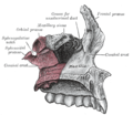
The orbital process of the palatine bone is placed on a higher level than the sphenoidal, and is directed upward and lateralward from the front of the vertical part, to which it is connected by a constricted neck. It presents five surfaces, which enclose an air cell. Of these surfaces, three are articular and two non-articular.

The articular surfaces are:

  1. the anterior or maxillary, directed forward, lateralward, and downward, of an oblong form, and rough for articulation with the maxilla
  2. the posterior or sphenoidal, directed backward, upward, and medialward; it presents the opening of the air cell, which usually communicates with the sphenoidal sinus; the margins of the opening are serrated for articulation with the sphenoidal concha
  3. the medial or ethmoidal, directed forward, articulates with the labyrinth of the ethmoid.

In some cases the air cell opens on this surface of the bone and then communicates with the posterior ethmoidal cells. More rarely it opens on both surfaces, and then communicates with the posterior ethmoidal cells and the sphenoidal sinus.

The non-articular surfaces are:

  1. the superior or orbital, directed upward and lateralward; it is triangular in shape, and forms the back part of the floor of the orbit; and
  2. the lateral, of an oblong form, directed toward the pterygopalatine fossa; it is separated from the orbital surface by a rounded border, which enters into the formation of the inferior orbital fissure.

Additional images

Related Research Articles

Ulna longer bone of the forearm

The ulna is a long bone found in the forearm that stretches from the elbow to the smallest finger, and when in anatomical position, is found on the medial side of the forearm. It runs parallel to the radius, the other long bone in the forearm, and is the larger and longer of the two.

Tibia larger of the two bones of the leg below the knee for vertebrates

The tibia, also known as the shinbone or shankbone, is the larger, stronger, and anterior (frontal) of the two bones in the leg below the knee in vertebrates, and it connects the knee with the ankle bones. The tibia is found on the medial side of the leg next to the fibula and closer to the median plane or centre-line. The tibia is connected to the fibula by the interosseous membrane of the leg, forming a type of fibrous joint called a syndesmosis with very little movement. The tibia is named for the flute tibia. It is the second largest bone in the human body next to the femur. The leg bones are the strongest long bones as they support the rest of the body.

Zygomatic bone Facial bone

In the human skull, the zygomatic bone is a paired irregular bone which articulates with the maxilla, the temporal bone, the sphenoid bone and the frontal bone. It is situated at the upper and lateral part of the face and forms the prominence of the cheek, part of the lateral wall and floor of the orbit, and parts of the temporal fossa and the infratemporal fossa. It presents a malar and a temporal surface; four processes, and four borders.

Ethmoid bone bone in the skull that separates the nasal cavity from the brain

The ethmoid bone is an unpaired bone in the skull that separates the nasal cavity from the brain. It is located at the roof of the nose, between the two orbits. The cubical bone is lightweight due to a spongy construction. The ethmoid bone is one of the bones that make up the orbit of the eye.

Sphenoid bone unpaired bone situated at the front middle of the skull in front of the temporal bone and basilar part of the occipital bone

The sphenoid bone is an unpaired bone of the neurocranium. It is situated in the middle of the skull towards the front, in front of the basilar part of the occipital bone. The sphenoid bone is one of the seven bones that articulate to form the orbit. Its shape somewhat resembles that of a butterfly or bat with its wings extended.

Palatine bone facial bone

The palatine bones are two irregular bones of the facial skeleton in many animal species. Together with the maxillae they comprise the hard palate.

Inferior nasal concha facial bone

The inferior nasal concha is one of the three paired nasal conchae in the nose. It extends horizontally along the lateral wall of the nasal cavity and consists of a lamina of spongy bone, curled upon itself like a scroll,. The inferior nasal conchae are considered a pair of facial bones. As the air passes through the turbinates, the air is churned against these mucosa-lined bones in order to receive warmth, moisture and cleansing. Superior to inferior nasal concha are the middle nasal concha and superior nasal concha which arise from the cranial portion of the skull. Hence, these two are considered as a part of the cranial bones.

Orbital part of frontal bone

The orbital or horizontal part of the frontal bone consists of two thin triangular plates, the orbital plates, which form the vaults of the orbits, and are separated from one another by a median gap, the ethmoidal notch.

Talus bone bone of the ankle

The talus, talus bone, astragalus, or ankle bone is one of the group of foot bones known as the tarsus. The tarsus forms the lower part of the ankle joint. It transmits the entire weight of the body from the lower legs to the foot.

Ethmoid sinus one of the four paired paranasal sinuses

The ethmoid sinuses or ethmoid air cells of the ethmoid bone are one of the four paired paranasal sinuses. The cells are variable in both size and number in the lateral mass of each of the ethmoid bones and cannot be palpated during an extraoral examination. They are divided into anterior and posterior groups. The ethmoid air cells are numerous thin-walled cavities situated in the ethmoidal labyrinth and completed by the frontal, maxilla, lacrimal, sphenoidal, and palatine bones. They lie between the upper parts of the nasal cavities and the orbits, and are separated from these cavities by thin bony lamellae.

Sphenoidal conchae

The sphenoidal conchae are two thin, curved plates, situated at the anterior and lower part of the body of the sphenoid. An aperture of variable size exists in the anterior wall of each, and through this the sphenoidal sinus opens into the nasal cavity.

Greater wing of sphenoid bone

The greater wing of the sphenoid bone, or alisphenoid, is a bony process of the sphenoid bone; there is one on each side, extending from the side of the body of the sphenoid and curving upward, laterally, and backward.

Ethmoidal labyrinth consists of a number of thin-walled cellular cavities

The ethmoidal labyrinth or lateral mass of the ethmoid bone consists of a number of thin-walled cellular cavities, the ethmoid air cells, arranged in three groups, anterior, middle, and posterior, and interposed between two vertical plates of bone; the lateral plate forms part of the orbit, the medial plate forms part of the nasal cavity. In the disarticulated bone many of these cells are opened into, but when the bones are articulated, they are closed in at every part, except where they open into the nasal cavity.

Lateral parts of occipital bone

The lateral parts of the occipital bone are situated at the sides of the foramen magnum; on their under surfaces are the condyles for articulation with the superior facets of the atlas.

Sphenoidal process of palatine bone thin, compressed plate, much smaller than the orbital, and directed upward and medialward

The sphenoidal process of the palatine bone is a thin, compressed plate, much smaller than the orbital, and directed upward and medialward.

Perpendicular plate of palatine bone vertical part of the palatine bone

The perpendicular plate of palatine bone is the vertical part of the palatine bone, and is thin, of an oblong form, and presents two surfaces and four borders.

Zygomatic process

Each Zygomatic process is the part of a bone which articulates with the zygomatic bone. The three processes are:

Frontal process of maxilla plate which forms part of the lateral boundary of the nose

The frontal process of maxilla is a strong plate, which projects upward, medialward, and backward from the maxilla, forming part of the lateral boundary of the nose.

Body of sphenoid bone

The body of the sphenoid bone, more or less cubical in shape, is hollowed out in its interior to form two large cavities, the sphenoidal sinuses, which are separated from each other by a septum.

Orbital lamina of ethmoid bone

The orbital lamina of ethmoid bone, is a smooth, oblong bone plate which forms the lateral surface of the labyrinth of the ethmoid bone in the skull. The plate covers in the middle and posterior ethmoidal cells and forms a large part of the medial wall of the orbit.

References

This article incorporates text in the public domain from page 168 of the 20th edition of Gray's Anatomy (1918)