
In anatomy, the temporomandibular joints (TMJ) are the two joints connecting the jawbone to the skull. It is a bilateral synovial articulation between the temporal bone of the skull above and the mandible below; it is from these bones that its name is derived. This joint is unique in that it is a bilateral joint that functions as one unit. Since the TMJ is connected to the mandible, the right and left joints must function together and therefore are not independent of each other.

The mandibular foramen is an opening on the internal surface of the ramus of the mandible for divisions of the mandibular nerve and blood vessels to pass through.

Alveolar osteitis, also known as dry socket, is inflammation of the alveolar bone. Classically, this occurs as a postoperative complication of tooth extraction.

The inferior alveolar nerve is a branch of the mandibular nerve, which is itself the third branch of the trigeminal nerve. The inferior alveolar nerves supply sensation to the lower teeth.

Prognathism is a positional relationship of the mandible or maxilla to the skeletal base where either of the jaws protrudes beyond a predetermined imaginary line in the coronal plane of the skull. In general dentistry, oral and maxillofacial surgery, and orthodontics, this is assessed clinically or radiographically (cephalometrics). The word prognathism derives from Greek πρό and γνάθος. One or more types of prognathism can result in the common condition of malocclusion, in which an individual's top teeth and lower teeth do not align properly.

Orthognathic surgery ; also known as corrective jaw surgery or simply jaw surgery, is surgery designed to correct conditions of the jaw and lower face related to structure, growth, airway issues including sleep apnea, TMJ disorders, malocclusion problems primarily arising from skeletal disharmonies, other orthodontic dental bite problems that cannot be easily treated with braces, as well as the broad range of facial imbalances, disharmonies, asymmetries and malproportions where correction can be considered to improve facial aesthetics and self esteem.
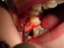
A dental extraction is the removal of teeth from the dental alveolus (socket) in the alveolar bone. Extractions are performed for a wide variety of reasons, but most commonly to remove teeth which have become unrestorable through tooth decay, periodontal disease, or dental trauma, especially when they are associated with toothache. Sometimes impacted wisdom teeth cause recurrent infections of the gum (pericoronitis), and may be removed when other conservative treatments have failed. In orthodontics if the teeth are crowded, healthy teeth may be extracted to create space so the rest of the teeth can be straightened.

The mental foramen is one of two foramina (openings) located on the anterior surface of the mandible. It transmits the terminal branches of the inferior alveolar nerve and vessels. The mental foramen descends slightly in toothless individuals.
The lingual nerve carries sensory innervation from the anterior two thirds of the tongue. It contains fibres from both the mandibular division of the trigeminal nerve (CN V3) and from the facial nerve (CN VII). The fibres from the mandibular nerve are for touch, pain and temperature (general sensation), and the ones from the facial nerve are for taste (special sensation).

The inferior alveolar artery is an artery of the face. It is a branch of the first portion of the maxillary artery.

In human anatomy, the mandibular canal is a canal within the mandible that contains the inferior alveolar nerve, inferior alveolar artery, and inferior alveolar vein. It runs obliquely downward and forward in the ramus, and then horizontally forward in the body, where it is placed under the alveoli and communicates with them by small openings.
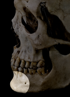
The human chin refers to the forward pointed part of the anterior mandible below the lower lip. A fully developed human skull has a chin of between 0.7 cm and 1.1 cm. While some more obese people may appear to have a “double chin”, this is only a facial feature of the neck where multiple layers of skin build up.

Mammalodontidae is a family of extinct whales known from the Oligocene of Australia and New Zealand.
Occlusion, in a dental context, means simply the contact between teeth. More technically, it is the relationship between the maxillary (upper) and mandibular (lower) teeth when they approach each other, as occurs during chewing or at rest.
Dental anesthesia is the application of anesthesia to dentistry. It includes local anesthetics, sedation, and general anesthesia.
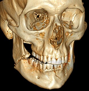
Mandibular fracture, also known as fracture of the jaw, is a break through the mandibular bone. In about 60% of cases the break occurs in two places. It may result in a decreased ability to fully open the mouth. Often the teeth will not feel properly aligned or there may be bleeding of the gums. Mandibular fractures occur most commonly among males in their 30s.

The mandibular incisive canal is a bony canal within the anterior mandible that runs bilaterally from the mental foramina usually to the region of the ipsilateral lateral incisor teeth. After branching into the mental nerve that exits the foramen of the same name, the inferior alveolar nerve continues anteriorly within the mandibular incisive canal as the incisive nerve, providing innervation to the mandibular first premolar, canine and lateral and central incisors. The mandibular incisive nerve either terminates as nerve endings within the anterior teeth or adjacent bone, or may join nerve endings that enter through the tiny lingual foramen.
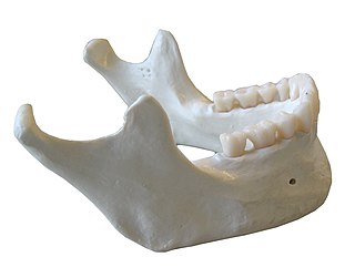
In anatomy, the mandible, lower jaw or jawbone is the largest, strongest and lowest bone in the human facial skeleton. It forms the lower jaw and holds the lower teeth in place. The mandible sits beneath the maxilla. It is the only movable bone of the skull. It is connected to the temporal bone by the temporomandibular joint.

In human anatomy, the mouth is the first portion of the alimentary canal that receives food and produces saliva. The oral mucosa is the mucous membrane epithelium lining the inside of the mouth.
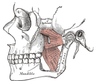
The pterygomandibular space is a fascial space of the head and neck. It is a potential space in the head and is paired on each side. It is located between the medial pterygoid muscle and the medial surface of the ramus of the mandible. The pterygomandibular space is one of the four compartments of the masticator space.

