Incisive canals

Last updated
Incisive canals
Gray160.png
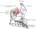
The bony palate and alveolar arch (incisive canals labeled at upper left)
Details
Identifiers
Latin canales incisivi
TA98 A02.1.00.061
TA2 788
FMA 59107
Anatomical terms of bone

The incisive canals (also: "nasopalatine canals") are two bony canals of the anterior hard palate connecting the nasal cavity and the oral cavity. An incisive canal courses through each maxilla. Below, the two incisive canals typically converge medially. [1]

Contents

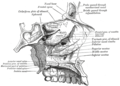
Each incisive canal transmits a nasopalatine nerve, and an anastomosis of the greater palatine artery and a posterior septal branch of the sphenopalatine artery. [1]

Anatomy

An incisive canal has an average length of 10 mm, and an average width of up to 6 mm at the incisive fossa (the dimensions of the canal change with age, trauma, and loss of teeth). [1]

Course and openings

The two incisive canals usually (in 60% of individuals) have a characteristic Y-shaped or V-shaped morphology: above, each incisive canal opens into the nasal cavity on either side of the nasal septum as the nasal foramina; below, the two incisive canals converge medially to open into the oral cavity at midline at the incisive fossa [1] as several incisive foramina. [2]

Variation

There are several alternative morphologies of the canals: the two canals may not converge at any point, may have multiple openings superiorly, or only a single canal (with one inferior as well as only one superior opening) may be present. [1]

Contents

Through each canal ascends the terminal branch of the greater palatine artery (to anastomose with the posterior septal branch of sphenopalatine artery) while the nasopalatine nerve descends (to anastomose with the greater palatine nerve). [3]

Additional images

See also

Related Research Articles

Articles related to anatomy include:

<span class="mw-page-title-main">Palatine bone</span> Bone of the facial skeleton

In anatomy, the palatine bones are two irregular bones of the facial skeleton in many animal species, located above the uvula in the throat. Together with the maxillae, they comprise the hard palate.

<span class="mw-page-title-main">Nasal cavity</span> Large, air-filled space above and behind the nose in the middle of the face

The nasal cavity is a large, air-filled space above and behind the nose in the middle of the face. The nasal septum divides the cavity into two cavities, also known as fossae. Each cavity is the continuation of one of the two nostrils. The nasal cavity is the uppermost part of the respiratory system and provides the nasal passage for inhaled air from the nostrils to the nasopharynx and rest of the respiratory tract.

<span class="mw-page-title-main">Orbit (anatomy)</span> Cavity or socket of the skull in which the eye and its appendages are situated

In anatomy, the orbit is the cavity or socket/hole of the skull in which the eye and its appendages are situated. "Orbit" can refer to the bony socket, or it can also be used to imply the contents. In the adult human, the volume of the orbit is 30 millilitres, of which the eye occupies 6.5 ml. The orbital contents comprise the eye, the orbital and retrobulbar fascia, extraocular muscles, cranial nerves II, III, IV, V, and VI, blood vessels, fat, the lacrimal gland with its sac and duct, the eyelids, medial and lateral palpebral ligaments, cheek ligaments, the suspensory ligament, septum, ciliary ganglion and short ciliary nerves.

<span class="mw-page-title-main">Maxillary nerve</span> Branch of the trigeminal nerve responsible for the mid-face

In neuroanatomy, the maxillary nerve (V2) is one of the three branches or divisions of the trigeminal nerve, the fifth (CN V) cranial nerve. It comprises the principal functions of sensation from the maxilla, nasal cavity, sinuses, the palate and subsequently that of the mid-face, and is intermediate, both in position and size, between the ophthalmic nerve and the mandibular nerve.

<span class="mw-page-title-main">Pterygopalatine fossa</span> Fossa in the skull

In human anatomy, the pterygopalatine fossa is a fossa in the skull. A human skull contains two pterygopalatine fossae—one on the left side, and another on the right side. Each fossa is a cone-shaped paired depression deep to the infratemporal fossa and posterior to the maxilla on each side of the skull, located between the pterygoid process and the maxillary tuberosity close to the apex of the orbit. It is the indented area medial to the pterygomaxillary fissure leading into the sphenopalatine foramen. It communicates with the nasal and oral cavities, infratemporal fossa, orbit, pharynx, and middle cranial fossa through eight foramina.

<span class="mw-page-title-main">Pterygoid processes of the sphenoid</span> Bone plates projecting from the sphenoid bone of the skull

The pterygoid processes of the sphenoid, one on either side, descend perpendicularly from the regions where the body and the greater wings of the sphenoid bone unite.

<span class="mw-page-title-main">Inferior orbital fissure</span> Gap between bones of the human eye socket

The inferior orbital fissure is a gap between the greater wing of sphenoid bone, and the maxilla. It connects the orbit (anteriorly) with the infratemporal fossa and pterygopalatine fossa (posteriorly).

<span class="mw-page-title-main">Nasopalatine nerve</span> Nerve of the head

The nasopalatine nerve (also long sphenopalatine nerve) is a nerve of the head. It is a sensory branch of the maxillary nerve (CN V2) that passes through the pterygopalatine ganglion (without synapsing) and then through the sphenopalatine foramen to enter the nasal cavity, and finally out of the nasal cavity through the incisive canal and then the incisive fossa to enter the hard palate. It provides sensory innervation to the posteroinferior part of the nasal septum, and gingiva just posterior to the upper incisor teeth.

<span class="mw-page-title-main">Incisive foramen</span> Opening of the incisive canals on the hard palate immediately behind the incisor teeth

In the human mouth, the incisive foramen is the opening of the incisive canals on the hard palate immediately behind the incisor teeth. It gives passage to blood vessels and nerves. The incisive foramen is situated within the incisive fossa of the maxilla.

<span class="mw-page-title-main">Sphenopalatine foramen</span>

The sphenopalatine foramen is a fissure of the skull that connects the nasal cavity and the pterygopalatine fossa. It gives passage to the sphenopalatine artery, nasopalatine nerve, and the superior nasal nerve.

<span class="mw-page-title-main">Sphenopalatine artery</span>

The sphenopalatine artery is an artery of the head, commonly known as the artery of epistaxis. It passes through the sphenopalatine foramen to reach the nasal cavity. It is the main artery of the nasal cavity.

The greater palatine artery is a branch of the descending palatine artery and contributes to the blood supply of the hard palate and nasal septum.

<span class="mw-page-title-main">Sphenoidal process of palatine bone</span> Thin, compressed plate

The sphenoidal process of palatine bone is a thin, superomedially directed plate of bone. It is smaller and more inferior compared to the orbital process of palatine bone.

<span class="mw-page-title-main">Perpendicular plate of palatine bone</span>

The perpendicular plate of palatine bone is the vertical part of the palatine bone, and is thin, of an oblong form, and presents two surfaces and four borders.

<span class="mw-page-title-main">Palatine process of maxilla</span> Thick, horizontal process of the maxilla

In human anatomy of the mouth, the palatine process of maxilla, is a thick, horizontal process of the maxilla. It forms the anterior three quarters of the hard palate, the horizontal plate of the palatine bone making up the rest.

<span class="mw-page-title-main">Greater palatine canal</span> Passage in the skull that transmits the descending palatine artery and vein

The greater palatine canal is a passage in the skull that transmits the descending palatine artery, vein, and greater and lesser palatine nerves between the pterygopalatine fossa and the oral cavity.

<span class="mw-page-title-main">Outline of human anatomy</span> Overview of and topical guide to human anatomy

The following outline is provided as an overview of and topical guide to human anatomy:

The Inferior posterior nasal branches of greater palatine nerve are small nerves which largely supply the posterior aspect of the nasal cavity. They pass through small foramina in the palatine canal to supply the lateral walls of the nasal cavity - including the superior, middle, and inferior nasal concha. These nerves run alongside the lateral posterior nasal arteries, which are branches of the sphenopalatine artery.

Crossing the under surface of the sphenoid the sphenopalatine artery ends on the nasal septum as the posterior septal branches; these anastomose with the ethmoidal arteries and the septal branch of the superior labial; one branch descends in a groove on the vomer to the incisive canal and anastomoses with the descending palatine artery.

References

  1. 1 2 3 4 5 Lake S, Iwanaga J, Kikuta S, Oskouian RJ, Loukas M, Tubbs RS (July 2018). "The Incisive Canal: A Comprehensive Review". Cureus. 10 (7): e3069. doi: 10.7759/cureus.3069 . PMC   6166911 . PMID   30280065.
  2. "incisive foramen". TheFreeDictionary.com. Retrieved 2022-05-25.
  3. Gray H (1918). Gray's Anatomy (20th ed.). p. 162.