Sphenopalatine foramen

Last updated
Sphenopalatine foramen
Gray192.png
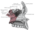
Medial wall of left orbit. (Sphenopalatine foramen labeled in upper right.)
Gray169.png
Left palatine bone. Posterior aspect. Enlarged. (Sphenopalatine foramen labeled in upper right.)
Details
Identifiers
Latin foramen sphenopalatinum
TA98 A02.1.00.097
TA2 502
FMA 53144
Anatomical terms of bone

The sphenopalatine foramen is a fissure of the skull that connects the nasal cavity and the pterygopalatine fossa. It gives passage to the sphenopalatine artery, nasopalatine nerve, and the superior nasal nerve (all passing from the pterygopalatine fossa into the nasal cavity). [1]

Contents

Structure

The processes of the superior border of the palatine bone are separated by the sphenopalatine notch, which is converted into the sphenopalatine foramen by the under surface of the body of the sphenoid.[ citation needed ]

The sphenopalatine foramen is situated posterior to the middle nasal meatus orbital process of palatine bone, anterior to the sphenoidal process of palatine bone, inferior to the body and concha[ clarification needed ] of the sphenoid bone, and superior to the superior margin of the perpendicular plate of palatine bone. [1]

Relations

The ethmoid crest (a reliable surgical landmark) is situated anterior to the sphenopalatine foramen. [1]

Additional images

Related Research Articles

Articles related to anatomy include:

<span class="mw-page-title-main">Sphenoid bone</span> Bone at the front of the skull

The sphenoid bone is an unpaired bone of the neurocranium. It is situated in the middle of the skull towards the front, in front of the basilar part of the occipital bone. The sphenoid bone is one of the seven bones that articulate to form the orbit. Its shape somewhat resembles that of a butterfly or bat with its wings extended.

<span class="mw-page-title-main">Palatine bone</span> Bone of the facial skeleton

In anatomy, the palatine bones are two irregular bones of the facial skeleton in many animal species, located above the uvula in the throat. Together with the maxillae, they comprise the hard palate.

<span class="mw-page-title-main">Pterygopalatine ganglion</span>

The pterygopalatine ganglion is a parasympathetic ganglion in the pterygopalatine fossa. It is one of four parasympathetic ganglia of the head and neck,.

<span class="mw-page-title-main">Maxillary nerve</span> Branch of the trigeminal nerve responsible for the mid-face

In neuroanatomy, the maxillary nerve (V2) is one of the three branches or divisions of the trigeminal nerve, the fifth (CN V) cranial nerve. It comprises the principal functions of sensation from the maxilla, nasal cavity, sinuses, the palate and subsequently that of the mid-face, and is intermediate, both in position and size, between the ophthalmic nerve and the mandibular nerve.

<span class="mw-page-title-main">Pterygopalatine fossa</span>

In human anatomy, the pterygopalatine fossa is a fossa in the skull. A human skull contains two pterygopalatine fossae—one on the left side, and another on the right side. Each fossa is a cone-shaped paired depression deep to the infratemporal fossa and posterior to the maxilla on each side of the skull, located between the pterygoid process and the maxillary tuberosity close to the apex of the orbit. It is the indented area medial to the pterygomaxillary fissure leading into the sphenopalatine foramen. It communicates with the nasal and oral cavities, infratemporal fossa, orbit, pharynx, and middle cranial fossa through eight foramina.

<span class="mw-page-title-main">Sphenoid sinus</span> One of the four paired paranasal sinuses

The sphenoid sinus is a paired paranasal sinus occurring within the body of the sphenoid bone. It represents one pair of the four paired paranasal sinuses. The pair of sphenoid sinuses are separated in the middle by a septum of sphenoid sinuses. Each sphenoid sinus communicates with the nasal cavity via the opening of sphenoidal sinus. The two sphenoid sinuses vary in size and shape, and are usually asymmetrical.

<span class="mw-page-title-main">Pterygoid processes of the sphenoid</span> Bone plates projecting from the sphenoid bone of the skull

The pterygoid processes of the sphenoid, one on either side, descend perpendicularly from the regions where the body and the greater wings of the sphenoid bone unite.

<span class="mw-page-title-main">Greater wing of sphenoid bone</span> Large part of the skull front behind the eye socket

The greater wing of the sphenoid bone, or alisphenoid, is a bony process of the sphenoid bone; there is one on each side, extending from the side of the body of the sphenoid and curving upward, laterally, and backward.

<span class="mw-page-title-main">Nasopalatine nerve</span> Nerve of the head

The nasopalatine nerve (long sphenopalatine nerve) is a nerve of the head. It is a sensory branch of the maxillary nerve (CN V2) that passes through the pterygopalatine ganglion (without synapsing) and then through the sphenopalatine foramen to enter the nasal cavity, and finally out of the nasal cavity thruogh the incisive canal. It provides sensory innervation to parts of the nasal septum and palate.

<span class="mw-page-title-main">Incisive canals</span> Two lateral canals in the incisive foramen

The incisive canals are two bony canals of the anterior hard palate connecting the nasal cavity and the oral cavity. An incisive canal courses through each maxilla. Below, the two incisive canals typically converge medially.

<span class="mw-page-title-main">Incisive foramen</span> Opening of the incisive canals on the hard palate immediately behind the incisor teeth

In the human mouth, the incisive foramen is the opening of the incisive canals on the hard palate immediately behind the incisor teeth. It gives passage to blood vessels and nerves. The incisive foramen is situated within the incisive fossa of the maxilla.

<span class="mw-page-title-main">Middle cranial fossa</span>

The middle cranial fossa is formed by the sphenoid bones, and the temporal bones. It lodges the temporal lobes, and the pituitary gland. It is deeper than the anterior cranial fossa, is narrow medially and widens laterally to the sides of the skull. It is separated from the posterior fossa by the clivus and the petrous crest.

<span class="mw-page-title-main">Anterior cranial fossa</span>

The anterior cranial fossa is a depression in the floor of the cranial base which houses the projecting frontal lobes of the brain. It is formed by the orbital plates of the frontal, the cribriform plate of the ethmoid, and the small wings and front part of the body of the sphenoid; it is limited behind by the posterior borders of the small wings of the sphenoid and by the anterior margin of the chiasmatic groove. The lesser wings of the sphenoid separate the anterior and middle fossae.

<span class="mw-page-title-main">Nerve of pterygoid canal</span>

The nerve of the pterygoid canal is formed by the union of the (parasympathetic) greater petrosal nerve and (sympathetic) deep petrosal nerve within the cartilaginous substance filling the foramen lacerum. From the foramen lacerum, the nerve of the pterygoid canal passes through the pterygoid canal to reach the pterygopalatine fossa, ending at the pterygopalatine ganglion.

<span class="mw-page-title-main">Palatovaginal canal</span>

The palatovaginal canal is a canal between the sphenoid bone and the palatine bone that connects the nasopharynx with the pterygopalatine fossa. It transmits the pharyngeal branch of the third part of the maxillary artery. An inconstant vomerovaginal canal may lie between the ala of the vomer and the vaginal process of the sphenoid bone, medial to the palatovaginal canal, and lead into the anterior end of the palatovaginal canal.

<span class="mw-page-title-main">Sphenoidal process of palatine bone</span> Thin, compressed plate

The sphenoidal process of the palatine bone is a thin, compressed plate, much smaller than the orbital, and directed upward and medialward.

<span class="mw-page-title-main">Perpendicular plate of palatine bone</span>

The perpendicular plate of palatine bone is the vertical part of the palatine bone, and is thin, of an oblong form, and presents two surfaces and four borders.

<span class="mw-page-title-main">Greater palatine canal</span> Passage in the skull that transmits the descending palatine artery and vein

The greater palatine canal is a passage in the skull that transmits the descending palatine artery, vein, and greater and lesser palatine nerves between the pterygopalatine fossa and the oral cavity.

<span class="mw-page-title-main">Outline of human anatomy</span> Overview of and topical guide to human anatomy

The following outline is provided as an overview of and topical guide to human anatomy:

References

  1. 1 2 3 Standring, Susan (2020). Gray's Anatomy: The Anatomical Basis of Clinical Practice (42th ed.). New York. p. 690. ISBN   978-0-7020-7707-4. OCLC   1201341621.

Sources

PD-icon.svgThis article incorporates text in the public domain from page 168 of the 20th edition of Gray's Anatomy (1918)