
The nasal cavity is a large, air-filled space above and behind the nose in the middle of the face. The nasal septum divides the cavity into two cavities, also known as fossae. Each cavity is the continuation of one of the two nostrils. The nasal cavity is the uppermost part of the respiratory system and provides the nasal passage for inhaled air from the nostrils to the nasopharynx and rest of the respiratory tract.

A nosebleed, also known as epistaxis, is an instance of bleeding from the nose. Blood can flow down into the stomach, and cause nausea and vomiting. In more severe cases, blood may come out of both nostrils. Rarely, bleeding may be so significant that low blood pressure occurs. Blood may also be forced to flow up and through the nasolacrimal duct and out of the eye, producing bloody tears.

The middle meningeal artery is typically the third branch of the first portion of the maxillary artery. After branching off the maxillary artery in the infratemporal fossa, it runs through the foramen spinosum to supply the dura mater and the calvaria. The middle meningeal artery is the largest of the three (paired) arteries that supply the meninges, the others being the anterior meningeal artery and the posterior meningeal artery.

The pterygopalatine ganglion is a parasympathetic ganglion in the pterygopalatine fossa. It is one of four parasympathetic ganglia of the head and neck,.
The greater petrosal nerve is a nerve of the head mainly containing pre-ganglionic parasympathetic fibres which ultimately synapse in the pterygopalatine ganglion.
In neuroanatomy, the maxillary nerve (V2) is one of the three branches or divisions of the trigeminal nerve, the fifth (CN V) cranial nerve. It comprises the principal functions of sensation from the maxilla, nasal cavity, sinuses, the palate and subsequently that of the mid-face, and is intermediate, both in position and size, between the ophthalmic nerve and the mandibular nerve.

Kiesselbach's plexus is an anastomotic arterial network (plexus) of four or five arteries in the nose supplying the nasal septum. It lies in the anterior inferior part of the septum known as Little's area, Kiesselbach's area, or Kiesselbach's triangle. It is a common site for anterior nosebleeds.

The obturator artery is a branch of the internal iliac artery that passes antero-inferiorly on the lateral wall of the pelvis, to the upper part of the obturator foramen, and, escaping from the pelvic cavity through the obturator canal, it divides into an anterior branch and a posterior branch.

The anterior ethmoidal artery is a branch of the ophthalmic artery in the orbit. It exits the orbit through the anterior ethmoidal foramen alongside the anterior ethmoidal nerve. It contributes blood supply to the ethmoid sinuses, frontal sinuses, the dura mater, lateral nasal wall, and nasal septum. It issues a meningeal branch, and nasal branches.
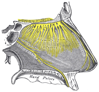
The nasopalatine nerve (also long sphenopalatine nerve) is a nerve of the head. It is a sensory branch of the maxillary nerve (CN V2) that passes through the pterygopalatine ganglion (without synapsing) and then through the sphenopalatine foramen to enter the nasal cavity, and finally out of the nasal cavity through the incisive canal and then the incisive fossa to enter the hard palate. It provides sensory innervation to the posteroinferior part of the nasal septum, and gingiva just posterior to the upper incisor teeth.
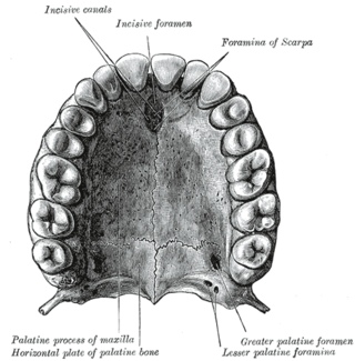
The incisive canals are two bony canals of the anterior hard palate connecting the nasal cavity and the oral cavity. An incisive canal courses through each maxilla. Below, the two incisive canals typically converge medially.

The sphenopalatine foramen is a foramen of the skull that connects the nasal cavity and the pterygopalatine fossa. It gives passage to the sphenopalatine artery, nasopalatine nerve, and the superior nasal nerve.
The greater palatine artery is a branch of the descending palatine artery and contributes to the blood supply of the hard palate and nasal septum.

The greater omentum is a large apron-like fold of visceral peritoneum that hangs down from the stomach. It extends from the greater curvature of the stomach, passing in front of the small intestines and doubles back to ascend to the transverse colon before reaching to the posterior abdominal wall. The greater omentum is larger than the lesser omentum, which hangs down from the liver to the lesser curvature. The common anatomical term "epiploic" derives from "epiploon", from the Greek epipleein, meaning to float or sail on, since the greater omentum appears to float on the surface of the intestines. It is the first structure observed when the abdominal cavity is opened anteriorly.
The posterior ethmoidal artery is an artery of the head which arises from the ophthalmic artery to supply the posterior ethmoidal air cells, and the meninges. It is smaller than the anterior ethmoidal artery.

The anterior superior alveolar nerve (or anterior superior dental nerve) is a branch of the infraorbital nerve (itself a branch of the maxillary nerve (CN V2)). It passes through the canalis sinuosus to reach and innervate upper front teeth. Through its nasal branch, it also innervates parts of the nasal cavity.

The greater palatine nerve is a branch of the pterygopalatine ganglion. This nerve is also referred to as the anterior palatine nerve, due to its location anterior to the lesser palatine nerve. It carries both general sensory fibres from the maxillary nerve, and parasympathetic fibers from the nerve of the pterygoid canal. It may be anaesthetised for procedures of the mouth and maxillary (upper) teeth.

The human nose is the first organ of the respiratory system. It is also the principal organ in the olfactory system. The shape of the nose is determined by the nasal bones and the nasal cartilages, including the nasal septum, which separates the nostrils and divides the nasal cavity into two.
The sphenopalatine artery passes through the sphenopalatine foramen into the cavity of the nose, at the back part of the superior meatus. Here it gives off its posterior lateral nasal branches which spread forward over the conchæ and meatuses, anastomose with the ethmoidal arteries and the nasal branches of the descending palatine, and assist in supplying the frontal, maxillary, ethmoidal, and sphenoidal sinuses.
The posterior septal artery is a branch of the sphenopalatine artery. The posterior septal artery supplies blood to the respiratory region of the nasal septum, which consists of the lower two-thirds of the nasal septum and most of the nasal floor. The path of the posterior septal artery can be understood by splitting it into three different segments: the pterygopalatine segment, the sphenoidal segment, and the septal segment.
