
The facial nerve, also known as the seventh cranial nerve, cranial nerve VII, or simply CN VII, is a cranial nerve that emerges from the pons of the brainstem, controls the muscles of facial expression, and functions in the conveyance of taste sensations from the anterior two-thirds of the tongue. The nerve typically travels from the pons through the facial canal in the temporal bone and exits the skull at the stylomastoid foramen. It arises from the brainstem from an area posterior to the cranial nerve VI and anterior to cranial nerve VIII.
Articles related to anatomy include:

Palatine tonsils, commonly called the tonsils and occasionally called the faucial tonsils, are tonsils located on the left and right sides at the back of the throat, which can often be seen as flesh-colored, pinkish lumps. Tonsils only present as "white lumps" if they are inflamed or infected with symptoms of exudates and severe swelling.

The external carotid artery is a major artery of the head and neck. It arises from the common carotid artery when it splits into the external and internal carotid artery. The external carotid artery supplies blood to the face, brain and neck.

In anatomy, the pharyngeal tonsil, also known as the nasopharyngeal tonsil or adenoid, is the superior-most of the tonsils. It is a mass of lymphatic tissue located behind the nasal cavity, in the roof of the nasopharynx, where the nose blends into the throat. In children, it normally forms a soft mound in the roof and back wall of the nasopharynx, just above and behind the uvula.
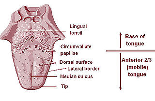
The lingual tonsils are a collection of lymphatic tissue located in the lamina propria of the root of the tongue. This lymphatic tissue consists of the lymphatic nodules rich in cells of the immune system (immunocytes). The immunocytes initiate the immune response when the lingual tonsils get in contact with invading microorganisms.

The facial artery is a branch of the external carotid artery that supplies structures of the superficial face.

The lingual artery arises from the external carotid artery between the superior thyroid artery and facial artery. It can be located easily in the tongue.

The ascending pharyngeal artery is an artery of the neck that supplies the pharynx.

The maxillary artery supplies deep structures of the face. It branches from the external carotid artery just deep to the neck of the mandible.
The ascending palatine artery is an artery is a branch of the facial artery which ascends along the neck before spliting into two terminal branches; one branch supplies the soft palate, and the other supplies the palatine tonsil and pharyngotympanic tube.

The lacrimal artery is an artery of the orbit. It is a branch of the ophthalmic artery. It accompanies the lacrimal nerve along the upper border of the lateral rectus muscle, travelling forward to reach the lacrimal gland. It supplies the lacrimal gland, two rectus muscles of the eye, the eyelids, and the conjunctiva.
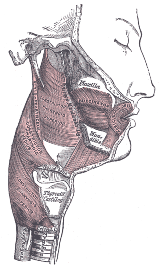
The pharyngeal muscles are a group of muscles that form the pharynx, which is posterior to the oral cavity, determining the shape of its lumen, and affecting its sound properties as the primary resonating cavity.
In anatomy, arterial tree is used to refer to all arteries and/or the branching pattern of the arteries. This article regards the human arterial tree. Starting from the aorta:

The submental artery is the largest branch of the facial artery in the neck. It first runs forward under the mouth, then turns upward upon reaching the chin.

This article describes the anatomy of the head and neck of the human body, including the brain, bones, muscles, blood vessels, nerves, glands, nose, mouth, teeth, tongue, and throat.
The pharyngeal artery is a branch of the ascending pharyngeal artery. The pharyngeal artery passes inferior-ward in between the superior margin of the superior pharyngeal constrictor muscle, and the levator veli palatini muscle. It issues branches to the constrictor muscles of the pharynx, the stylopharyngeus muscle, the pharyngotympanic tube, and palatine tonsil; a palatine branch may sometimes be present, replacing the ascending palatine branch of facial artery.

The fauces, isthmus of fauces, or the oropharyngeal isthmus is the opening at the back of the mouth into the throat. It is a narrow passage between the velum and the base of the tongue.

The following outline is provided as an overview of and topical guide to human anatomy:

The pharynx is the part of the throat behind the mouth and nasal cavity, and above the esophagus and trachea. It is found in vertebrates and invertebrates, though its structure varies across species. The pharynx carries food to the esophagus and air to the larynx. The flap of cartilage called the epiglottis stops food from entering the larynx.
