
A spinal nerve is a mixed nerve, which carries motor, sensory, and autonomic signals between the spinal cord and the body. In the human body there are 31 pairs of spinal nerves, one on each side of the vertebral column. These are grouped into the corresponding cervical, thoracic, lumbar, sacral and coccygeal regions of the spine. There are eight pairs of cervical nerves, twelve pairs of thoracic nerves, five pairs of lumbar nerves, five pairs of sacral nerves, and one pair of coccygeal nerves. The spinal nerves are part of the peripheral nervous system.

In human anatomy, the subclavian arteries are paired major arteries of the upper thorax, below the clavicle. They receive blood from the aortic arch. The left subclavian artery supplies blood to the left arm and the right subclavian artery supplies blood to the right arm, with some branches supplying the head and thorax. On the left side of the body, the subclavian comes directly off the aortic arch, while on the right side it arises from the relatively short brachiocephalic artery when it bifurcates into the subclavian and the right common carotid artery.

The internal carotid artery is an artery in the neck which supplies the anterior circulation of the brain.

The levator scapulae is a slender skeletal muscle situated at the back and side of the neck. It originates from the transverse processes of the four uppermost cervical vertebrae; it inserts onto the upper portion of the medial border of the scapula. It is innervated by the cervical nerves C3-C4, and frequently also by the dorsal scapular nerve. As the Latin name suggests, its main function is to lift the scapula.

In tetrapods, cervical vertebrae are the vertebrae of the neck, immediately below the skull. Truncal vertebrae lie caudal of cervical vertebrae. In sauropsid species, the cervical vertebrae bear cervical ribs. In lizards and saurischian dinosaurs, the cervical ribs are large; in birds, they are small and completely fused to the vertebrae. The vertebral transverse processes of mammals are homologous to the cervical ribs of other amniotes. Most mammals have seven cervical vertebrae, with the only three known exceptions being the manatee with six, the two-toed sloth with five or six, and the three-toed sloth with nine.

The obliquus capitis superior muscle is a small muscle in the upper back part of the neck. It is one of the suboccipital muscles. It attaches inferiorly at the transverse process of the atlas ; it attaches superiorly at the external surface of the occipital bone. The muscle is innervated by the suboccipital nerve.

The vertebral arteries are major arteries of the neck. Typically, the vertebral arteries originate from the subclavian arteries. Each vessel courses superiorly along each side of the neck, merging within the skull to form the single, midline basilar artery. As the supplying component of the vertebrobasilar vascular system, the vertebral arteries supply blood to the upper spinal cord, brainstem, cerebellum, and posterior part of brain.
In anatomy, the left and right common carotid arteries (carotids) are arteries that supply the head and neck with oxygenated blood; they divide in the neck to form the external and internal carotid arteries.

The semispinalis muscles are a group of three muscles belonging to the transversospinales. These are the semispinalis capitis, the semispinalis cervicis and the semispinalis thoracis.

The occipital artery is a branch of the external carotid artery that provides arterial supply to the back of the scalp, sternocleidomastoid muscles, and deep muscles of the back and neck.

The posterior triangle is a region of the neck.

The vertebral vein is formed in the suboccipital triangle, from numerous small tributaries which spring from the internal vertebral venous plexuses and issue from the vertebral canal above the posterior arch of the atlas.

The transverse cervical artery is an artery in the neck and a branch of the thyrocervical trunk, running at a higher level than the suprascapular artery.
The deep cervical vein is the vena comitans of the deep cervical artery. The vein is formed in the suboccipital region by the convergence of communicating branches of the occipital vein, veins draining the suboccipital muscles, and veins from the venous plexuses that surround cervical nerves. The vein and corresponding artery then pass in between the semispinalis capitis muscle and the semispinalis colli muscle. The vein passes anterior-ward in between the transverse process of the 7th cervical vertebra and the neck of the first rib to terminate in the vertebral vein.

The deep cervical fascia lies under cover of the platysma, and invests the muscles of the neck; it also forms sheaths for the carotid vessels, and for the structures situated in front of the vertebral column. Its attachment to the hyoid bone prevents the formation of a dewlap.
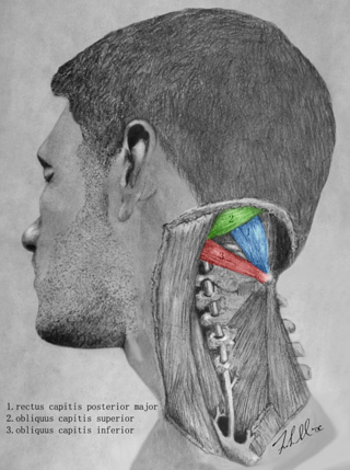
The suboccipital triangle is a region of the neck bounded by the following three muscles of the suboccipital group of muscles:

The middle cervical ganglion is the smallest of the three cervical sympathetic ganglia. It presumably represents the merging of the sympathetic ganglia of cervical segments C5–C6. It is usually situated at the level of the sixth cervical vertebra.
The inferior cervical ganglion is one of the three cervical sympathetic ganglia. It is situated between the base of the transverse process of the last cervical vertebra and the neck of the first rib, on the medial side of the costocervical artery.
The superior cardiac nerve arises by two or more branches from the superior cervical ganglion, and occasionally receives a filament from the trunk between the first and second cervical ganglia. It runs down the neck behind the common carotid artery, and in front of the Longus colli muscle; and crosses in front of the inferior thyroid artery, and recurrent nerve. The course of the nerves on the two sides then differs.

Each vertebra is an irregular bone with a complex structure composed of bone and some hyaline cartilage, that make up the vertebral column or spine, of vertebrates. The proportions of the vertebrae differ according to their spinal segment and the particular species.
