| Left coronary artery | |
|---|---|
 Heart viewed from above, atria removed, base of ventricles exposed. Left coronary artery visible at left. | |
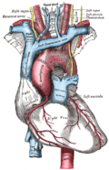
 Heart viewed from the front. Coronary arteries (labeled in red text) and other major landmarks (in blue text). Left coronary artery is at upper right in the image. | |
| Details | |
| Source | Ascending aorta |
| Branches |
|
| Identifiers | |
| Latin | arteria coronaria sinistra |
| TA98 | A12.2.03.201 |
| TA2 | 4142 |
| FMA | 50040 |
| Anatomical terminology | |

The left coronary artery (LCA, also known as the left main coronary artery, or left main stem coronary artery) is a coronary artery that arises from the aorta above the left cusp of the aortic valve, and supplies blood to the left side of the heart muscle.[ citation needed ] The left coronary artery typically runs for 10–25 mm, then bifurcates into the left anterior descending artery, and the left circumflex artery. [1]
Contents
The part that is between the aorta and the bifurcation only is known as the left main artery (LM), while the term "LCA" might refer to just the left main, or to the left main and all its eventual branches.[ citation needed ]