Related Research Articles

The rectus abdominis muscle, also known as the "abdominal muscle" or simply the "abs", is a pair of segmented skeletal muscle on the ventral aspect of a person's abdomen. The paired muscle is separated at the midline by a band of dense connective tissue called the linea alba, and the connective tissue defining each lateral margin of the rectus abdominus is the linea semilunaris. The muscle extends from the pubic symphysis, pubic crest and pubic tubercle inferiorly, to the xiphoid process and costal cartilages of the 5th–7th ribs superiorly.

The transverse abdominal muscle (TVA), also known as the transverse abdominis, transversalis muscle and transversus abdominis muscle, is a muscle layer of the anterior and lateral abdominal wall, deep to the internal oblique muscle. It is thought by most fitness instructors to be a significant component of the core.

The transversus thoracis muscle, also known as triangularis sterni, lies internal to the thoracic cage, anteriorly. It is usually a thin plane of muscular and tendinous fibers, however on athletic individuals it can be a thick 'slab of meat', situated upon the inner surface of the front wall of the chest. It is in the same layer as the subcostal muscles and the innermost intercostal muscles.

The abdominal internal oblique muscle, also internal oblique muscle or interior oblique, is an abdominal muscle in the abdominal wall that lies below the external oblique muscle and just above the transverse abdominal muscle.
The abdominal external oblique muscle is the largest and outermost of the three flat abdominal muscles of the lateral anterior abdomen.

The quadratus lumborum muscle, informally called the QL, is a paired muscle of the left and right posterior abdominal wall. It is the deepest abdominal muscle, and commonly referred to as a back muscle. Each is irregular and quadrilateral in shape.

The intercostal nerves are part of the somatic nervous system, and arise from the anterior rami of the thoracic spinal nerves from T1 to T11. The intercostal nerves are distributed chiefly to the thoracic pleura and abdominal peritoneum, and differ from the anterior rami of the other spinal nerves in that each pursues an independent course without plexus formation.

The internal iliac artery is the main artery of the pelvis.

In human anatomy, the inferior epigastric artery is an artery that arises from the external iliac artery. It is accompanied by the inferior epigastric vein; inferiorly, these two inferior epigastric vessels together travel within the lateral umbilical fold The inferior epigastric artery then traverses the arcuate line of rectus sheath to enter the rectus sheath, then anastomoses with the superior epigastric artery within the rectus sheath.

The femoral nerve is a nerve in the thigh that supplies skin on the upper thigh and inner leg, and the muscles that extend the knee. It is the largest branch of the lumbar plexus.

The iliohypogastric nerve is a nerve that originates from the lumbar plexus that supplies sensation to skin over the lateral gluteal and hypogastric regions and motor to the internal oblique muscles and transverse abdominal muscles.

The inferior phrenic artery is a bilaterally paired artery of the abdominal cavity which represents the main source of arterial supply to the diaphragm. Each artery usually arises either from the coeliac trunk or the abdominal aorta, however, their origin is highly variable and the different sites of origin are different for the left artery and right artery. The superior suprarenal artery is a branch of the inferior phrenic artery.
The median sacral artery is a small artery that arises posterior to the abdominal aorta and superior to its bifurcation.

The lumbar arteries are arteries located in the lower back or lumbar region. The lumbar arteries are in parallel with the intercostals.
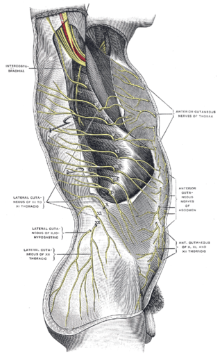
The subcostal nerve is a mixed motor and sensory nerve contributing to the lumbar plexus. It runs along the lower border of the twelfth rib, often gives a communicating branch to the first lumbar nerve, and passes under the lateral lumbocostal arch.

The costocervical trunk arises from the upper and back part of the second part of subclavian artery, behind the scalenus anterior on the right side, and medial to that muscle on the left side.

The intercostal arteries are a group of arteries passing within an intercostal space. There are 9 anterior and 11 posterior intercostal arteries on each side of the body. The anterior intercostal arteries are branches of the internal thoracic artery and its terminal branch - the musculophrenic artery. The posterior intercostal arteries are branches of the supreme intercostal artery and thoracic aorta.
The lumbar veins are four pairs of veins running along the inside of the posterior abdominal wall, and drain venous blood from parts of the abdominal wall. Each lumbar vein accompanies a single lumbar artery. The lower two pairs of lumbar veins all drain directly into the inferior vena cava, whereas the fate of the upper two pairs is more variable.
The lumbar fascia is the lumbar portion of the thoracolumbar fascia. It consists of three fascial layers - posterior, middle, and anterior - that enclose two muscular compartments. The anterior and middle layers occur only in the lumbar region, whereas the posterior layer extends superiorly to the inferior part of the neck, and the inferiorly to the dorsal surface of the sacrum. The quadratus lumborum is contained in the anterior muscular compartment, and the erector spinae in the posterior compartment. Psoas major lies anterior to the anterior layer. Various superficial muscles of the posterior thorax and abdomen arise from the posterior layer - namely the latissimus dorsi, and serratus posterior inferior.

The following outline is provided as an overview of and topical guide to human anatomy:
References
- 1 2 3 Sinnatamby, Chummy (2011). Last's Anatomy (12th ed.). p. 395. ISBN 978-0-7295-3752-0.
- 1 2 3 Gray, Henry (1918). Gray's Anatomy (20th ed.). p. 601.
![]() This article incorporates text in the public domain from page 601 of the 20th edition of Gray's Anatomy (1918)
This article incorporates text in the public domain from page 601 of the 20th edition of Gray's Anatomy (1918)