Lumbar fascia

Last updated

The lumbar fascia is the lumbar portion of the thoracolumbar fascia. It consists of three fascial layers - posterior, middle, and anterior - that enclose two (anterior and posterior) muscular compartments. The anterior and middle layers occur only in the lumbar region, whereas the posterior layer (as part of the thoracolumbar fascia) extends superiorly to the inferior part of the neck, and the inferiorly to the dorsal surface of the sacrum. The quadratus lumborum is contained in the anterior muscular compartment (between anterior and middle layers), and the erector spinae in the posterior compartment (between middle and posterior layers). [1] :274 Psoas major lies anterior to the anterior layer. Various superficial muscles of the posterior thorax and abdomen arise from the posterior layer - namely the latissimus dorsi, and serratus posterior inferior.[ citation needed ]

Contents

Anatomy

The posterior layer and middle layer unite at the lateral margin of erector spinae, forming a though raphe; all three layers then unite at the lateral margin of quadratus lumborum, thus forming the aponeurotic origin of the transversus abdominis muscle. [2] :814–815

Layers

Posterior layer

The posterior layer is thick, being reinforced by the aponeurosis (origin) of the latissimus dorsi muscle. [1] :274 The posterior layer is attached to the spinous processes of lumbar and sacral vertebrae, and to the supraspinous ligament. [2] :814–815

At sacral levels, the posterior layer attaches to the posterior superior iliac spines, and posterior iliac crest, fuses with the underlying erector spinae muscle aponeurosis, [3] :814–815 and extends along the transverse tubercles of the sacrum. [1] :274

In the thoracic region, the posterior layer (i.e. thoracic part of thoracolumbar fascia) attaches to the supraspinous ligament and spinous processes of all thoracic vertebrae, and to the costal angles of all ribs. It extends as far superiorly as the inferior part of the neck. [1] :274

Middle layer

The middle layer is attached superiorly to the inferior margin of the 12th rib, and the lumbocostal ligament, medially to the tips of transverse processes of lumbar vertebrae, and inferiorly to [2] :814–815 the posterior part of the intermediate area of [4] the iliac crest. [2] :814–815

Anterior layer

The anterior layer is the thinnest of the three layers.[ citation needed ] The anterior layer forms the lateral arcuate ligament superiorly, is attached medially to anterior surfaces of transverse processes of lumbar vertebra, and is attached inferiorly to the iliolumbar ligament and adjoining iliac crest. [2] :814–815

Relations

The ascending colon [1] :257 and descending colon [1] :258 lie upon the lumbar fascia.

The lumbar fascia gives origin to the latissimus dorsi muscle (which arises from posterior layer of lumbar fascia), [1] :40 abdominal internal oblique muscle (which arises along the entire length of the lumbar fascia), transversus abdominis muscle, [1] :222 and gluteus maximus muscle. [1] :125

The psoas fascia (a part of the iliac fascia) laterally blends with the lumbar fascia. [1] :272 The lateral arcuate ligament arises as a thickening of the lumbar fascia. [1] :185 The superior band of the iliolumbar ligament is continuous with the anterior layer of lumbar fascia. [1] :325 The inferior portion of the supraspinous ligament becomes indistinct amid the lumbar fascia. [1] :424

Each subcostal artery passes across the anterior surface of the lumbar fascia before reaching the anterior abdominal wall. [1] :276 Each subcostal nerve passes across the anterior aspect of the anterior layer of lumbar fascia. [1] :278 The ilioinguinal nerve pierces the anterior layer of lumbar fascia posterior to the kidney to come to pass inferoanteriorly anterior to the anterior layer of lumbar fascia. [1] :279

Function

The lumbar fascia binds the deep muscles of the back to the spine and to the transverse processes of the vertebrae. Additionally the lumbar fascia is an attachment for the gluteus maximus muscle and the latissimus dorsi muscle.[ citation needed ]

Related Research Articles

Articles related to anatomy include:

<span class="mw-page-title-main">Latissimus dorsi muscle</span> Large, flat back muscle

The latissimus dorsi is a large, flat muscle on the back that stretches to the sides, behind the arm, and is partly covered by the trapezius on the back near the midline. The word latissimus dorsi comes from Latin and means "broadest [muscle] of the back", from "latissimus" ' and "dorsum". The pair of muscles are commonly known as "lats", especially among bodybuilders. The latissimus dorsi is the largest muscle in the upper body.

<span class="mw-page-title-main">Spinal nerve</span> Nerve that carries signals between the spinal cord and the body

A spinal nerve is a mixed nerve, which carries motor, sensory, and autonomic signals between the spinal cord and the body. In the human body there are 31 pairs of spinal nerves, one on each side of the vertebral column. These are grouped into the corresponding cervical, thoracic, lumbar, sacral and coccygeal regions of the spine. There are eight pairs of cervical nerves, twelve pairs of thoracic nerves, five pairs of lumbar nerves, five pairs of sacral nerves, and one pair of coccygeal nerves. The spinal nerves are part of the peripheral nervous system.

<span class="mw-page-title-main">Aponeurosis</span> Tissue which connects muscles to other organs

An aponeurosis is a type or a variant of the deep fascia, in the form of a sheet of pearly-white fibrous tissue that attaches sheet-like muscles needing a wide area of attachment. Their primary function is to join muscles and the body parts they act upon, whether bone or other muscles. They have a shiny, whitish-silvery color, are histologically similar to tendons, and are very sparingly supplied with blood vessels and nerves. When dissected, aponeuroses are papery and peel off by sections. The primary regions with thick aponeuroses are in the ventral abdominal region, the dorsal lumbar region, the ventriculus in birds, and the palmar (palms) and plantar (soles) regions.

<span class="mw-page-title-main">Serratus posterior inferior muscle</span> Muscle of the mid-low back

The serratus posterior inferior muscle, also known as the posterior serratus muscle, is a muscle of the human body.

<span class="mw-page-title-main">Abdominal external oblique muscle</span> Skeletal muscle in the abdomen

The abdominal external oblique muscle is the largest and outermost of the three flat abdominal muscles of the lateral anterior abdomen.

<span class="mw-page-title-main">Quadratus lumborum muscle</span> Muscle in the lower back

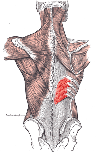
The quadratus lumborum muscle, informally called the QL, is a paired muscle of the left and right posterior abdominal wall. It is the deepest abdominal muscle, and commonly referred to as a back muscle. Each is irregular and quadrilateral in shape.

<span class="mw-page-title-main">Erector spinae muscles</span> Human muscle group

The erector spinae or spinal erectors is a set of muscles that straighten and rotate the back. The spinal erectors work together with the glutes to maintain stable posture standing or sitting.

<span class="mw-page-title-main">Thoracolumbar fascia</span> Anatomical Feature

The thoracolumbar fascia a complex, multilayer arrangement of fasciae and aponeurotic sheets forming a separation between the paraspinal muscles on one hand, and the muscles of the posterior abdominal wall on the other. It spans the length if the back, extending between the neck superiorly and the sacrum inferiorly. It entails the fasciae and aponeuroses of the latissimus dorsi muscle, serratus posterior inferior muscle, abdominal internal oblique muscle, and transverse abdominal muscle.

<span class="mw-page-title-main">Iliocostalis</span>

Iliocostalis muscle is the muscle immediately lateral to the longissimus that is the nearest to the furrow that separates the epaxial muscles from the hypaxial. It lies very deep to the fleshy portion of the serratus posterior muscle. It laterally flexes the vertebral column to the same side.

<span class="mw-page-title-main">Longissimus</span>

The longissimus is the muscle lateral to the semispinalis muscles. It is the longest subdivision of the erector spinae muscles that extends forward into the transverse processes of the posterior cervical vertebrae.

<span class="mw-page-title-main">Wing of ilium</span> Flat portion of the hip bone

The wing(ala)of ilium is the large expanded portion of the ilium, the bone which bounds the greater pelvis laterally. It presents for examination two surfaces—an external and an internal—a crest, and two borders—an anterior and a posterior.

<span class="mw-page-title-main">Iliac crest</span> Top border of the hip

The crest of the ilium is the superior border of the wing of ilium and the superiolateral margin of the greater pelvis.

<span class="mw-page-title-main">Iliolumbar ligament</span>

The iliolumbar ligament is a strong ligament passing from the tip of the transverse process of the fifth lumbar vertebra to the posterior part of the inner lip of the iliac crest.

<span class="mw-page-title-main">Deep circumflex iliac artery</span> Artery in the pelvis

The deep circumflex iliac artery is an artery in the pelvis that travels along the iliac crest of the pelvic bone.

<span class="mw-page-title-main">Outline of human anatomy</span> Overview of and topical guide to human anatomy

The following outline is provided as an overview of and topical guide to human anatomy:

<span class="mw-page-title-main">Hip bone</span> Bone of the pelvis

The hip bone is a large flat bone, constricted in the center and expanded above and below. In some vertebrates it is composed of three parts: the ilium, ischium, and the pubis.

<span class="mw-page-title-main">Pelvis</span> Lower part of the trunk of the human body between the abdomen and the thighs

The pelvis is the lower part of the trunk, between the abdomen and the thighs, together with its embedded skeleton.

<span class="mw-page-title-main">Vertebra</span> Bone in the vertebral column

The spinal column, a defining synapomorphy shared by nearly all vertebrates, is a moderately flexible series of vertebrae, each constituting a characteristic irregular bone whose complex structure is composed primarily of bone, and secondarily of hyaline cartilage. They show variation in the proportion contributed by these two tissue types; such variations correlate on one hand with the cerebral/caudal rank, and on the other with phylogenetic differences among the vertebrate taxa.

References

  1. 1 2 3 4 5 6 7 8 9 10 11 12 13 14 15 16 Sinnatamby, Chummy (2011). Last's Anatomy (12th ed.). ISBN   978-0-7295-3752-0.
  2. 1 2 3 4 5 Standring, Susan (2020). Gray's Anatomy: The Anatomical Basis of Clinical Practice (42nd ed.). New York. ISBN   978-0-7020-7707-4. OCLC   1201341621.
  3. Standring, Susan (2020). Gray's Anatomy: The Anatomical Basis of Clinical Practice (42nd ed.). New York. ISBN   978-0-7020-7707-4. OCLC   1201341621.
  4. Vleeming, Andry; Pool-Goudzwaard, Annelies L.; Stoeckart, Rob; van Wingerden, Jan-Paul; Snijders, Chris J. (April 1995). "The Posterior Layer of the Thoracolumbar Fascia|Its Function in Load Transfer From Spine to Legs". Spine. 20 (7): 753. ISSN   0362-2436.