Quadratus lumborum muscle

Last updated
Quadratus lumborum muscle
Quadratuslumborum.png
The left quadratus lumborum, one of the posterior abdominal muscles, is depicted in red.
Details
Origin Posterior border of iliac crest
Insertion Inferior border of 12th rib and L1-L4
Artery Lumbar arteries, lumbar branch of iliolumbar artery
Nerve The twelfth thoracic and first through fourth ventral rami of lumbar nerves (T12, L1-L4)
Actions Alone(unilateral), lateral flexion of vertebral column; Together (bilateral), depression of thoracic rib cage
Identifiers
Latin musculus quadratus lumborum
TA98 A04.5.01.027
TA2 2382
FMA 15569
Anatomical terms of muscle

The quadratus lumborum muscle, informally called the QL, is a paired muscle of the left and right posterior abdominal wall. It is the deepest abdominal muscle, and commonly referred to as a back muscle. Each is irregular and quadrilateral in shape.

Contents

The quadratus lumborum muscles originate from the wings of the ilium; their insertions are on the transverse processes of the upper four lumbar vertebrae plus the lower posterior border of the twelfth rib. Contraction of one of the pair of muscles causes lateral flexion of the lumbar spine, elevation of the pelvis, or both. Contraction of both causes extension of the lumbar spine.

A disorder of the quadratus lumborum muscles is pain due to muscle fatigue from constant contraction due to prolonged sitting, such as at a computer or in a car. [1] Kyphosis and weak gluteal muscles can also contribute to the likelihood of quadratus lumborum pain.

Structure

The quadratus lumborum muscle originates by aponeurotic fibers into the iliolumbar ligament and the internal lip of the iliac crest for about 5 centimetres (2.0 in). It inserts from the lower border of the last rib for about half its length and by four small tendons from the apices of the transverse processes of the upper four lumbar vertebrae.

The number of attachments to the vertebræ, and the extent of its attachment to the last rib, may vary. Also, occasionally, a second portion of this muscle is found in front of the preceding. It arises from the upper borders of the transverse processes of the lower three or four lumbar vertebræ, and is inserted into the lower margin of the last rib.

Relationships

Anterior to the quadratus lumborum are the colon, the kidney, the psoas major muscle, (if present) the psoas minor muscle, and the diaphragm; between the fascia and the muscle are the twelfth thoracic, ilioinguinal, and iliohypogastric nerves. The quadratus lumborum muscle is a continuation of transverse abdominal muscle.

Nerve supply

Anterior branches of the ventral rami of T12 to L4.

Functions

The quadratus lumborum can perform four actions:

  1. Lateral flexion of vertebral column, with ipsilateral contraction
  2. Extension of lumbar vertebral column, with bilateral contraction (based on line of force passing ~3.5 cm posterior L3 rotation axis [2] )
  3. Fixes the 12th rib during forced expiration. The quadratus lumborum assists the diaphragm in inhalation
  4. Elevates the Ilium (bone), with ipsilateral contraction ("hip hiking")

Additional functions:

Clinical significance

The quadratus lumborum muscles can be the source of back pain when overused, or in association with scoliosis or weak gluteal muscles.

Mechanism

The quadratus lumborum is a common source of unilateral or bilateral lower back pain, including localized pain and tenderness over the wing of the ilium. [3] Because quadratus lumborum connects the pelvis to the spine and is therefore capable of extending the lower back when contracting bilaterally, the two quadratus lumborum muscles pick up the slack, as it were, when the lower fibers of the erector spinae are weak or inhibited (as they often are in the case of habitual seated computer use and/or the use of a lower back support in a chair). Given their comparable mechanical disadvantage, constant contraction while seated can overuse the quadratus lumborum, resulting in muscle fatigue. [1] A constantly contracted quadratus lumborum, like any other muscle, will experience decreased blood flow, and, in time, adhesions in the muscle and fascia may develop, the end point of which is muscle spasm.

Association with kyphosis

This chain of events can be and often is accelerated by kyphosis, which is invariably accompanied by rounded shoulders, both of which place greater stress on the quadratus lumborum by shifting body weight forward, forcing the erector spinae, quadratus lumborum, multifidi, and especially the levator scapulae to work harder in both seated and standing positions to maintain an erect torso and neck. The experience of "productive pain" or pleasure by a patient upon palpation of the quadratus lumborum is indicative of such a condition.

Association with weak gluteal muscles

Hip abduction is performed primarily by the hip abductors (gluteus medius and minimus). When the gluteus medius/minimus are weak or inhibited, the tensor fasciae latae or quadratus lumborum will compensate by becoming the prime mover. The most impaired movement pattern of hip abduction is when the quadratus lumborum initiates the movement, which results in hip hiking during swing phase of gait. Hip hiking places excessive side-bending compressive stresses on the lumbar segments. Thus, a tight quadratus lumborum may be another hidden cause of low back pain (Janda 1987).

When the hip adductors are tight or hypertonic, their antagonist (gluteus medius) may experience reciprocal inhibition. The gluteus medius will become weak and inhibited. This in turn may cause hypertonicity of ipsilateral quadratus lumborum. Chronic hypertonicity of quadratus lumborum tends to cause low back pain due to its ability to create compressive stress on lumbar segment.

Treatment

While stretching and strengthening the quadratus lumborum are indicated for unilateral lower back pain, heat or ice applications as well as massage should be considered as part of any comprehensive rehabilitation regimen.

Current studies show that application of heat or ice, massage, and estim will not leave long-term benefits. Careful assessment of muscular imbalances and movement impairments by a therapist is recommended in order to address the underlying issues mentioned. [4]

Related Research Articles

<span class="mw-page-title-main">Human leg</span> Lower extremity or limb of the human body (foot, lower leg, thigh and hip)

The human leg, in the general word sense, is the entire lower limb of the human body, including the foot, thigh or sometimes even the hip or gluteal region. However, the definition in human anatomy refers only to the section of the lower limb extending from the knee to the ankle, also known as the crus or, especially in non-technical use, the shank. Legs are used for standing, and all forms of locomotion including recreational such as dancing, and constitute a significant portion of a person's mass. Female legs generally have greater hip anteversion and tibiofemoral angles, but shorter femur and tibial lengths than those in males.

<span class="mw-page-title-main">Sacrum</span> Triangular-shaped bone at the bottom of the spine

The sacrum, in human anatomy, is a large, triangular bone at the base of the spine that forms by the fusing of the sacral vertebrae (S1–S5) between ages 18 and 30.

<span class="mw-page-title-main">Spinal nerve</span> Nerve that carries signals between the spinal cord and the body

A spinal nerve is a mixed nerve, which carries motor, sensory, and autonomic signals between the spinal cord and the body. In the human body there are 31 pairs of spinal nerves, one on each side of the vertebral column. These are grouped into the corresponding cervical, thoracic, lumbar, sacral and coccygeal regions of the spine. There are eight pairs of cervical nerves, twelve pairs of thoracic nerves, five pairs of lumbar nerves, five pairs of sacral nerves, and one pair of coccygeal nerves. The spinal nerves are part of the peripheral nervous system.

<span class="mw-page-title-main">Lordosis</span> Medical condition

Lordosis is historically defined as an abnormal inward curvature of the lumbar spine. However, the terms lordosis and lordotic are also used to refer to the normal inward curvature of the lumbar and cervical regions of the human spine. Similarly, kyphosis historically refers to abnormal convex curvature of the spine. The normal outward (convex) curvature in the thoracic and sacral regions is also termed kyphosis or kyphotic. The term comes from the Greek lordōsis, from lordos.

<span class="mw-page-title-main">Hip</span> Anatomical region between the torso and the legs, holding the buttocks and genital region

In vertebrate anatomy, hip refers to either an anatomical region or a joint.

<span class="mw-page-title-main">Psoas major muscle</span> Long fusiform muscle located in the lumbar region

The psoas major is a long fusiform muscle located in the lateral lumbar region between the vertebral column and the brim of the lesser pelvis. It joins the iliacus muscle to form the iliopsoas. In animals, this muscle is equivalent to the tenderloin.

<span class="mw-page-title-main">Abdomen</span> Part of the body between the chest and pelvis

The abdomen is the part of the body between the thorax (chest) and pelvis, in humans and in other vertebrates. The abdomen is the front part of the abdominal segment of the torso. The area occupied by the abdomen is called the abdominal cavity. In arthropods it is the posterior tagma of the body; it follows the thorax or cephalothorax.

<span class="mw-page-title-main">Muscles of the hip</span> Causes movement in the hip.

In human anatomy, the muscles of the hip joint are those muscles that cause movement in the hip. Most modern anatomists define 17 of these muscles, although some additional muscles may sometimes be considered. These are often divided into four groups according to their orientation around the hip joint: the gluteal group; the lateral rotator group; the adductor group; and the iliopsoas group.

<span class="mw-page-title-main">Lumbar plexus</span>

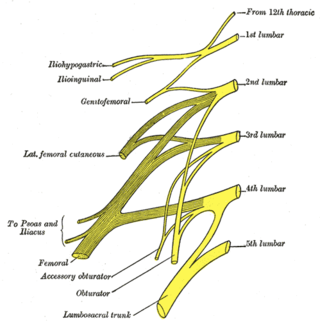
The lumbar plexus is a web of nerves in the lumbar region of the body which forms part of the larger lumbosacral plexus. It is formed by the divisions of the first four lumbar nerves (L1-L4) and from contributions of the subcostal nerve (T12), which is the last thoracic nerve. Additionally, the ventral rami of the fourth lumbar nerve pass communicating branches, the lumbosacral trunk, to the sacral plexus. The nerves of the lumbar plexus pass in front of the hip joint and mainly support the anterior part of the thigh.

<span class="mw-page-title-main">Lumbar nerves</span>

The lumbar nerves are the five pairs of spinal nerves emerging from the lumbar vertebrae. They are divided into posterior and anterior divisions.

<span class="mw-page-title-main">Erector spinae muscles</span> Human muscle group

The erector spinae or spinal erectors is a set of muscles that straighten and rotate the back. The spinal erectors work together with the glutes to maintain stable posture standing or sitting.

<span class="mw-page-title-main">Thoracolumbar fascia</span> Anatomical Feature

The thoracolumbar fascia is a deep investing membrane throughout most of the posterior thorax and abdomen although it is a thin fibrous lamina in the thoracic region. Above, it is continuous with a similar investing layer on the back of the neck—the nuchal fascia.

<span class="mw-page-title-main">Lumbar arteries</span>

The lumbar arteries are arteries located in the lower back or lumbar region. The lumbar arteries are in parallel with the intercostals.

<span class="mw-page-title-main">Iliac crest</span> Top border of the hip

The crest of the ilium is the superior border of the wing of ilium and the superiolateral margin of the greater pelvis.

<span class="mw-page-title-main">Iliolumbar ligament</span>

The iliolumbar ligament is a strong ligament passing from the tip of the transverse process of the fifth lumbar vertebra to the posterior part of the inner lip of the iliac crest.

The lumbar fascia is an anatomic structure of the lumbar region. It consists of a band or sheet of connective tissue fibres, primarily collagen, that attaches, stabilizes, encloses and separates muscles not limited to the lumbar region as the name suggests, but extending upwards over the thorax to the neck and downwards over the muscles covering the sacrum. The upper thoracic portion is thin while the lumbar and sacral regions are thicker and stronger.

<span class="mw-page-title-main">Outline of human anatomy</span> Overview of and topical guide to human anatomy

The following outline is provided as an overview of and topical guide to human anatomy:

<span class="mw-page-title-main">Hip bone</span> Bone of the pelvis

The hip bone is a large flat bone, constricted in the center and expanded above and below. In some vertebrates it is composed of three parts: the ilium, ischium, and the pubis.

<span class="mw-page-title-main">Pelvis</span> Lower part of the trunk of the human body between the abdomen and the thighs

The pelvis is the lower part of the trunk, between the abdomen and the thighs, together with its embedded skeleton.

References

PD-icon.svgThis article incorporates text in the public domain from page 420 of the 20th edition of Gray's Anatomy (1918)

  1. 1 2 Core Topics in Pain, p. 131, Anita Holdcraft and Sian Jaggar, 2005.
  2. McGill SM, Santaguida L, Stevens J: Measurement of the trunk musculature from T5 to L5 using MRI scans of 15 young males corrected for muscle fibre orientation, Clin Biomech (Bristol, Avon) 8:171-178, 1993.
  3. Clinical Orthopaedic Examination, Ronald McRae, 2004 (5th ed.).
  4. Assessment and Treatment of Muscle Imbalances, p. 100, Phil Page, 2010.