Abdominal external oblique muscle

Last updated
Abdominal external oblique muscle
Illu trunk muscles.jpg
Muscles of the trunk.
Gray392.png
The abdominal external oblique muscle
Details
Origin Ribs 5-12
Insertion Xiphoid process, outer lip of the iliac crest, pubic crest, pubic tubercle, linea alba, inguinal ligament, anterior superior iliac spine (ASIS)
Nerve Thoraco-abdominal nerves (T7-11) and subcostal nerve (T12)
Actions Flexion of the torso and contralateral rotation of torso
Identifiers
Latin musculus obliquus externus abdominis
TA98 A04.5.01.008
TA2 2364
FMA 13335
Anatomical terms of muscle

The abdominal external oblique muscle (also external oblique muscle or exterior oblique) is the largest and outermost of the three flat abdominal muscles of the lateral anterior abdomen.

Contents

Structure

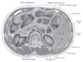
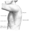
The external oblique is situated on the lateral and anterior parts of the abdomen. It is broad, thin, and irregularly quadrilateral, its muscular portion occupying the side, its aponeurosis the anterior wall of the abdomen. In most humans, the oblique is not visible, due to subcutaneous fat deposits and the small size of the muscle.

It arises from eight fleshy digitations, each from the external surfaces and inferior borders of the fifth to twelfth ribs (lower eight ribs). These digitations are arranged in an oblique line which runs inferiorly and anteriorly, with the upper digitations being attached close to the cartilages of the corresponding ribs, the lowest to the apex of the cartilage of the last rib, the intermediate ones to the ribs at some distance from their cartilages.

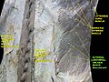
The five superior serrations increase in size from above downward, and are received between corresponding processes of the serratus anterior muscle; the three lower ones diminish in size from above downward and receive between them corresponding processes from the latissimus dorsi. From these attachments the fleshy fibers proceed in various directions. Its posterior fibers from the ribs to the iliac crest form a free posterior border.

Those from the lowest ribs pass nearly vertically downward, and are inserted into the anterior half of the outer lip of the iliac crest; the middle and upper fibers, directed downward (inferiorly) and forward (anteriorly), become aponeurotic at approximately the midclavicular line and form the anterior layer of the rectus sheath. This aponeurosis formed from fibres from either side of the external oblique decussates at the linea alba.

The aponeurosis of the external oblique muscle forms the inguinal ligament. The muscle also contributes to the inguinal canal.

The internal oblique muscle is just deep to the external oblique muscle. [1]

Nerve supply

The external oblique muscle is supplied by ventral branches of the lower six thoracoabdominal nerves and the subcostal nerve on each side.

Blood supply

The cranial portion of the muscle is supplied by the lower intercostal arteries, whereas the caudal portion is supplied by a branches of either the deep circumflex iliac artery or the iliolumbar artery.

Function

The external oblique functions to pull the chest downwards and compress the abdominal cavity, which increases the intra-abdominal pressure as in a Valsalva maneuver. It also performs ipsilateral (same side) side-bending and contralateral (opposite side) rotation: the right external oblique would side-bend to the right and rotate to the left, and vice versa. The internal oblique muscle functions similarly except it rotates ipsilaterally.

Society and culture

Oblique strain

The oblique strain is a common baseball injury, particularly in pitchers. In both batters and pitchers it can affect the contralateral (leading) side external oblique, or the trailing internal oblique. [2]

Training

Additional images

Related Research Articles

Articles related to anatomy include:

<span class="mw-page-title-main">Inguinal canal</span> Human abdominal anatomy

The inguinal canal is a passage in the anterior abdominal wall on each side of the body, which in males, convey the spermatic cords and in females, the round ligament of the uterus. The inguinal canals are larger and more prominent in males.

<span class="mw-page-title-main">Rectus abdominis muscle</span> Paired straight muscle

The rectus abdominis muscle, also known as the "abdominal muscle" or simply the "abs", is a pair of segmented skeletal muscle on the ventral aspect of a person's abdomen. The paired muscle is separated at the midline by a band of dense connective tissue called the linea alba, and the connective tissue defining each lateral margin of the rectus abdominus is the linea semilunaris. The muscle extends from the pubic symphysis, pubic crest and pubic tubercle inferiorly, to the xiphoid process and costal cartilages of the 5th–7th ribs superiorly.

<span class="mw-page-title-main">Inguinal ligament</span> Band running from the pubic tubercle to the anterior superior iliac spine

The inguinal ligament, also known as Poupart's ligament or groin ligament, is a band running from the pubic tubercle to the anterior superior iliac spine. It forms the base of the inguinal canal through which an indirect inguinal hernia may develop.

<span class="mw-page-title-main">Transverse abdominal muscle</span> Muscle of the abdominal area

The transverse abdominal muscle (TVA), also known as the transverse abdominis, transversalis muscle and transversus abdominis muscle, is a muscle layer of the anterior and lateral abdominal wall, deep to the internal oblique muscle. It is thought by most fitness instructors to be a significant component of the core.

<span class="mw-page-title-main">External iliac artery</span> Arteries of the pelvis

The external iliac arteries are two major arteries which bifurcate off the common iliac arteries anterior to the sacroiliac joint of the pelvis.

<span class="mw-page-title-main">Abdominal internal oblique muscle</span> Muscle in the abdominal wall

The abdominal internal oblique muscle, also internal oblique muscle or interior oblique, is an abdominal muscle in the abdominal wall that lies below the external oblique muscle and just above the transverse abdominal muscle.

<span class="mw-page-title-main">Abdomen</span> Part of the body between the chest and pelvis

The abdomen is the part of the body between the thorax (chest) and pelvis, in humans and in other vertebrates. The abdomen is the front part of the abdominal segment of the torso. The area occupied by the abdomen is called the abdominal cavity. In arthropods, it is the posterior tagma of the body; it follows the thorax or cephalothorax.

<span class="mw-page-title-main">Internal iliac artery</span> Main artery of the pelvis

The internal iliac artery is the main artery of the pelvis.

<span class="mw-page-title-main">Inferior epigastric artery</span> Blood vessel

In human anatomy, the inferior epigastric artery is an artery that arises from the external iliac artery. It is accompanied by the inferior epigastric vein; inferiorly, these two inferior epigastric vessels together travel within the lateral umbilical fold The inferior epigastric artery then traverses the arcuate line of rectus sheath to enter the rectus sheath, then anastomoses with the superior epigastric artery within the rectus sheath.

<span class="mw-page-title-main">Abdominal wall</span> Boundary of abdominal cavity

In anatomy, the abdominal wall represents the boundaries of the abdominal cavity. The abdominal wall is split into the anterolateral and posterior walls.

<span class="mw-page-title-main">Lumbar plexus</span> Web of nerves in the lower spine

The lumbar plexus is a web of nerves in the lumbar region of the body which forms part of the larger lumbosacral plexus. It is formed by the divisions of the first four lumbar nerves (L1-L4) and from contributions of the subcostal nerve (T12), which is the last thoracic nerve. Additionally, the ventral rami of the fourth lumbar nerve pass communicating branches, the lumbosacral trunk, to the sacral plexus. The nerves of the lumbar plexus pass in front of the hip joint and mainly support the anterior part of the thigh.

<span class="mw-page-title-main">Iliohypogastric nerve</span>

The iliohypogastric nerve is a nerve that originates from the lumbar plexus that supplies sensation to skin over the lateral gluteal and hypogastric regions and motor to the internal oblique muscles and transverse abdominal muscles.

<span class="mw-page-title-main">Conjoint tendon</span> Medial part of the posterior wall of the inguinal canal

The conjoint tendon is a sheath of connective tissue formed from the lower part of the common aponeurosis of the abdominal internal oblique muscle and the transversus abdominis muscle, joining the muscle to the pelvis. It forms the medial part of the posterior wall of the inguinal canal.

The subcostal arteries, so named because they lie below the last ribs, constitute the lowest pair of branches derived from the thoracic aorta, and are in series with the intercostal arteries.

<span class="mw-page-title-main">Deep circumflex iliac artery</span> Artery in the pelvis

The deep circumflex iliac artery is an artery in the pelvis that travels along the iliac crest of the pelvic bone.

<span class="mw-page-title-main">Aponeurosis of the abdominal external oblique muscle</span> Membranous structure

The aponeurosis of the abdominal external oblique muscle is a thin but strong membranous structure, the fibers of which are directed downward and medially.

The lumbar fascia is the lumbar portion of the thoracolumbar fascia. It consists of three fascial layers - posterior, middle, and anterior - that enclose two muscular compartments. The anterior and middle layers occur only in the lumbar region, whereas the posterior layer extends superiorly to the inferior part of the neck, and the inferiorly to the dorsal surface of the sacrum. The quadratus lumborum is contained in the anterior muscular compartment, and the erector spinae in the posterior compartment. Psoas major lies anterior to the anterior layer. Various superficial muscles of the posterior thorax and abdomen arise from the posterior layer - namely the latissimus dorsi, and serratus posterior inferior.

<span class="mw-page-title-main">Outline of human anatomy</span> Overview of and topical guide to human anatomy

The following outline is provided as an overview of and topical guide to human anatomy:

<span class="mw-page-title-main">Hip bone</span> Bone of the pelvis

The hip bone is a large flat bone, constricted in the center and expanded above and below. In some vertebrates it is composed of three parts: the ilium, ischium, and the pubis.

References

PD-icon.svgThis article incorporates text in the public domain from page 409 of the 20th edition of Gray's Anatomy (1918)

  1. Leblanc, Eric; Frumovitz, Michael (2018-01-01), Ramirez, Pedro T.; Frumovitz, Michael; Abu-Rustum, Nadeem R. (eds.), "Chapter 8 - Surgical Staging for Treatment Planning", Principles of Gynecologic Oncology Surgery, Elsevier, pp. 116–126, doi:10.1016/b978-0-323-42878-1.00008-0, ISBN   978-0-323-42878-1 , retrieved 2020-11-23
  2. Conte, SA; Thompson, MM; Marks, MA; Dines, JS (March 2012). "Abdominal muscle strains in professional baseball: 1991-2010". The American Journal of Sports Medicine. 40 (3): 650–6. doi:10.1177/0363546511433030. PMID   22268233. S2CID   29014372.