Iliac crest

Last updated
Iliac crest
Pelvic girdle illustration.svg
Pelvic girdle.
Pelvis diagram.png
Overview of Ilium as largest bone of the pelvis
Details
Identifiers
Latin crista iliaca
TA98 A02.5.01.106
TA2 1322
FMA 16914
Anatomical terms of bone

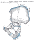
The crest of the ilium (or iliac crest) is the superior border of the wing of ilium and the superiolateral margin of the greater pelvis.

Contents

Structure

The iliac crest stretches posteriorly from the anterior superior iliac spine (ASIS) to the posterior superior iliac spine (PSIS). Behind the ASIS, it divides into an outer and inner lip separated by the intermediate zone. The outer lip bulges laterally into the iliac tubercle. [1] Palpable in its entire length, the crest is convex superiorly but is sinuously curved, being concave inward in front, concave outward behind. [2]

It is thinner at the center than at the extremities.

Development

The iliac crest is derived from endochondral bone.

Function

To the external lip are attached the Tensor fasciae latae , Obliquus externus abdominis , and Latissimus dorsi , and along its whole length the fascia lata ; to the intermediate line, the Obliquus internus abdominis .

To the internal lip, the iliac fascia , the Transversus abdominis , Quadratus lumborum, Sacrospinalis, and Iliacus .

Clinical significance

A fracture of the left iliac wing IliacCrestFrac.png
A fracture of the left iliac wing

The iliac crest has a large amount of red bone marrow, and thus it is the site of bone marrow harvests (from both sides) to collect the stem cells used in bone marrow transplantation. The iliac crest is also considered the best donor site for bone grafting when a large quantity of bone is needed. For example, oral and maxillofacial surgeons will often use iliac crest bone to fill in large osseous defects of the oral cavity caused by severe periodontal disease, excess bone resorption following tooth loss, trauma, or congenital defects including alveolar clefts. [4]

The top of the iliac crests also marks the level of the fourth lumbar vertebral body (L4), above or below which lumbar puncture may be performed. Furthermore, said level is often referred to as the "intercristal line".

Additional images

See also

Related Research Articles

<span class="mw-page-title-main">Latissimus dorsi muscle</span> Large, flat back muscle

The latissimus dorsi is a large, flat muscle on the back that stretches to the sides, behind the arm, and is partly covered by the trapezius on the back near the midline. The word latissimus dorsi comes from Latin and means "broadest [muscle] of the back", from "latissimus" and "dorsum". The pair of muscles are commonly known as "lats", especially among bodybuilders. The latissimus dorsi is the largest muscle in the upper body.

<span class="mw-page-title-main">Aponeurosis</span> Tissue which connects muscles to other organs

An aponeurosis is a flattened tendon by which muscle attaches to bone or fascia. Aponeuroses exhibit an ordered arrangement of collagen fibres, thus attaining high tensile strength in a particular direction while being vulnerable to tensional or shear forces in other directions. They have a shiny, whitish-silvery color, are histologically similar to tendons, and are very sparingly supplied with blood vessels and nerves. When dissected, aponeuroses are papery and peel off by sections. The primary regions with thick aponeuroses are in the ventral abdominal region, the dorsal lumbar region, the ventriculus in birds, and the palmar (palms) and plantar (soles) regions.

<span class="mw-page-title-main">Quadratus lumborum muscle</span> Muscle in the lower back

The quadratus lumborum muscle, informally called the QL, is a paired muscle of the left and right posterior abdominal wall. It is the deepest abdominal muscle, and commonly referred to as a back muscle. Each is irregular and quadrilateral in shape.

<span class="mw-page-title-main">Tensor fasciae latae muscle</span> Muscle of the thigh

The tensor fasciae latae is a muscle of the thigh. Together with the gluteus maximus, it acts on the iliotibial band and is continuous with the iliotibial tract, which attaches to the tibia. The muscle assists in keeping the balance of the pelvis while standing, walking, or running.

<span class="mw-page-title-main">Muscles of the hip</span> Causes movement in the hip

In human anatomy, the muscles of the hip joint are those muscles that cause movement in the hip. Most modern anatomists define 17 of these muscles, although some additional muscles may sometimes be considered. These are often divided into four groups according to their orientation around the hip joint: the gluteal group; the lateral rotator group; the adductor group; and the iliopsoas group.

<span class="mw-page-title-main">Lumbar plexus</span> Web of nerves in the lower spine

The lumbar plexus is a web of nerves in the lumbar region of the body which forms part of the larger lumbosacral plexus. It is formed by the divisions of the first four lumbar nerves (L1-L4) and from contributions of the subcostal nerve (T12), which is the last thoracic nerve. Additionally, the ventral rami of the fourth lumbar nerve pass communicating branches, the lumbosacral trunk, to the sacral plexus. The nerves of the lumbar plexus pass in front of the hip joint and mainly support the anterior part of the thigh.

<span class="mw-page-title-main">Iliohypogastric nerve</span>

The iliohypogastric nerve is a nerve that originates from the lumbar plexus that supplies sensation to skin over the lateral gluteal and hypogastric regions and motor to the internal oblique muscles and transverse abdominal muscles.

<span class="mw-page-title-main">Ilioinguinal nerve</span> Branch of the first lumbar nerve

The ilioinguinal nerve is a branch of the first lumbar nerve (L1). It separates from the first lumbar nerve along with the larger iliohypogastric nerve. It emerges from the lateral border of the psoas major just inferior to the iliohypogastric, and passes obliquely across the quadratus lumborum and iliacus. The ilioinguinal nerve then perforates the transversus abdominis near the anterior part of the iliac crest, and communicates with the iliohypogastric nerve between the transversus and the internal oblique muscle.

<span class="mw-page-title-main">Thoracolumbar fascia</span> Anatomical Feature

The thoracolumbar fascia is a complex, multilayer arrangement of fascial and aponeurotic layers forming a separation between the paraspinal muscles on one side, and the muscles of the posterior abdominal wall on the other. It spans the length of the back, extending between the neck superiorly and the sacrum inferiorly. It entails the fasciae and aponeuroses of the latissimus dorsi muscle, serratus posterior inferior muscle, abdominal internal oblique muscle, and transverse abdominal muscle.

<span class="mw-page-title-main">Lumbar arteries</span> Arteries within the lower back

The lumbar arteries are arteries located in the lower back or lumbar region. The lumbar arteries are in parallel with the intercostals.

<span class="mw-page-title-main">Wing of ilium</span> Flat portion of the hip bone

The wing(ala)of ilium is the large expanded portion of the ilium, the bone which bounds the greater pelvis laterally. It presents for examination two surfaces—an external and an internal—a crest, and two borders—an anterior and a posterior.

<span class="mw-page-title-main">Iliac fascia</span> Fascia of the pelvis

The iliac fascia is the fascia overlying the iliacus muscle.

<span class="mw-page-title-main">Iliolumbar ligament</span>

The iliolumbar ligament is a strong ligament which attaches medially to the transverse process of the 5th lumbar vertebra, and laterally to back of the inner lip of the iliac crest.

<span class="mw-page-title-main">Lumbar triangle</span>

The lumbar triangle can refer to either the inferior lumbar (Petit) triangle, which lies superficially, or the superior lumbar (Grynfeltt) triangle, which is deep and superior to the inferior triangle. Of the two, the superior triangle is the more consistently found in cadavers and is more commonly the site of herniation; however, the inferior lumbar triangle is often simply called the lumbar triangle, perhaps owing to its more superficial location and ease in demonstration.

<span class="mw-page-title-main">Deep circumflex iliac artery</span> Artery in the pelvis

The deep circumflex iliac artery is an artery in the pelvis that travels along the iliac crest of the pelvic bone.

The lumbar fascia is the lumbar portion of the thoracolumbar fascia. It consists of three fascial layers - posterior, middle, and anterior - that enclose two muscular compartments. The anterior and middle layers occur only in the lumbar region, whereas the posterior layer extends superiorly to the inferior part of the neck, and the inferiorly to the dorsal surface of the sacrum. The quadratus lumborum is contained in the anterior muscular compartment, and the erector spinae in the posterior compartment. Psoas major lies anterior to the anterior layer. Various superficial muscles of the posterior thorax and abdomen arise from the posterior layer - namely the latissimus dorsi, and serratus posterior inferior.

<span class="mw-page-title-main">Outline of human anatomy</span> Overview of and topical guide to human anatomy

The following outline is provided as an overview of and topical guide to human anatomy:

<span class="mw-page-title-main">Hip bone</span> Bone of the pelvis

The hip bone is a large flat bone, constricted in the center and expanded above and below. In some vertebrates it is composed of three parts: the ilium, ischium, and the pubis.

<span class="mw-page-title-main">Pelvis</span> Lower torso of the human body

The pelvis is the lower part of the trunk, between the abdomen and the thighs, together with its embedded skeleton.

References

PD-icon.svgThis article incorporates text in the public domain from page 234 of the 20th edition of Gray's Anatomy (1918)

  1. Platzer (2004), p 186
  2. Palastanga (2006), p 243
  3. Bogduk (2005), p 106
  4. Rose, Louis F.. Periodontics: Medicine, Surgery and Implants. Mosby, 072004. 26.2.2.3