Abdominal internal oblique muscle

Last updated
Abdominal internal oblique muscle
Gray395.png
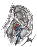
The abdominal internal oblique muscle.
Illu trunk muscles.jpg
Muscles of the trunk.
Details
Origin Inguinal ligament, iliac crest and the lumbodorsal fascia
Insertion Linea alba, pectineal line of pubis (via conjoint tendon) and ribs 10-12.
Artery Subcostal arteries
Nerve Thoracoabdominal nn. (T7-T11), subcostal n. (T12), iliohypogastric n. (L1) and ilioinguinal n. (L1)
Actions Bilateral: Compresses abdomen Unilateral: Ipsilateral trunk rotation
Identifiers
Latin musculus obliquus internus abdominis
TA98 A04.5.01.017
TA2 2373
FMA 13891
Anatomical terms of muscle

The abdominal internal oblique muscle, also internal oblique muscle or interior oblique, is an abdominal muscle in the abdominal wall that lies below the external oblique muscle and just above the transverse abdominal muscle.

Contents

Structure

Its fibers run perpendicular to the external oblique muscle, beginning in the thoracolumbar fascia of the lower back, the anterior 2/3 of the iliac crest (upper part of hip bone) and the lateral half of the inguinal ligament. The muscle fibers run from these points superomedially (up and towards midline) to the muscle's insertions on the inferior borders of the 10th through 12th ribs and the linea alba.

In males, the cremaster muscle is also attached to the internal oblique.

Nerve supply

The internal oblique is supplied by the lower intercostal nerves, as well as the iliohypogastric nerve and the ilioinguinal nerve.

Function

The internal oblique performs two major functions. Firstly as an accessory muscle of respiration, it acts as an antagonist (opponent) to the diaphragm, helping to reduce the volume of the chest cavity during exhalation. When the diaphragm contracts, it pulls the lower wall of the chest cavity down, increasing the volume of the lungs which then fill with air. Conversely, when the internal obliques contract they compress the organs of the abdomen, pushing them up into the diaphragm which intrudes back into the chest cavity reducing the volume of the air-filled lungs, producing an exhalation.

Secondly, its contraction causes ipsilateral rotation and side-bending. It acts with the external oblique muscle of the opposite side to achieve this torsional movement of the trunk. For example, the right internal oblique and the left external oblique contract as the torso flexes and rotates to bring the left shoulder towards the right hip. For this reason, the internal obliques are referred to as "same-side rotators."

Additional images

See also

Related Research Articles

<span class="mw-page-title-main">Levator ani</span> Broad, thin muscle group, situated on either side of the pelvis

The levator ani is a broad, thin muscle group, situated on either side of the pelvis. It is formed from three muscle components: the pubococcygeus, the iliococcygeus, and the puborectalis.

<span class="mw-page-title-main">Phrenic nerve</span> Nerve controlling the diaphragm

The phrenic nerve is a mixed motor/sensory nerve that originates from the C3-C5 spinal nerves in the neck. The nerve is important for breathing because it provides exclusive motor control of the diaphragm, the primary muscle of respiration. In humans, the right and left phrenic nerves are primarily supplied by the C4 spinal nerve, but there is also a contribution from the C3 and C5 spinal nerves. From its origin in the neck, the nerve travels downward into the chest to pass between the heart and lungs towards the diaphragm.

<span class="mw-page-title-main">Thoracic diaphragm</span> Sheet of internal skeletal muscle

The thoracic diaphragm, or simply the diaphragm, is a sheet of internal skeletal muscle in humans and other mammals that extends across the bottom of the thoracic cavity. The diaphragm is the most important muscle of respiration, and separates the thoracic cavity, containing the heart and lungs, from the abdominal cavity: as the diaphragm contracts, the volume of the thoracic cavity increases, creating a negative pressure there, which draws air into the lungs. Its high oxygen consumption is noted by the many mitochondria and capillaries present; more than in any other skeletal muscle.

<span class="mw-page-title-main">Inguinal canal</span> Human abdominal anatomy

The inguinal canal is a passage in the anterior abdominal wall on each side of the body, which in males, convey the spermatic cords and in females, the round ligament of the uterus. The inguinal canals are larger and more prominent in males.

<span class="mw-page-title-main">Intercostal muscles</span> Muscle groups between the ribs which form and move the chest wall during breathing

The intercostal muscles comprise many different groups of muscles that run between the ribs, and help form and move the chest wall. The intercostal muscles are mainly involved in the mechanical aspect of breathing by helping expand and shrink the size of the chest cavity.

<span class="mw-page-title-main">Rectus abdominis muscle</span> Paired straight muscle

The rectus abdominis muscle, also known as the "abdominal muscle" or simply the "abs", is a pair of segmented skeletal muscle on the ventral aspect of a person's abdomen. The paired muscle is separated at the midline by a band of dense connective tissue called the linea alba, and the connective tissue defining each lateral margin of the rectus abdominus is the linea semilunaris. The muscle extends from the pubic symphysis, pubic crest and pubic tubercle inferiorly, to the xiphoid process and costal cartilages of the 5th–7th ribs superiorly.

<span class="mw-page-title-main">Inguinal ligament</span> Band running from the pubic tubercle to the anterior superior iliac spine

The inguinal ligament, also known as Poupart's ligament or groin ligament, is a band running from the pubic tubercle to the anterior superior iliac spine. It forms the base of the inguinal canal through which an indirect inguinal hernia may develop.

<span class="mw-page-title-main">Serratus anterior muscle</span> Muscle on the surface of the ribs

The serratus anterior is a muscle of the chest. It originates at the side of the chest from the upper 8 or 9 ribs; it inserts along the entire length of the anterior aspect of the medial border of the scapula. It is innervated by the long thoracic nerve from the brachial plexus. The serratus anterior acts to pull the scapula forward around the thorax.

<span class="mw-page-title-main">Transverse abdominal muscle</span> Muscle of the abdominal area

The transverse abdominal muscle (TVA), also known as the transverse abdominis, transversalis muscle and transversus abdominis muscle, is a muscle layer of the anterior and lateral abdominal wall, deep to the internal oblique muscle. It is thought by most fitness instructors to be a significant component of the core.

<span class="mw-page-title-main">Abdominal external oblique muscle</span> Skeletal muscle in the abdomen

The abdominal external oblique muscle is the largest and outermost of the three flat abdominal muscles of the lateral anterior abdomen.

<span class="mw-page-title-main">Abdomen</span> Part of the body between the chest and pelvis

The abdomen is the part of the body between the thorax (chest) and pelvis, in humans and in other vertebrates. The abdomen is the front part of the abdominal segment of the torso. The area occupied by the abdomen is called the abdominal cavity. In arthropods, it is the posterior tagma of the body; it follows the thorax or cephalothorax.

<span class="mw-page-title-main">Inferior epigastric artery</span> Blood vessel

In human anatomy, the inferior epigastric artery is an artery that arises from the external iliac artery. It is accompanied by the inferior epigastric vein; inferiorly, these two inferior epigastric vessels together travel within the lateral umbilical fold The inferior epigastric artery then traverses the arcuate line of rectus sheath to enter the rectus sheath, then anastomoses with the superior epigastric artery within the rectus sheath.

<span class="mw-page-title-main">Iliohypogastric nerve</span>

The iliohypogastric nerve is a nerve that originates from the lumbar plexus that supplies sensation to skin over the lateral gluteal and hypogastric regions and motor to the internal oblique muscles and transverse abdominal muscles.

<span class="mw-page-title-main">Transversalis fascia</span> Aponeurosis between the transverse abdominal muscle and the extraperitoneal fat

The transversalis fascia is the fascial lining of the anterolateral abdominal wall situated between the inner surface of the transverse abdominal muscle, and the preperitoneal fascia. It is directly continuous with the iliac fascia, the internal spermatic fascia, and pelvic fascia.

<span class="mw-page-title-main">Arcuate line of rectus sheath</span> Line of demarcation in the human abdomen

The arcuate line of rectus sheath is a line of demarcation corresponding to the free inferior margin of the posterior layer of the rectus sheath inferior to which only the anterior layer of the rectus sheath is present and the rectus abdominis muscle is therefore in direct contact with the transversalis fascia. The arcuate line is concave inferior-wards.

<span class="mw-page-title-main">Rectus sheath</span> Laminas around abdominal muscles

The rectus sheath is a tough fibrous compartment formed by the aponeuroses of the transverse abdominal muscle, and the internal and external oblique muscles. It contains the rectus abdominis and pyramidalis muscles, as well as vessels and nerves.

<span class="mw-page-title-main">Aponeurosis of the abdominal external oblique muscle</span> Membranous structure

The aponeurosis of the abdominal external oblique muscle is a thin but strong membranous structure, the fibers of which are directed downward and medially.

<span class="mw-page-title-main">Muscles of respiration</span> Muscles involved in breathing

The muscles of respiration are the muscles that contribute to inhalation and exhalation, by aiding in the expansion and contraction of the thoracic cavity. The diaphragm and, to a lesser extent, the intercostal muscles drive respiration during quiet breathing. The elasticity of these muscles is crucial to the health of the respiratory system and to maximize its functional capabilities.

<span class="mw-page-title-main">Outline of human anatomy</span> Overview of and topical guide to human anatomy

The following outline is provided as an overview of and topical guide to human anatomy:

<span class="mw-page-title-main">Vaginal support structures</span> Structures that maintain the position of the vagina within the pelvic cavity

The vaginal support structures are those muscles, bones, ligaments, tendons, membranes and fascia, of the pelvic floor that maintain the position of the vagina within the pelvic cavity and allow the normal functioning of the vagina and other reproductive structures in the female. Defects or injuries to these support structures in the pelvic floor leads to pelvic organ prolapse. Anatomical and congenital variations of vaginal support structures can predispose a woman to further dysfunction and prolapse later in life. The urethra is part of the anterior wall of the vagina and damage to the support structures there can lead to incontinence and urinary retention.

References