Transverse abdominal muscle

Last updated
Transverse abdominal muscle
Illu trunk muscles.jpg
Muscles of the trunk.
Transversus abdominis.png
Details
Origin Iliac crest, inguinal ligament, thoracolumbar fascia, and costal cartilages 7-12
Insertion Xiphoid process, linea alba, pubic crest and pecten pubis via conjoint tendon
Artery subcostal arteries.
Nerve Thoracoabdominal nn. (T6-T11), Subcostal n. (T12), iliohypogastric nerve (L1), and ilioinguinal nerve (L1).
Actions Compresses abdominal contents
Identifiers
Latin musculus transversus abdominis
TA98 A04.5.01.019
TA2 2375
FMA 15570
Anatomical terms of muscle

The transverse abdominal muscle (TVA), also known as the transverse abdominis, transversalis muscle and transversus abdominis muscle, is a muscle layer of the anterior and lateral (front and side) abdominal wall , deep to (layered below) the internal oblique muscle. It is thought by most fitness instructors to be a significant component of the core.

Contents

Structure

The transverse abdominal, so called for the direction of its fibers, is the innermost of the flat muscles of the abdomen. It is positioned immediately deep to the internal oblique muscle.

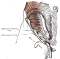
The transverse abdominal arises as fleshy fibers, from the lateral third of the inguinal ligament, from the anterior three-fourths of the inner lip of the iliac crest, from the inner surfaces of the cartilages of the lower six ribs, interdigitating with the diaphragm, and from the thoracolumbar fascia. It ends anteriorly in a broad aponeurosis (the Spigelian fascia), the lower fibers of which curve inferomedially (medially and downward), and are inserted, together with those of the internal oblique muscle, into the crest of the pubis and pectineal line, forming the inguinal conjoint tendon also called the aponeurotic falx. In layman's terms, the muscle ends in the middle line of a person's abdomen. [1] :248–250

Throughout the rest of its extent the aponeurosis passes horizontally to the middle line, and is inserted into the linea alba; its upper three-fourths lie behind the rectus muscle and blend with the posterior lamella of the aponeurosis of the internal oblique; its lower fourth is in front of the rectus abdominis.

Innervation

The transverse abdominal is innervated by the lower intercostal nerves (thoracoabdominal, nerve roots T7-T11), as well as the iliohypogastric nerve and the ilioinguinal nerve.

Function

The transverse abdominal helps to compress the ribs and viscera, providing thoracic and pelvic stability. This is explained further here. The transverse abdominal also helps a pregnant woman to deliver her child.

Without a stable spine, one aided by proper contraction of the TVA, the nervous system fails to recruit the muscles in the extremities efficiently, and functional movements cannot be properly performed. [2] The transverse abdominal and the segmental stabilizers (e.g. the multifidi) of the spine have evolved to work in tandem.

It is true that the TVA is vital to back and core health, the muscle also has the effect of pulling in what would otherwise be a protruding abdomen (hence its nickname, the “corset muscle”). Training the rectus abdominis muscles alone will not and can not give one a "flat" belly; this effect is achieved only through training the TVA. [3] Thus, to the extent that traditional abdominal exercises (e.g. crunches) or more advanced abdominal exercises tend to "flatten" the belly, this is owed to the tangential training of the TVA inherent in such exercises. Recently, the transverse abdominal has become the subject of debate between biokineticists, kinesiologists, strength trainers, and physical therapists. The two positions on the muscle are (1) that the muscle is effective and capable of bracing the human core during extremely heavy lifts and (2) that it is not. Specifically, one recent systematic review has found that the baseline dysfunction of TVA cannot predict the clinical outcomes of low back pain. [4] Similarly, another systematic review has revealed that the changes in TVA function or morphology after different nonsurgical treatments are unrelated to the improvement of pain intensity or low back pain related-disability. [5] These findings have challenged the traditional emphasis of using TVA-targeted intervention to treat low back pain.

Exercise

The most well known method of strengthening the TVA is the vacuum exercise. The TVA also (involuntarily) contracts during many lifts; it is the body's natural weight-lifting belt, stabilizing the spine and pelvis during lifting movements. It has been estimated that the contraction of the TVA and other muscles reduces the vertical pressure on the intervertebral discs by as much as 40%. [6] Failure to engage the TVA during higher intensity lifts is dangerous and encourages injury to the spine[ citation needed ]. The TVA acts as a girdle or corset by creating hoop tension around the midsection.

Additional images

Related Research Articles

<span class="mw-page-title-main">Inguinal canal</span> Human abdominal anatomy

The inguinal canal is a passage in the anterior abdominal wall on each side of the body, which in males, convey the spermatic cords and in females, the round ligament of the uterus. The inguinal canals are larger and more prominent in males.

<span class="mw-page-title-main">Aponeurosis</span> Tissue which connects muscles to other organs

An aponeurosis is a flattened tendon by which muscle attaches to bone or fascia. Aponeuroses exhibit an ordered arrangement of collagen fibres, thus attaining high tensile strength in a particular direction while being vulnerable to tensional or shear forces in other directions. They have a shiny, whitish-silvery color, are histologically similar to tendons, and are very sparingly supplied with blood vessels and nerves. When dissected, aponeuroses are papery and peel off by sections. The primary regions with thick aponeuroses are in the ventral abdominal region, the dorsal lumbar region, the ventriculus in birds, and the palmar (palms) and plantar (soles) regions.

<span class="mw-page-title-main">Rectus abdominis muscle</span> Paired straight muscle

The rectus abdominis muscle, also known as the "abdominal muscle" or simply the "abs", is a pair of segmented skeletal muscle on the ventral aspect of a person's abdomen. The paired muscle is separated at the midline by a band of dense connective tissue called the linea alba, and the connective tissue defining each lateral margin of the rectus abdominus is the linea semilunaris. The muscle extends from the pubic symphysis, pubic crest and pubic tubercle inferiorly, to the xiphoid process and costal cartilages of the 5th–7th ribs superiorly.

<span class="mw-page-title-main">Inguinal ligament</span> Band running from the pubic tubercle to the anterior superior iliac spine

The inguinal ligament, also known as Poupart's ligament or groin ligament, is a band running from the pubic tubercle to the anterior superior iliac spine. It forms the base of the inguinal canal through which an indirect inguinal hernia may develop.

<span class="mw-page-title-main">Abdominal internal oblique muscle</span> Muscle in the abdominal wall

The abdominal internal oblique muscle, also internal oblique muscle or interior oblique, is an abdominal muscle in the abdominal wall that lies below the external oblique muscle and just above the transverse abdominal muscle.

<span class="mw-page-title-main">Abdominal external oblique muscle</span> Skeletal muscle in the abdomen

The abdominal external oblique muscle is the largest and outermost of the three flat abdominal muscles of the lateral anterior abdomen.

<span class="mw-page-title-main">Abdomen</span> Part of the body between the chest and pelvis

The abdomen is the part of the body between the thorax (chest) and pelvis, in humans and in other vertebrates. The abdomen is the front part of the abdominal segment of the torso. The area occupied by the abdomen is called the abdominal cavity. In arthropods, it is the posterior tagma of the body; it follows the thorax or cephalothorax.

<span class="mw-page-title-main">Inferior epigastric artery</span> Blood vessel

In human anatomy, the inferior epigastric artery is an artery that arises from the external iliac artery. It is accompanied by the inferior epigastric vein; inferiorly, these two inferior epigastric vessels together travel within the lateral umbilical fold The inferior epigastric artery then traverses the arcuate line of rectus sheath to enter the rectus sheath, then anastomoses with the superior epigastric artery within the rectus sheath.

<span class="mw-page-title-main">Abdominal wall</span> Boundary of abdominal cavity

In anatomy, the abdominal wall represents the boundaries of the abdominal cavity. The abdominal wall is split into the anterolateral and posterior walls.

<span class="mw-page-title-main">Iliohypogastric nerve</span>

The iliohypogastric nerve is a nerve that originates from the lumbar plexus that supplies sensation to skin over the lateral gluteal and hypogastric regions and motor to the internal oblique muscles and transverse abdominal muscles.

<span class="mw-page-title-main">Ilioinguinal nerve</span> Branch of the first lumbar nerve

The ilioinguinal nerve is a branch of the first lumbar nerve (L1). It separates from the first lumbar nerve along with the larger iliohypogastric nerve. It emerges from the lateral border of the psoas major just inferior to the iliohypogastric, and passes obliquely across the quadratus lumborum and iliacus. The ilioinguinal nerve then perforates the transversus abdominis near the anterior part of the iliac crest, and communicates with the iliohypogastric nerve between the transversus and the internal oblique muscle.

<span class="mw-page-title-main">Conjoint tendon</span> Medial part of the posterior wall of the inguinal canal

The conjoint tendon is a sheath of connective tissue formed from the lower part of the common aponeurosis of the abdominal internal oblique muscle and the transversus abdominis muscle, joining the muscle to the pelvis. It forms the medial part of the posterior wall of the inguinal canal.

<span class="mw-page-title-main">Transversalis fascia</span> Aponeurosis between the transverse abdominal muscle and the extraperitoneal fat

The transversalis fascia is the fascial lining of the anterolateral abdominal wall situated between the inner surface of the transverse abdominal muscle, and the preperitoneal fascia. It is directly continuous with the iliac fascia, the internal spermatic fascia, and pelvic fascia.

The subcostal arteries, so named because they lie below the last ribs, constitute the lowest pair of branches derived from the thoracic aorta, and are in series with the intercostal arteries.

<span class="mw-page-title-main">Arcuate line of rectus sheath</span>

The arcuate line of rectus sheath is a line of demarcation corresponding to the free inferior margin of the posterior layer of the rectus sheath inferior to which only the anterior layer of the rectus sheath is present and the rectus abdominis muscle is therefore in direct contact with the transversalis fascia. The arcuate line is concave inferior-wards.

<span class="mw-page-title-main">Deep circumflex iliac artery</span> Artery in the pelvis

The deep circumflex iliac artery is an artery in the pelvis that travels along the iliac crest of the pelvic bone.

<span class="mw-page-title-main">Linea semilunaris</span>

The linea semilunaris is a curved tendinous intersection found on either side of the rectus abdominis muscle.

<span class="mw-page-title-main">Rectus sheath</span> Laminas around abdominal muscles

The rectus sheath is a tough fibrous compartment formed by the aponeuroses of the transverse abdominal muscle, and the internal and external oblique muscles. It contains the rectus abdominis and pyramidalis muscles, as well as vessels and nerves.

<span class="mw-page-title-main">Thoraco-abdominal nerves</span>

The anterior divisions of the seventh, eighth, ninth, tenth, and eleventh thoracic intercostal nerves are continued anteriorly from the intercostal spaces into the abdominal wall; hence they are named thoraco-abdominal nerves.

<span class="mw-page-title-main">Aponeurosis of the abdominal external oblique muscle</span>

The aponeurosis of the abdominal external oblique muscle is a thin but strong membranous structure, the fibers of which are directed downward and medially.

References

PD-icon.svgThis article incorporates text in the public domain from page 414 of the 20th edition of Gray's Anatomy (1918)

  1. Drake, Richard L.; Vogl, Wayne; Tibbitts, Adam W.M. Mitchell; illustrations by Richard; Richardson, Paul (2005). Gray's anatomy for students (Pbk. ed.). Philadelphia: Elsevier/Churchill Livingstone. ISBN   978-0-443-06612-2.
  2. Aruin, Alexanders; Latash, Markl (1995). "Directional specificity of postural muscles in feed-forward postural reactions during fast voluntary arm movements". Experimental Brain Research. 103 (2): 323–32. doi:10.1007/BF00231718. PMID   7789439. S2CID   24706658.
  3. Dart, R.A. (March 1947). "The Double-Spiral Arrangement Of The Voluntary Musculature In The Human Body". Surgeons' Hall Journal. 10 (2).
  4. Wong, Arnold Y.L; Parent, Eric C; Funabashi, Martha; Stanton, Tasha R; Kawchuk, Gregory N (2013). "Do various baseline characteristics of transversus abdominis and lumbar multifidus predict clinical outcomes in nonspecific low back pain? A systematic review". Pain. 154 (12): 2589–602. doi:10.1016/j.pain.2013.07.010. PMID   23867731. S2CID   26095069.
  5. Wong, Arnold Y.L; Parent, Eric C; Funabashi, Martha; Kawchuk, Gregory N (2014). "Do Changes in Transversus Abdominis and Lumbar Multifidus During Conservative Treatment Explain Changes in Clinical Outcomes Related to Nonspecific Low Back Pain? A Systematic Review". The Journal of Pain. 15 (4): 377.e1–35. doi: 10.1016/j.jpain.2013.10.008 . PMID   24184573.
  6. Hodges, Paul W; Richardson, Carolyn A (1997). "Contraction of the Abdominal Muscles Associated with Movement of the Lower Limb". Physical Therapy. 77 (2): 132–42, discussion 142–4. doi: 10.1093/ptj/77.2.132 . PMID   9037214.