Iliacus muscle

Last updated
Iliacus muscle
Iliacus muscle06.png
Position of iliacus muscle (shown in red)
Anterior Hip Muscles 2.PNG
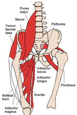
The iliacus and nearby muscles
Details
Pronunciation /ɪˈl.əkəs/
Origin Upper two-thirds of the iliac fossa
Insertion Base of the lesser trochanter of femur
Artery Medial femoral circumflex artery, iliac branch of iliolumbar artery
Nerve Femoral nerve
Actions Flexes and rotates medially thigh [ citation needed ]
Antagonist Gluteus maximus
Identifiers
Latin musculus iliacus
TA98 A04.7.02.003
TA2 2594
FMA 22310
Anatomical terms of muscle

The iliacus is a flat, triangular muscle which fills the iliac fossa. It forms the lateral portion of iliopsoas, providing flexion of the thigh and lower limb at the acetabulofemoral joint.

Contents

Structure

The iliacus arises from the iliac fossa on the interior side of the hip bone, and also from the region of the anterior inferior iliac spine (AIIS). It joins the psoas major to form the iliopsoas. [1] It proceeds across the iliopubic eminence through the muscular lacuna to its insertion on the lesser trochanter of the femur. [1] Its fibers are often inserted in front of those of the psoas major and extend distally over the lesser trochanter. [2]

Nerve supply

The iliopsoas is innervated by the femoral nerve and direct branches from the lumbar plexus. [3]

Function

In open-chain exercises, as part of the iliopsoas, the iliacus is important for lifting (flexing) the femur forward (e.g. front scale). In closed-chain exercises, the iliopsoas bends the trunk forward and can lift the trunk from a lying posture (e.g. sit-ups, back scale) because the psoas major crosses several vertebral joints and the sacroiliac joint. From its origin in the lesser pelvis the iliacus acts exclusively on the hip joint. [2]

Additional images

Notes

  1. 1 2 Davenport, Kathleen L. (2019-01-01), Elson, Lauren E. (ed.), "Chapter 9 - The Professional Dancer's Hip", Performing Arts Medicine, Philadelphia: Elsevier, pp. 77–87, ISBN   978-0-323-58182-0 , retrieved 2021-01-17
  2. 1 2 Platzer (2004), p 234
  3. Thieme Atlas of Anatomy (2006), p 422

References